34 | P a g e
SOME STUDIES ON INVESTIGATING THE CONCEPT
OF REPRESENTATION & FABRICATION OF
ZINC-OXIDE NANOPARTICLES OF VARIOUS SIZES AND
ANALYSIS OF SAMPLE ARE DONE BY SCANNING
ELECTRON MICROSCOPE & X-RAY DIFFRACTION
Shubham Sharma
1, Dr. Paramjit Singh Bedi
2, Shalab Sharma
31
P.G. Research Scholar, Mechanical Engineering, D.A.V University (India)
2P.H.D Research Scholar, Chemistry Department, C.T Group of institutions, (India)
3U.G. Research Scholar, Mechanical Engineering, C.T Institute of Technology, (India)
ABSTRACT
This study presents the analyses of various different methods of fabrication of Zinc Nanoparticles along with the representation of Phase composition and structure properties. Using soluble starch, Zinc Nanoparticles were fabricated from Zinc Nitrate solution. Further growth & formation of these nanoparticles are only possible by using Precipitation method. Further XRD & SEM quantitative analyses & Non-Destructive testing techniques are used to study various Morphology properties such as size, shape, phase composition, structure of the sample of this white fabricated Zinc Nanoparticles. SEM & XRD analyses are made possible to ensure uniformly dispersion & suspended stability of this formed Zinc Nanoparticles of an average particle size (APS) ranges from 30-45 nm. Further results revealed that in order to enhance various thermo-physical properties & Heat transfer characteristics of Nanoparticles, we need to alter size of paticles rather than their composition. Also result shows that with increase in the size of Nanoparticles, growth of micro-organisms decreases or restricted and vice-versa.
Keywords:
Morphology, Micro-organisms, SEM, Precipitation, XRD.
Abbreviations: XRD (X-ray Diffraction), SEM (Scanning electron microscope).
I INTRODUCTION
Zinc oxide is an organic compound that exhibits semiconducting [5], piezoelectric & pyroelectric properties. Zinc
oxide occurs rarely in nature & most available in crystalline form. Zinc oxide are usually white at room temperature
but when heated it changes it’s colour from white to yellow colour which further leads to change in energy level
properties of materials. Although zinc oxide have ability to form broad diverse structures whose position as well as
orientation are much more abundant as compared to other Nanoparticles & Nanomaterials. This synthetically
35 | P a g e
This Zinc oxide nanoparticles have wide range of an applications because all nanoparticles as well as nanomaterials
properties are mostly dependent upon Morphology & Nano-dimensions. Although it has been observed that by
reducing the dimension of Zinc Oxide structures to nanoscale ranges photonic performance, photon to electron
conversion efficiency & gas sensing ability increases. This enhancement occurs due to increase in surface area as
well as Quantum confinement affect where electrons are confined in small space rather than space of bulk material.
Zinc oxide generally occurs as rare oxide mineral of Hexagonal crystal structure, named Zincite. This Zinc oxide
material is an inorganic compound that can react both acid as well as base, depend upon its oxidation state. This
Zinc oxide material attains Cubic & Hexagonal crystal lattice structure [1] in which Hexagonal structure is most
stable & commonly used even at an ambient conditions. But in case of Zinc oxide cubic lattice structure,
stabilization is the main issue. This issue is resolved by forming Zinc oxide particles & then put this Zinc oxide
material on the bulk of semiconductor material i.e, substrate where growth & development of Zinc oxide particles
takes place with Cubic Lattice structure. Usually chemical & physical properties of any nanomaterial can be
determined by using behaviour of electrons in material & atoms arrangement, which further decided by Electronic
Band structure, i.e, by using Energy band diagrams. In this case, Zinc oxide material is a n-type semiconductor
material where forbidden gap is very small & ranges of energy i.e, Band Gap are extremely wide.
II PROPERTIES OF ZINC OXIDE NANOMATERIALS
2.1 Physical Properties
Chemical Name - ZnO Nanopowder
Appearance - White Dusty powder
Morphology - Hexagonal crystal lattice structure
Purity - >99.84%
Average particles size - (30 to 45 nm)
Density - 5.658 g/cm3
Thermal conductivity - 50 W/mK
Crystal Lattice structure - Hexagonal (Stable), Cubic (Unstable)
Melting Point temperature - 1970°C
Dielectric constant - 8.64
Molecular weight - 81.379 g/mol [2]
2.2 Optical Properties
1. Presence of oxide has made an adverse effect on photoresponse during photoconductivity measurement of Zinc
36 | P a g e
2. Zinc oxide nanoparticles improves spectral shift of an optical region and this zinc oxide nanostructures are used as
building blocks for integrated optoelectronic circuits [4].
2.3 Chemical Properties
1. Zinc oxide exhibits solid inorganic chemical compound property in which some small percentage of atoms are
missed or too many atoms are packed into crystal lattice structure.
2. Zinc oxide exhibits the property of reacting with both acids as well as bases. This reaction ensures separation of
different cations, depend upon it’s oxidation state.
3. Zinc oxide when blends with an Aqueous solution forms cement like product.
4. Zinc oxide produces salt of carboxylic acid by reacting slowly with fatty acids in the presence of oil [2].
2.4 Mechanical Properties
1. Zinc oxide has excellent thermal conductivity, specific heat, overall heat capacity properties.
2. Zinc oxide has hardness of 4.5 & has low value of coefficient of thermal expansion.
3. Zinc oxide with n-type semiconductor material have low value of an elastic constants that are Poisson’s ratio,
Young’s modulus, Bulk modulus & Modulus of rigidity as compared to crystalline semiconductor material with
high degree of stoichometry.
4. Zinc oxide crystalline structure exhibits the property of Piezoelectricity i.e, movement of electrons resulting from
an applied mechanical stress or pressure.
2.5 Electrical Properties
1. Ranges for energy in electronic band structure for Zinc oxide at room temperature is about 3.2 electron volt (eV).
2. Movement of electrons through Zinc oxide resulting by an applied electric field strongly depends upon
temperature & at 80K attains maximum value of 1970-2000 cm2/(V.S.).
3. Zinc oxide exhibits property to sustain large electric fields.
4. Random fluctuation of an electrical signal in Zinc oxide is less [3].
III LITERATURE REVIEW
1. Kakaç et al. reviewed that with addition of nanoparticles heat transfer characteristics of conventional heat
transfer fluids such as ethylene glycol, water and oil increases. They highlight the necessity of heat transfer
fundamentals for advancement in nanotechnology in broader area. Experimental and theoretical analysis of
particle mechanism at microscopic level is crucial.
2. Hong et al. performed experiments on Fe/ethylene glycol to investigate the effect of clustering of nanoparticles.
Results revealed that thermal conductivity of nanoparticles increases as time of sonication increases. At high
37 | P a g e
3. Wang et al. performed experiments by preparing Zinc oxide nanoparticles. Reagents used for fabricating zinc
oxide nanoparticles includes an aqueous solution of zinc sulphate and ammonium bicarbonate. Surface active
element used in order to reduce agglomeration of nanoparticles is CTAB. Results revealed that antimicrobial
activity increases with decrease in size of particles.
4. Cao et al. performed experiments by preparing or fabricating Cellulose hydrolysis using aqueous zinc chloride
solution and then adding sodium hydroxide. Results revealed that this aqueous zinc chloride solution acts as
swelling agent for cellulose.
5. Nafchi et al. performed experiments by preparing zinc oxide nanorod incorporated with soluble starch and further
characterization is done for bionanocomposite films. Results revealed that dielectric property, antibacterial activity
& UV absorbance increases for zinc oxide nanoparticles.
IV FABRICATION OF ZINC OXIDE NANOPARTICLES
Commonly used reagents used for fabricating the zinc oxide nanoparticles were purchased from Intelligent solutions
private limited, Panchkula. Experimental procedure for the synthesis of Zinc oxide nanoparticles includes Zinc
nitrate which acts as a precursors & starch as a stabilizing agent. Zinc oxide nanoparticles were fabricated from zinc
nitrate solution & further acid-base precipitation is done for the growth and development of zinc oxide
nanoparticles. Reagents used for fabricating zinc oxide nanoparticles are zinc nitrate solution, zinc acetate dihydrate
chemical & an alkaline aqueous solution of sodium hydroxide in the presence of de-ionised water. Although
surfactants & organic compounds are used to control growth and development of this newly formed precipitated
particles but one adverse affect is observed with the use of this organic compound is that colloids came out of
suspension in the form of flakes & also these small particles merge together to form large particles in solution &
thus ultimately affects supersaturation of the growth species and nuclei. Above mentioned limitation is resolved with
addition of macromolecules. Further operating parameters such as temperature, pH value are varied to control the
precipitation process.
V EXPERIMENTAL PROCEDURE FOR SYNTHESIS OF ZINC OXIDE NANOPARTICLES
1. Firstly take 0.13M bulk of zinc acetate solution which is prepared by adding 50 ml methanol & stirring is done by
magnetic stirrer or mixer that employs rotating magnetic field to cause stir bar immersed in liquid to spin very
quickly.
2. Now add 26 ml of 0.35M sodium hydroxide solution which was dispersed in methanol under continous stirring.
This sodium hydroxide solution was added in order to determine basic pH value of reactants. This pH test was
carried out with pH tester.
3. Now this dispersed solution was put onto stainless steel pressure vessel apparatus called “Autoclave”. This
autoclave was used because they have ability to withstand high pressure (100 Bar) & temperature (100°C-300°C) for
38 | P a g e
4. Maintain temperature gradient at the opposite ends of chamber which results in hotter end dissolves the species of
zinc oxide nanoparticles & cooler end causes further growth of zinc oxide nanoparticles species at room
temperature.
5. Now aqueous solution is transported to the upper part of an autoclave due to convective motion of solution.
6. Finally crystallization process takes place when solution became supersaturated i.e, when temperature of an
equilibrium mixture reduces.
7. Now wash out this white dusty solid nanoparticles or products with ethanol in order to remove excessive starch &
other By-products or slag impurities.
8. Now particles are dried in air in laboratory oven at 45°C-65°C.
9. Finally Characterization or representation of Zinc oxide nanoparticles is done by using SEM & XRD
non-destructive testing technique to analyses morphology & structural properties.
Figure 1: Fabrication of Zinc oxide Nanoparticles
VI CHARACTERIZATION OR REPRESENTATION OF ZINC OXIDE NANOPARTICLES
6.1 X-Ray Diffraction technique (XRD)
For quantitative analyses of crystal structure & to investigate bravais lattice crystal structure of zinc oxide
nanomaterials, diffraction technique using X-rays is carried out. XRD is one of the non-destructive testing technique
39 | P a g e
crystal lattice solid structure [1]. XRD technique was used to analyse phase distribution of sample of zinc oxide
nanoparticles. X-rays are diffracted by atoms in crystal lattice structure when X-rays have wavelength in order of
magnitude of few Angstroms & secondly distance between atoms in crystal structure is approximately same as that
of wavelength of X-rays wave. X-ray Diffractometer was used to measure X-rays diffraction patterns of Zinc oxide
nanoparticles sample which was recorded from 20° to 90° with PANalytical system Diffractometer model named
DY-1656 using more intense CuK alpha radiation & 40 KV accelerating voltage. XRD patterns data was collected at
1 degree/minute with respect to direction of incident beam, diffracted X-rays makes 2Ɵ angle at detector. An
accurate determination of crystal structure of zinc oxide nanoparticles are made by moving both detector & zinc
oxide nanoparticles sample around sample axis through centre. Thus the problem of Kdoublet radiation was sort out.
Now analyses of zinc oxide nanoparticles can be obtained by Debye Scherrer Diffractometer formulae. This
formulae was used to evaluate average particles size (A.P.S) less than 100 nm.
A.P.S = 0.9 λ / β cos θB where λ is wavelength,
β is full maxima half width caused by nanoparticles size.
θB is Bragg’s diffraction angle.
After analyses it has been observed that average particles size (A.P.S) of zinc oxide nanoparticles sample
crystallization structure is exactly same as size measured by using particle size analyzer (PSA). Also it has been
observed that 2-D image produced by Diffractometer is almost same. But one limitation was observed i.e, due to
presence of impurities in zinc oxide nanoparticles sample, there were some undesired picks or slag.
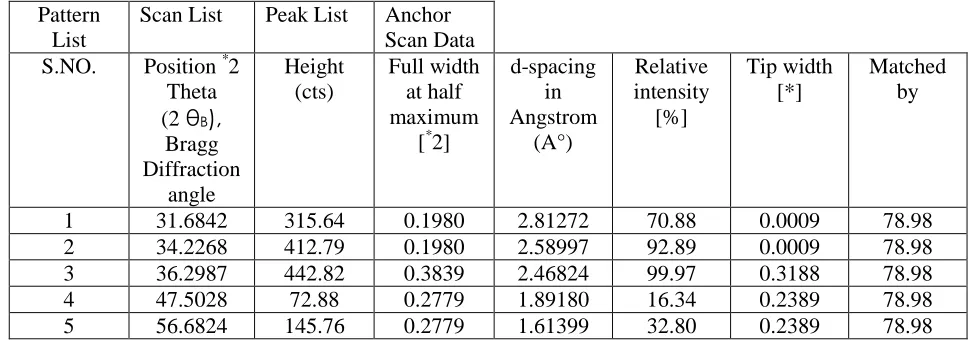
6.2 X-Rays Diffractometer analysis graphical characterization
40 | P a g e
Figure 3: X-rays Diffractometer peak list structure analyses representing curve for Zinc oxide
nanoparticles.
Pattern
List
Scan List
Peak List
Anchor
Scan Data
S.NO.
Position
*2
Theta
(2
θ
B),
Bragg
Diffraction
angle
Height
(cts)
Full width
at half
maximum
[
*2]
d-spacing
in
Angstrom
(A°)
Relative
intensity
[%]
Tip width
[*]
Matched
by
1
31.6842
315.64
0.1980
2.81272
70.88
0.0009
78.98
2
34.2268
412.79
0.1980
2.58997
92.89
0.0009
78.98
3
36.2987
442.82
0.3839
2.46824
99.97
0.3188
78.98
4
47.5028
72.88
0.2779
1.89180
16.34
0.2389
78.98
5
56.6824
145.76
0.2779
1.61399
32.80
0.2389
78.98
Table 1: X-ray Diffraction analyses representation for zinc oxide nanoparticles.
6.3 Scanning Electron Microscope
In SEM technique, electrons are used (which sometimes behave as a particles or sometimes as wave) of approximate
wavelength scattered from sample which further are detected by using electrostatic lenses. In order to produce high
resonance image this technique adopt or uses backscattered electrons which are reflevcted by elastic scattering. The
reason behind use of backscattered electrons is it’s signal intensity which relates atomic number of sample &
secondly element or phase distribution revealed by backscattered electrons is accurate.SEM is also one of the
41 | P a g e
image surface of thick sample. High energy electron beam are used to produce variety of signal from scan generator
alongwith amplified signal from electron collector at the surface of zinc oxide sample.
Usually image resolution upto 2-5 mm can be achieved at an accelerating voltage of 18-30 KeV. Thus SEM
technique is used for the analyses of crystalline structure, chemical composition, external appearance of specimen or
morphology properties, configuration i.e, position or orientation of nanomaterials, surface topography & electrical
conductivity. Energy dispersive analyses of X-rays technique (EDXA) is used to determine sample chemical
composition & also measures impurities level or presence of elements in the sample. High magnified resonance
image is obtained by reducing the specimen size only in the case of display screen of fixed size. Magnification or
high resonance image of specimen is controlled by:
a). Passing current only to scanning coils.
b). Passing voltage to x, y deflector plates.
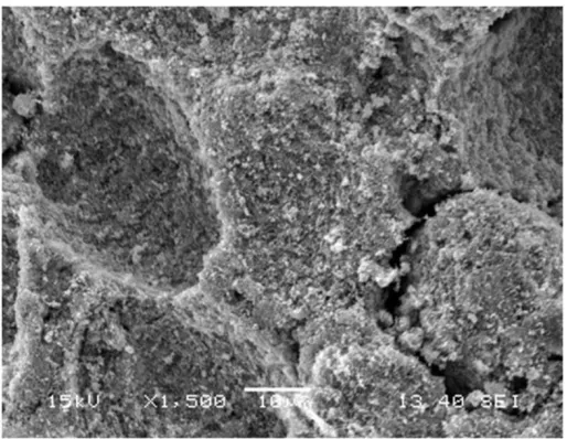
Figure 4: Analyses of SEM showing surface texture, chemical composition & other Morphology
properties of Zinc oxide nanoparticles.
6.4 Absorption spectrum analyses
Using absorption spectrum graphical representation, analyses is done which determines an energy gap as well as
absorption peak position. This energy band gap is determined by using formulae as written follows:
E = h c / λ
Where, h is Plancks constant,
C is speed of light
42 | P a g e
Excitation peak position determines energy gap & it is found out to be around 3.287 eV. Moreover this energy band
gap can also be used to determine an Average particles size (A.P.S) by using effective mass tight banding formulae
as written below:
E = Eg + h 2π2
{1/me+1/mh}/2R 2
- 1.8e2/RƐ
where E = Band gap of fabricated particles
Eg = Bulk band gap of zinc oxide nanoparticles
R = Radius of particle
me & mh = effective mass of electron & hole.
Ɛ = Dielectric constant of zinc oxide nanomaterial i.e, around 9.13.
h = Plancks constant
e = number of electrons
Results revealed that absorption peak curve for zinc oxide nanoparticles are observed at 359.88 nm.
Figure 5: UV absorption spectrum for zinc oxide nanoparticles
VII CONCLUSION
Zinc oxide nanoparticles was fabricated by using Hydrothermal synthesis [6,7] from zinc nitrate solution & soluble
starch using sodium hydroxide & further growth and development of white dusty nanoparticles are done by using
precipitation method. Characterization or representation are done by using XRD & SEM non-destructive testing
techniques. XRD analyses peak list graphical representation are used which determines the Bravais lattice crystal
structure, phase distribution & amount of impurities in the sample. Results revealed that zinc oxide nanoparticles
attains Hexagonal crystal lattice structure.
Further Average particles size (APS) of Zinc oxide nanoparticles is obtained by using Scherrer formulae i.e,
43 | P a g e
Results revealed that average particles size obtained is nearly about 21.689 nm. XRD analyses peak list graphical
representation also shows that the peak values of zinc oxide nanoparticles indicates crystalline structure & rest other
are amorphous structures. Another non-destructive testing technique used for characterization of microstructure is
SEM, which determines chemical composition & presence of impurities in the sample or agglomeration. Results
revealed that zinc oxide nanoparticles are suspended stably & uniformly dispersed, hence agglomeration observed is
little or less.
VIII FUTURE OUTLOOK
This paper intends to show that firstly zinc oxide nanoparticles are not only restricted to mechanical, optical,
pyroelectrical, semiconductors & piezoelectrical properties but zinc oxide nanoparticles also acts as an effective
anti-odour agent & anti-bacterial agent i.e, possesses good biological properties. Secondly under favourable
conditions, this paper presents focus on an innovative zinc oxide nanoparticles fabrication procedure with
representation results.
REFERENCES
[1] Jayanta Kumar Behera. Synthesis and Characterization of Nano-particles. M.Tech Thesis, NIT Rourkela [2] http://www.znoxide.org/properties.html Physical Properties of Zinc Oxide - CAS 1314-13-2 International zinc Association.
[3] http://www.aadet.com/article/nanoparticle, Nanoparticle Data. [4] http://www.scribd.com/doc/43567667/Next-Generation-Technology-1.
[5] Parmanand Sharma, Amita Gupta, K.V. Rao, Frank J. Owens, Renu Sharma, Rajeev Ahuja,J. M. Osorio
Guillen, BörjeJohansson and G. A. Gehring, Ferromagnetism above room temperature in bulk and transparent
thin films of Mn-doped ZnO,nature materials, 2, 673 (2003).
[6] K. Suresh Babu and V. Narayanan, Hydrothermal Synthesis of Hydrated Zinc Oxide Nanoparticles and its
Characterization,Chem. Sci. Trans. 2(S1),S33 (2013).
[7] Chen S, Kumar RV, Gedanken A, Zaban A, Sonochemical Synthesis of crystalline nanoporous Zinc Oxide