Eye and Brain
Dove
press
R E V I E W
open access to scientific and medical research
Open Access Full Text Article
The development of human visual cortex
and clinical implications
Caitlin R Siu
1Kathryn M Murphy
1,21McMaster Integrative Neuroscience
Discovery and Study (MiNDS) Program, McMaster University, Hamilton, ON, Canada; 2Department
of Psychology, Neuroscience & Behaviour, McMaster University, Hamilton, ON, Canada
Abstract: The primary visual cortex (V1) is the first cortical area that processes visual infor-mation. Normal development of V1 depends on binocular vision during the critical period, and age-related losses of vision are linked with neurobiological changes in V1. Animal studies have provided important details about the neurobiological mechanisms in V1 that support normal vision or are changed by visual diseases. There is very little information, however, about those neurobiological mechanisms in human V1. That lack of information has hampered the transla-tion of biologically inspired treatments from preclinical models to effective clinical treatments. We have studied human V1 to characterize the expression of neurobiological mechanisms that regulate visual perception and neuroplasticity. We have identified five stages of development for human V1 that start in infancy and continue across the life span. Here, we describe these stages, compare them with visual and anatomical milestones, and discuss implications for translating treatments for visual disorders that depend on neuroplasticity of V1 function.
Keywords: development, human visual cortex, amblyopia, synaptic plasticity, glutamatergic, GABAergic, receptors
Introduction
The human brain has
>
20 cortical areas that receive strong visually driven activity and
process that information to support all aspects of our visual perceptions. Changes in
any of those cortical areas can affect visual perception, and abnormal visual
experi-ence, especially in childhood, often disrupts the maturation of visual cortical circuits
causing poor vision. The role of the visual cortex in processing visual perception and
plasticity has been well studied in animal models,
1–9but there are few studies about
the neurobiology of human visual cortex
10–19and even fewer examine how it develops
and changes across the life span.
20–24Brain imaging studies are beginning to address
structural and functional development of the human cortex,
25but the lack of information
about cellular and molecular mechanisms has slowed the translation of biologically
inspired treatments for visual disorders.
Over the past decade, our laboratory has focused on studying the neurobiology of
human visual cortex by measuring the expression of molecular markers that regulate
neural function and plasticity, and characterizing a series of neurobiological milestones.
Perhaps the most striking finding from our studies has been the prolonged
develop-ment of those markers in human primary visual cortex (V1). We have found that
development of the human V1
20–22,24mirrors the long process of visual maturation and
age-related changes in perception.
26–29In this review, we will focus on the five stages
that we identified for human V1 and link them with visual and anatomical milestones.
Correspondence: Kathryn M Murphy Department of Psychology, Neuroscience & Behaviour, McMaster University, 1280 Main Street West, Hamilton, ON L8S 4K1, Canada
Tel +1 905 525 9140 ext 24264 Email [email protected]
Journal name: Eye and Brain Article Designation: REVIEW Year: 2018
Volume: 10
Running head verso: Siu and Murphy
Running head recto: Development of human visual cortex and clinical implications DOI: http://dx.doi.org/10.2147/EB.S130893
Eye and Brain downloaded from https://www.dovepress.com/ by 118.70.13.36 on 22-Aug-2020
For personal use only.
Dovepress
Siu and Murphy
Finally, we will discuss the implications of these new findings
for translation of biologically inspired neuroplasticity-based
therapeutic approaches for treating visual disorders.
To study the development of human visual cortex, we
chose to measure the expression of synaptic and non-synaptic
proteins because these mechanisms link structure and
func-tion.
20–22,24Imagine a Venn diagram with the anatomical
structure of V1 on one side and the physiological and visual
functions on the other side. The neural proteins sit at the
interface joining structure with function, they regulate how
synapses and circuits develop, they respond to plasticity, and
they control neural communication. Furthermore, measuring
neural proteins in postmortem tissue from human cortex is a
robust methodology that provides high-quality reliable data
about these rare and valuable human tissue samples.
Stage 1: the first year, early
maturation of vision and the
structure of V1 neurobiology
Visual milestones
Early visual development is characterized by
progres-sive improvements in functions such as acuity,
30,31contrast
sensitivity,
32orientation selectivity,
33and motion sensitivity.
34None of those visual abilities, however, attain adult levels at
this early stage. In contrast, binocular functions such as fusion,
stereopsis, and stereoacuity emerge abruptly around 3 months
of age.
35By 2 months of age, infants can discriminate some
color from white light,
36and by 3 months evidence for
trichro-macy emerges.
37,38Infants develop the ability to individuate
objects by shape and size by 4.5 months,
39while the ability to
integrate contours or edges emerges later around 6 months.
40,41By 5 months most infants have fusion and stereopsis, followed
by rapid development to reach adult levels by 6–7 months of
age;
35meanwhile, the development of spatial acuity continues
to improve well past infancy (Figure 1).
31,42Normal time course
for the development of spatial visual functions is not
prepro-grammed but instead is experience-dependent and abnormal
vision can have a profound effect on the maturation of these
functions.
43,44Infancy marks the onset of the sensitive period
for developing amblyopia, as the average age of diagnosis
is about 1.2 years.
45Early treatment of cataracts in infancy
shows rapid improvement in visual acuity even within 1 hour
of cataract removal.
46Many studies and clinical experience
have shown that early treatment for amblyopia, even starting
in infancy, improves the chance of developing normal acuity.
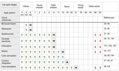
47Figure 1 Summary chart for development of human visual milestones.
Notes: A summary of the development of key visual perceptual milestones across the life span. The top panel shows the stages of human development (infants, young children, older children, teens, young adults, older adults), and associated ages in months and years (as presented by Siu et al22). The rows below illustrate the approximate timing of onset and emergence (green arrows), adult-like levels (gray shade with black arrows), and loss of function (red arrows). References linked to each milestone are provided in the right column.
Abbreviations: V1,primary visual cortex; mo, months.
/LIHVSDQVWDJHV
,QIDQWV
PR PR PR
5HIHUHQFHV
±
<RXQJ
FKLOGUHQ FKLOGUHQ2OGHU 7HHQV <RXQJDGXOWV 2OGHUDGXOWV
$JHV\HDUV
9LVXDO PLOHVWRQHV %LQRFXODUIXVLRQ
6WHUHRSVLV
6SDWLDODFXLW\
&RQWUDVW VHQVLWLYLW\
2ULHQWDWLRQ
0RWLRQ
&RORUSHUFHSWLRQ
&RQWRXU LQWHJUDWLRQ )DFHSHUFHSWLRQ
Eye and Brain downloaded from https://www.dovepress.com/ by 118.70.13.36 on 22-Aug-2020
Dovepress Development of human visual cortex and clinical implications
Anatomical milestones
Many of the anatomical features of human V1 develop
prenatally. Neurogenesis begins around embryonic day 33
and is complete by birth,
48–50while the thalamic input to
layer 4 in V1,
51,52bipolar, and pyramidal cells with long
and thin dendritic spines forms distinct laminar patterns at
around 20–30 weeks gestation.
16,53Other aspects of cortical
development that begin prenatally continue to mature after
birth. Cytochrome oxidase expression is present at 26 weeks
gestation, and is organized into clearly visible “puffs” by 24
days postnatal, and becomes well organized by 4 months
postnatal.
19Vertical interlaminar connections form between
26 and 29 weeks gestation, while long-range horizontal
connections in layers 4B and 5 emerge at around 37 weeks
gestation, and show adult-like patchiness by 8 weeks
postna-tal. Layer 2/3 horizontal connections emerge later at around
16 weeks postnatal and become adult-like by 15 months of
age.
15By 4 months of age, feedforward connections from
V1 to extrastriate area V2 have formed mature connections,
while feedback connections are still immature only reaching
adult levels at around 2 years of age.
14Synaptogenesis in
human V1 increases to reach a peak between 8 months and
2 years and is followed by a longer period of synaptic
prun-ing to reach adult levels later in childhood.
13The number of
dendritic spines in V1 follows a similar trajectory that peaks
at around 5 months of age and then decreases to adult levels
by 2 years.
54Many anatomical features are already adult-like
by the end of this stage (Figure 2); however, vision continues
to mature well beyond the first year of life.
Neurobiological milestones
We have found that the first stage of human V1 development
is characterized by rapid changes in neurobiological
mecha-nisms that will support the emergence of visual function and
synaptic plasticity. There are some early changes to both
excitatory glutamatergic and inhibitory gamma-aminobutyric
acid (GABA)ergic synaptic receptors (Figure 3),
22,24and a
shift toward a balance between these excitatory and inhibitory
(E-I) receptors.
20The immature GABA
A
receptors subunits,
GABA
Aα
2 and GABA
Aα
3, dominate expression in the first
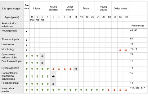
Figure 2 Summary chart for development of human V1 anatomical milestones.
Notes: A summary of the development of key neuroanatomical milestones in human V1 across the life span. The top panel shows the stages of human development (prenatal, infants, young children, older children, teens, young adults, older adults), and associated ages in months and years (as presented by Siu et al22). The rows below illustrate the approximate timing of onset and emergence (green arrows), adult-like levels and structure (gray shade with black arrows), and loss of expression (red arrows). Black dots refer to anatomical milestones that are completed before birth. References linked to each milestone are provided in the right column.
Abbreviations: V1,primary visual cortex; mo, months.
/LIHVSDQVWDJHV
$JHV\HDUV
$QDWRPLFDO9 PLOHVWRQHV 1HXURJHQHVLV
7KDODPLFLQSXWV /DPLQDWLRQ 0RUSKRORJ\ &\WRFKURPH R[LGDVHEOREV )HHGIRUZDUGLQSXW
6\QDSWRJHQHVLV
)HHGEDFNLQSXW ,QWUDFRUWLFDO P\HOLQ +RUL]RQWDODQG LQWHUODPLQD FRQQHFWLRQV
,QIDQWV
PR PR PR 3UH
QDWDO FKLOGUHQ<RXQJ FKLOGUHQ2OGHU 7HHQV <RXQJDGXOWV 2OGHUDGXOWV
5HIHUHQFHV
Eye and Brain downloaded from https://www.dovepress.com/ by 118.70.13.36 on 22-Aug-2020
Dovepress
Siu and Murphy
year, but quickly show signs of maturation as those subunits
are replaced by GABA
Aα
1 (Figure 4) so that there is relatively
more GABA
Aα
1 by about 1 year of age, that peaks later in
adolescence (Figure 3).
24For glutamatergic synapses, the
N-methyl-
d-aspartate (NMDA) receptor subunit GluN1 is
highly expressed at birth, then rapidly declines to reach adult
levels at around 1 year. That loss is balanced by an increase
in GluA2 containing AMPA receptor (AMPAR) expression,
and the shift from more NMDA to more AMPA signals the
loss of NMDA-dominated silent glutamatergic synapses to be
replaced by active AMPA containing synapses (Figure 3).
22The maturation of both GABA
Areceptor subunits and the
AMPA:NMDA balance speed up responses at those receptors
and trigger an environment that supports
experience-depen-dent plasticity (Figure 4).
55–57For example, the maturation of
GABA
Areceptor regulates the critical period for plasticity,
56as the mature
α
1 subunit is necessary for ocular dominance
plasticity.
55Moreover, the insertion of AMPAR is driven by
visual experience and is an important step in initiating the
critical period.
58Stage 2: preschool children have
high variability in V1 development
(1–4 years)
Visual milestones
Many aspects of visual perception continue to improve through
the first few years of development (Figure 1). Young children
have experience-dependent improvements in visual acuity,
42bio-logical motion perception,
59and contrast sensitivity,
60,61but those
abilities are still not adult-like.
62During the first 2 years of this
stage (
~
1–3 years), children are most susceptible to abnormal
binocular vision
63that can cause amblyopia. Alternatively, if
abnormal vision is identified and treated in children under
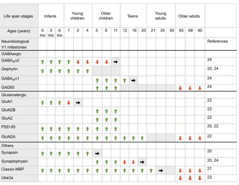
Figure 3 Summary chart for development of human V1 neurobiological milestones.
Notes: A summary of the development of key neurobiological milestones in human V1 across the life span. The top panel shows the stages of human development (infants, young children, older children, teens, young adults, older adults), and associated ages in months and years (as presented by Siu et al22). The rows below illustrate the approximate timing of onset and emergence (green arrows), peak expression (gray shades), adult-like levels (gray shade with black arrows), and loss of expression (red arrows). References linked to each milestone are provided in the right column.
Abbreviations: V1,primary visual cortex; mo, months; MBP, myelin basic protein.
/LIHVSDQVWDJHV ,QIDQWV FKLOGUHQ<RXQJ FKLOGUHQ2OGHU 7HHQV <RXQJDGXOWV 2OGHUDGXOWV
5HIHUHQFHV
$JHV\HDUV
1HXURELRORJLFDO
*OXWDPDWHUJLF *OX1 *OX1% *OX$ 36'
*OX1$
2WKHUV 6\QDSVLQ 6\QDSWRSK\VLQ &ODVVLF0%3 8EHD 9PLOHVWRQHV *$%$HUJLF
*HSK\ULQ *$%$$α
*$' *$%$$α
PR PR PR
Eye and Brain downloaded from https://www.dovepress.com/ by 118.70.13.36 on 22-Aug-2020
Dovepress Development of human visual cortex and clinical implications
5 years of age, there is the greatest likelihood for recovery,
64for
both high and low spatial frequencies.
65For example, binocular
iPad treatment for amblyopia shows improvement in visual
acu-ity at this stage for amblyopic children.
66Anatomical milestones
In young children, V1 undergoes synaptic and dendritic
refinement to reach adult appearance at around 2 years of age
(Figure 2).
54Other aspects of human cortical development are
characterized as “adult-like” by this stage, including cortical
thickness
67and the appearance of feedback connections from
extrastriate areas to V1.
14Neurobiological milestones
Although this period of experience-driven visual
develop-ment points to significant increases in visual plasticity, we
have found little evidence that neural plasticity mechanisms
complete maturation during this stage (Figure 3). Instead, we
found a novel aspect of human cortical development that is
characterized by waves of high interindividual variability in
the expression of neural plasticity markers in human V1.
20–23Interindividual variability in human V1 can be characterized
across development using the variance-to-mean ratio of protein
expression across a moving window of three age-adjacent
cases. Using this, we found a period of high interindividual
variability, or a “wave”, during young childhood for many of
the synaptic proteins, but not during the other stages of
devel-opment.
22This variability may signify either interindividual
differences in the rate of development, or it may identify
intraindividual fluctuations in the expression of plasticity
mechanisms.
22Nevertheless, this large dynamic range in
protein expression likely contributes to increased plasticity or
learning for optimal behavioral performance.
68Interestingly,
this stage of interindividual variability comes just after the E-I
balance has been reached in human V1.
20Balanced excitation
and inhibition in the cortex establish cortical criticality, defined
as a dynamic range of spontaneous activity that maximizes the
processing of input activity.
69Thus, the waves of variability in
cortical development may be an important stage of
develop-ment when visual circuits “learn” complex processing by using
the variability to fine-tune optimal neural circuits.
22,70Stage 3: experience-dependent
visual development in school aged
children (5–11 years)
Visual milestones
Many visual abilities finish maturation in older children
(Figure 1). However, the precise age of maturation may vary
significantly, and usually, depends on the type of measure
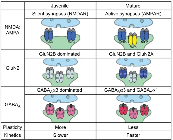
Figure 4 Summary chart of glutamatergic and GABAergic receptor subunits.
Notes: This figure presents a summary of some key glutamate (AMPAR and NMDAR) and GABA (GABAA) receptor subunit compositions that regulate neuroplasticity
in the primary visual cortex. The columns represent functional significance of the balance of NMDA:AMPA (top), GluN2A:GluN2B (middle), and GABAAα1:GABAAα3 (bottom). More juvenile synapses are dominated by more NMDAR, GluN2B containing NMDAR, and GABAAα3 containing GABAA receptors that allow for LTP in excitatory synapses127 and slower kinetics through the receptors. More mature synapses are dominated by more AMPAR, GluN2A containing NMDAR, and GABA
Aα1 containing GABAA receptors that allow for more LTD in excitatory synapses,127 and faster kinetics through the receptors.
Abbreviations: GABA, gamma-aminobutyric acid; LTD, long-term depression; AMPAR, AMPA receptor; NMDAR, N-methyl-d-aspartate receptor; LTP, long-term potentiation.
-XYHQLOH
10'$ $03$
*OX1
*OX1%GRPLQDWHG
10'$ 10'$ 10'$
$03$ 10'$
*$%$$αGRPLQDWHG *$%$$αDQG*$%$$α
*OX1%DQG*OX1$
3ODVWLFLW\ 0RUH 6ORZHU
/HVV )DVWHU .LQHWLFV
*$%$$
6LOHQWV\QDSVHV10'$5 $FWLYHV\QDSVHV$03$5 0DWXUH
Eye and Brain downloaded from https://www.dovepress.com/ by 118.70.13.36 on 22-Aug-2020
Dovepress
Siu and Murphy
used to assess vision, or the type of patterned visual input
that a child has experienced.
71For example, visual acuity can
mature between 5 and 15 years, while contrast sensitivity can
mature anywhere between 6 and 19 years of age.
26,72While
some studies suggest that motion perception can mature in
older children,
73–76others suggest that it continues to improve
beyond childhood into adolescence and adulthood.
34,77–79Children aged 6–10 years are just beyond the period of
susceptibility for developing amblyopia
65,80,81that is
associ-ated with the end of the critical period for ocular dominance
plasticity in animal models.
20Despite this, there is evidence
for significant visual plasticity at this stage in both children
with amblyopia and children with normal vision.
82–84The
neurobiological mechanisms that regulate this
experience-dependent plasticity, including triggers proteins that promote
neuroplasticity, and brakes that limit it are well studied in
animal models.
5,85,86Neurobiological milestones
The expression of some neural plasticity mechanisms peaks
during this stage of development (Figure 3). These include
peaks in the expression of the glutamatergic receptor
scaf-folding protein PSD-95 and AMPA receptor subunit GluA2.
22Peaks in the expression for both of these proteins have been
linked with ending the critical period for experience-dependent
plasticity in the visual cortex.
58,87The maturation of those
proteins is experience-dependent
87and contributes to
stabiliz-ing synapses.
88Furthermore, GluA2 is necessary for a form
of plasticity, homeostatic synaptic scaling, which regulates
synaptic strength over fluctuations in synaptic activity.
89The
homeostatic scaling up or down of AMPAR expression is
dependent on synaptic activity and cooperates with
NMDA-dependent Hebbian plasticity to refine cortical connectivity and
promote synaptic stability during the development of V1.
90–92In human V1, expression of the GABA receptor
scaf-folding protein gephyrin also matures during late childhood
development (Figure 3).
20,24Gephyrin is directly related to
the strength and stability of inhibitory synapses
93and this
peak suggests a developmental balance between excitatory
(eg, PSD-95, GluA2) and inhibitory synaptic mechanisms.
20It is interesting to note that the AMPAR subunit GluA2 is
highly expressed on parvalbumin-positive (PV
+
) inhibitory
interneurons,
94and PV
+
cell activity regulates critical period
plasticity.
95It will be important for future experiments to
address the development of PV
+
inhibitory interneurons in
human V1 to fully understand the maturation of these
plas-ticity mechanisms during this important stage of childhood
visual development.
Stage 4: prolonged visual
development in adolescence and
adulthood (12–55 years)
Visual milestones
A series of studies characterizing visual development have
shown that “higher-order” visual abilities continue to mature
through the teen and young adult years (Figure 1). For
example, global and biological motion
34,77,96–98and spatial
integration of contours
27mature during adolescence (eg,
14–15 years of age). Face perception has an even slower pace
of maturation, with continuous improvements into adulthood,
as face learning and recognition improve into the third decade
of life.
29,99,100Expertise in face perception depends on visual
experience, and abnormal early vision has a “sleeper effect”
on the development of the neural circuits and perceptual
processing that support normal face perception.
101–103Children older than 7 years of age are less responsive to
amblyopia treatment,
104but some forms of treatment may
be effective in teens and adults and suggest that plasticity
persists in the visual cortex.
105–107For example, perceptual
training for low-level perceptual abilities like contrast
sensi-tivity and letter-recognition can improve the vision of some
amblyopic patients.
108These training-induced improvements
are often small and not clinically significant.
109Perceptual
learning studies have shown that there is plasticity in
adult-hood that can support recovery from amblyopia.
110Adults
with amblyopia can improve visual acuity with extensive
perceptual training,
111and succeed in refining contrast
sensitivity,
83orientation selectivity,
112stereopsis,
113spatial
discrimination,
114and face learning.
115,116Anatomical milestones
Structural imaging studies of humans show that
intracorti-cal myelin in the visual cortex continues to increase well
into adulthood, peaking between 30 and 40 years of age
( Figure 2).
117Anatomical analysis of postmortem human
visual cortex also indicates the prolonged development of
cortical myelination that continues into the third decade of
life.
118Gray matter density mapping shows a slow linear
decline of cortical thickness with age in the occipital
cor-tex,
119,120and that regression appears to mature sequentially
across the cortical areas, with primary areas maturing before
higher-order association areas.
121Neurobiological milestones
Our studies of neuroplasticity mechanisms in human V1
provide new evidence that many aspects of V1 continue to
Eye and Brain downloaded from https://www.dovepress.com/ by 118.70.13.36 on 22-Aug-2020
Dovepress Development of human visual cortex and clinical implications
develop into the adult years (Figure 3). Our findings include
measures of myelin expression as a brake on plasticity
classic-myelin basic protein [MBP]),
122and of the balance between
NMDA receptor subunits 2A and 2B that supports plasticity
when 2B dominates and reduces plasticity when 2A
domi-nates (2A:2B) (Figure 4).
123–127Myelin expression peaks in
young adults
21and the shift from more 2B to more 2A also
ends at around 35 years of age.
22Some GABAergic proteins
also continue developing into adulthood. The enzyme that
makes the on-demand pool of GABA (GAD65) and the
GABA
Aα
1 receptor subunit continues to increase into
adult-hood (Figure 3).
24Each of these late maturing mechanisms is
important for regulating neural transmission and plasticity.
For example, the bidirectional regulation of the 2A:2B
bal-ance can facilitate plasticity when 2B is favored, or reduce
experience-dependent plasticity when 2A is favored.
123–127All
of these findings show that the progressive shift in human
V1 from a very plastic environment in childhood to a less
plastic environment in adults continues into the third decade
of life (Figure 3). That pace of neurobiological development
is much slower than predicted by vision studies or animal
research.
20–22,24The very slow maturation of human V1 may keep a
sliver of the plasticity window open that both normal visual
development and some types of vision treatments can use.
Stage 5: loss of plasticity
mechanisms during aging of human
V1 (
>
55 years)
Visual milestones
Certain visual losses in aging have been interpreted as part
of normal aging that changes the receptive field properties
of neurons in V1 (Figure 1).
28During normal aging, there is
an increase in the population receptive field size for neurons
in V1 and in the extrastriate area V2 that serve the foveal
representations.
128These neural changes and others contribute
to age-related losses of low-level visual functions like visual
acuity,
129contrast sensitivity,
130and orientation selectivity.
131In addition, there are age-related losses for many
higher-order visual perceptions,
132including face perception,
99,133–135motion processing,
136–140and reading speed.
28There are also acquired causes for age-related vision loss
that include diseases such as diabetic retinopathy, macular
degeneration, cataracts, and glaucoma. All of these
dis-eases affect the eye and either directly or indirectly reduce
retinal functioning either because the cataract has degraded
the image or the disease has caused degeneration of the
retina.
141These retinal changes impact the information that
is transmitted to the central visual pathway and many
stud-ies have shown changes in the visual areas of the brain.
142Often, these eye diseases are described as neurodegeneration
spreading that starts in the eye and progresses to affect the
visual cortex.
143Thus, it is likely that vision changes in normal
aging and adult-acquired visual diseases may involve
neuro-degenerative processes in V1 in addition to optical changes.
Anatomical milestones
Aging in the visual cortex is characterized by specific
microstructural changes (Figure 2). These include significant
changes in the morphology of pyramidal cell bodies in human
V1, a loss of dendrite number, and reduced complexity of the
dendritic arborizations.
10In primate V1, aging changes the
process of cortical demyelination and remyelination so that
remyelinated axons have shorter segments and the myelin
sheaths are less tightly compacted around the axons thereby
affecting the efficiency of axonal conduction.
144–146In human
V1, there is a progressive loss of intracortical myelin content
in the stria of Gennari that begins around 30 years of age and
continues to decline into the late 90s.
147Some animal studies
have also found a loss of intracortical inhibition in V1 that
leads to poor orientation selectivity,
148–150and treating V1
of old monkeys with the neurotransmitter GABA sharpens
orientation selectivity of V1 neurons so they are similar to
the selectivity found in young adults.
150Neurobiological milestones
Our studies of human V1 have found that the expression of
many synaptic and non-synaptic proteins decreases in older
adults (Figure 3). There are losses for GAD65, the enzyme
that makes the on-demand pool of GABA,
24Ube3A, an E3
ubiquitin ligase that is necessary for experience-dependent
plasticity,
23classic-MBP that is necessary for normal axonal
myelination,
21and GluN2A, the mature subunit of the NMDA
receptor that regulates certain forms of plasticity.
22Not all
synaptic proteins change on aging; for example, expression
of the GABA
Areceptor subunit does not decline.
24Interest-ingly, most of the proteins that do change shift toward the
more juvenile-like partner. For example, the age-related loss
of GluN2A shifts the 2A:2B balance toward more 2B, and
that may reinstate a more plastic environment.
127Also, the
loss of classic-MBP shifts the composition of MBP to favor
the immature oligodendrocyte protein Golli-MBP
21which
can give rise to various developmental regulated isoforms
of MBP.
151These shifts toward more juvenile-like neural
proteins raise the possibility that there is support for a more
plastic environment in aging. However, not all of the changes
Eye and Brain downloaded from https://www.dovepress.com/ by 118.70.13.36 on 22-Aug-2020
Dovepress
Siu and Murphy
point to greater plasticity since there is a loss of Ube3A, a
protein that is necessary for experience-dependent
plastic-ity in the visual system.
152Perhaps, the age-related losses
of these important neural proteins reflect an internal drive
to maintain stable functioning of visual cortical circuits in
the face of degraded inputs due to optical changes
153,154and
neurodegenerative processes.
143Conclusions
We have demonstrated lifelong changes in the
neurobio-logical mechanisms found in human V1 that support neural
development, plasticity, and processing of visual information.
These changes can be described in five stages: very early
establishment of mechanisms for E-I transmission; a novel
stage of variability in young children; maturation of
low-level mechanisms in older children; continued fine-tuning
through teens and young adults; and age-related losses. How
far these five stages of V1 maturation will generalize across
the 20 cortical areas that process visual information remains
unknown. This is an important question to address, especially
for developing new cortically inspired treatments for
adult-acquired vision loss, since plasticity in the extrastriate area
may prove to be important for supporting maintenance or
recovery of vision caused by a retinal disease.
The first three figures summarize key milestones for
visual system development and illustrate compelling
similarities between the timing of visual, anatomical, and
neurobiological milestones in human V1. Tapping into these
neurobiological mechanisms is going to be key for the next
generation of treatments for visual disorders. For example, a
wide range of potential new therapies has been developed in
animal models for amblyopia. The treatments include
every-thing from fine-tuning of traditional patching therapy
155,156to
drug treatments,
157to novel visual stimulation paradigms
158and visual environments.
159–161Also, it is likely that normal
age-related changes in human V1 interact with the
spread-ing neurodegeneration caused by diseases like glaucoma. In
contrast to the excitement from preclinical models, no new
plasticity-based treatments have crossed the chasm into
clini-cal practice. One of the impediments has been the lack of
information about neurobiological mechanisms in the human
visual cortex, but our studies are beginning to fill that gap.
Although our approach to studying neurobiology in human
V1 does not provide information about circuitry, synaptic
function, cellular type, or laminar localization, these data
are valuable as the first steps for identifying neurobiological
mechanisms that underlie visual perception and plasticity
in humans. In addition, these data will help to guide future
human and animal studies by making it easier to make more
direct links between the neurobiological developments of V1
in humans and animal models, that can pave the way for the
translation of biologically inspired vision treatments.
Acknowledgments
We thank Kyle Hornby and Drs Kate Williams, Joshua Pinto,
and Simon Beshara for technical help and Justin Balsor for
comments on the manuscript. An NSERC grant
RGPIN-2015-06215 was awarded to KMM, and Woodburn Heron
OGS awarded to CRS.
Disclosure
The authors report no conflicts of interest in this work.
References
1. Hubel DH, Wiesel TN. Receptive fields of single neurones in the cat’s striate cortex. J Physiol. 1959;148(3):574–591.
2. Hubel DH, Wiesel TN. The period of susceptibility to the physi-ological effects of unilateral eye closure in kittens. J Physiol. 1970;206(2):419–436.
3. Levelt CN, Hübener M. Critical-period plasticity in the visual cortex. Annu Rev Neurosci. 2012;35(1):309–330.
4. Sengpiel F. Plasticity of the visual cortex and treatment of amblyopia. Curr Biol. 2014;24(18):R936–R940.
5. Hensch TK. Critical period regulation. Annu Rev Neurosci. 2004;27(1):549–579.
6. Turrigiano GG. The dialectic of Hebb and homeostasis. Philos Trans R Soc Lond B Biol Sci. 2017;372(1715):20160258.
7. Smith GB, Heynen AJ, Bear MF. Bidirectional synaptic mechanisms of ocular dominance plasticity in visual cortex. Philos Trans R Soc Lond B Biol Sci. 2009;364(1515):357–367.
8. Cooke SF, Bear MF. How the mechanisms of long-term synaptic potentiation and depression serve experience-dependent plastic-ity in primary visual cortex. Philos Trans R Soc Lond B Biol Sci. 2014;369(1633):20130284.
9. Kiorpes L. Visual development in primates: neural mechanisms and critical periods. Dev Neurobiol. 2015;75(10):1080–1090.
10. Mavroudis IA, Manani MG, Petrides F, et al. Age-related dendritic and spinal alterations of pyramidal cells of the human visual cortex. Folia Neuropathol. 2015;53(2):100–110.
11. Eickhoff SB, Rottschy C, Kujovic M, Palomero-Gallagher N, Zilles K. Organizational principles of human visual cortex revealed by receptor mapping. Cereb Cortex. 2008;18(11):2637–2645.
12. Huttenlocher PR. Morphometric study of human cerebral-cortex development. Neuropsychologia. 1990;28(6):517–527.
13. Huttenlocher PR, de Courten C, Garey LJ, Van der Loos H. Synap-togenesis in human visual cortex – evidence for synapse elimination during normal development. Neurosci Lett. 1982;33(3):247–252. 14. Burkhalter A. Development of forward and feedback connections
between areas V1 and V2 of human visual cortex. Cereb Cortex. 1993;3(5):476–487.
15. Burkhalter A, Bernardo KL, Charles V. Development of local circuits in human visual cortex. J Neurosci. 1993;13(5):1916–1931. 16. Takashima S, Chan F, Becker LE, Armstrong DL. Morphology of the
developing visual-cortex of the human infant – a quantitative and quali-tative Golgi-study. J Neuropathol Exp Neurol. 1980;39(4):487–501. 17. Leuba G, Garey LJ. Evolution of neuronal numerical density in
the developing and aging human visual cortex. Hum Neurobiol. 1987;6(1):11–18.
Eye and Brain downloaded from https://www.dovepress.com/ by 118.70.13.36 on 22-Aug-2020
Dovepress Development of human visual cortex and clinical implications
18. Burkhalter A, Bernardo KL. Organization of corticocortical connec-tions in human visual cortex. Proc Natl Acad Sci U S A. 1989;86(3): 1071–1075.
19. Wong-Riley MT, Hevner RF, Cutlan R, et al. Cytochrome oxidase in the human visual cortex: distribution in the developing and the adult brain. Vis Neurosci. 1993;10(1):41–58.
20. Pinto JGA, Jones DG, Williams CK, Murphy KM. Characterizing synaptic protein development in human visual cortex enables align-ment of synaptic age with rat visual cortex. Front Neural Circuits. 2015;9:3.
21. Siu CR, Balsor JL, Jones DG, Murphy KM. Classic and Golli myelin basic protein have distinct developmental trajectories in human visual cortex. Front Neurosci. 2015;9:138.
22. Siu CR, Beshara SP, Jones DG, Murphy KM. Development of glutama-tergic proteins in human visual cortex across the lifespan. J Neurosci. 2017;37(25):6031–6042.
23. Williams K, Irwin DA, Jones DG, Murphy KM. Dramatic loss of Ube3A expression during aging of the mammalian cortex. Front Aging Neurosci. 2010;2:18.
24. Pinto JGA, Hornby KR, Jones DG, Murphy KM. Developmental changes in GABAergic mechanisms in human visual cortex across the lifespan. Front Cell Neurosci. 2010;4:16.
25. Gilmore JH, Knickmeyer RC, Gao W. Imaging structural and func-tional brain development in early childhood. Nat Rev Neurosci. 2018;19(3):123–137.
26. Ellemberg D, Lewis TL, Liu CH, Maurer D. Development of spatial and temporal vision during childhood. Vision Res. 1999;39(14):2325–2333. 27. Kovacs I, Kozma P, Fehér A, Benedek G. Late maturation of
visual spatial integration in humans. Proc Natl Acad Sci U S A. 1999;96(21):12204–12209.
28. Owsley C. Aging and vision. Vision Res. 2011;51(13):1610–1622. 29. Hartshorne JK, Germine LT. When does cognitive functioning peak?
The asynchronous rise and fall of different cognitive abilities across the life span. Psychol Sci. 2015;26(4):433–443.
30. Hamer RD, Norcia AM, Tyler CW, Hsu-Winges C. The development of monocular and binocular VEP acuity. Vision Res. 1989;29(4):397–408. 31. Norcia AM, Tyler CW. Spatial frequency sweep VEP: visual acuity
during the first year of life. Vision Res. 1985;25(10):1399–1408. 32. Pirchio M, Spinelli D, Fiorentini A, Maffei L. Infant contrast sensitivity
evaluated by evoked potentials. Brain Res. 1978;141(1):179–184. 33. Morrone MC, Burr DC. Evidence for the existence and development
of visual inhibition in humans. Nature. 1986;321(6067):235–237. 34. Hadad B, Schwartz S, Maurer D, Lewis TL. Motion perception: a
review of developmental changes and the role of early visual experi-ence. Front Integr Neurosci. 2015;9(583):5532.
35. Birch E, Petrig B. FPL and VEP measures of fusion, stereopsis and stereoacuity in normal infants. Vision Res. 1996;36(9):1321–1327. 36. Teller DY, Peeples DR, Sekel M. Discrimination of chromatic from
white light by two-month-old human infants. Vision Res. 1978;18(1): 41–48.
37. Bornstein MH. Infants are trichromats. J Exp Child Psychol. 1976;21(3):425–445.
38. Boothe RG, Dobson V, Teller DY. Postnatal development of vision in human and nonhuman primates. Annu Rev Neurosci. 1985;8(1):495–545.
39. Wilcox T. Object individuation: infants’ use of shape, size, pattern, and color. Cognition. 1999;72(2):125–166.
40. Baker TJ, Tse J, Gerhardstein P, Adler SA. Contour integration by 6-month-old infants: discrimination of distinct contour shapes. Vision Res. 2008;48(1):136–148.
41. Taylor G, Hipp D, Moser A, Dickerson K, Gerhardstein P. The development of contour processing: evidence from physiology and psychophysics. Front Psychol. 2014;5:719.
42. Lai Y-H, Wang H-Z, Hsu H-T. Development of visual acuity in pre-school children as measured with Landolt C and Tumbling E charts. J AAPOS. 2011;15(3):251–255.
43. Birch EE. Amblyopia and binocular vision. Prog Retin Eye Res. 2013;33:67–84.
44. Jando G, Miko-Barath E, Marko K, Hollody K, Toeroek B, Kovacs I. Early-onset binocularity in preterm infants reveals experience-dependent visual development in humans. Proc Natl Acad Sci U S A. 2012;109(27):11049–11052.
45. Birch EE, Holmes JM. The clinical profile of amblyopia in children younger than 3 years of age. J AAPOS. 2010;14(6):494–497. 46. Maurer D, Lewis TL, Brent HP, Levin AV. Rapid improvement in the
acuity of infants after visual input. Science. 1999;286(5437):108–110. 47. Birch EE, Swanson WH, Stager DR, Woody M, Everett M. Outcome after very early treatment of dense congenital unilateral cataract. Invest Ophthalmol Vis Sci. 1993;34(13):3687–3699.
48. Rakic P. Neuroscience. No more cortical neurons for you. Science. 2006;313(5789):928–929.
49. Clowry G, Molnár Z, Rakic P. Renewed focus on the developing human neocortex. J Anat. 2010;217(4):276–288.
50. Bhardwaj RD, Curtis MA, Spalding KL, et al. Neocortical neurogen-esis in humans is restricted to development. Proc Natl Acad Sci U S A. 2006;103(33):12564–12568.
51. Flower MJ. Neuromaturation of the human fetus. J Med Philos. 1985;10(3):237–251.
52. Kostovic I, Rakic P. Development of prestriate visual projections in the monkey and human fetal cerebrum revealed by transient cholinesterase staining. J Neurosci. 1984;4(1):25–42.
53. Sauer B. Quantitative analysis of the laminae of the striate area in man. An application of automatic image analysis. J Hirnforsch. 1983;24(1):89–97.
54. Michel AE, Garey LJ. The development of dendritic spines in the human visual cortex. Hum Neurobiol. 1984;3(4):223–227.
55. Fagiolini M, Fritschy J-M, Löw K, Möhler H, Rudolph U, Hensch TK. Specific GABAA circuits for visual cortical plasticity. Science. 2004;303(5664):1681–1683.
56. Chen L, Yang C, Mower GD. Developmental changes in the expression of GABA(A) receptor subunits (alpha(1), alpha(2), alpha(3)) in the cat visual cortex and the effects of dark rearing. Brain Res Mol Brain Res. 2001;88(1–2):135–143.
57. Liao D, Scannevin RH, Huganir R. Activation of silent synapses by rapid activity-dependent synaptic recruitment of AMPA receptors. J Neurosci. 2001;21(16):6008–6017.
58. Huang X, Stodieck SK, Goetze B, et al. Progressive maturation of silent synapses governs the duration of a critical period. Proc Natl Acad Sci U S A. 2015;112(24):E3131–E3140.
59. Sweeny TD, Wurnitsch N, Gopnik A, Whitney D. Sensitive perception of a person’s direction of walking by 4-year-old children. Dev Psychol. 2013;49(11):2120–2124.
60. Richman JE, Lyons S. A forced choice procedure for evaluation of contrast sensitivity function in preschool children. J Am Optom Assoc. 1994;65(12):859–864.
61. Scharre JE, Cotter SA, Block SS, Kelly SA. Normative contrast sen-sitivity data for young children. Optom Vis Sci. 1990;67(11):826–832. 62. Yu T-Y, Jacobs RJ, Anstice NS, et al. Global motion perception in
2-year-old children: a method for psychophysical assessment and rela-tionships with clinical measures of visual function. Invest Ophthalmol Vis Sci. 2013;54(13):8408–8419.
63. Banks MS, Aslin RN, Letson RD. Sensitive period for the development of human binocular vision. Science. 1975;190(4215):675–677. 64. US Preventive Services Task Force; Grossman DC, Curry SJ, Owens
DK, et al. Vision screening in children aged 6 months to 5 years: US Preventive Services Task Force recommendation statement. JAMA. 2017;318(9):836–844.
65. Lewis TL, Maurer D. Multiple sensitive periods in human visual devel-opment: evidence from visually deprived children. Dev Psychobiol. 2005;46(3):163–183.
66. Birch EE, Li SL, Jost RM, et al. Binocular iPad treatment for amblyopia in preschool children. J AAPOS. 2015;19(1):6–11.
Eye and Brain downloaded from https://www.dovepress.com/ by 118.70.13.36 on 22-Aug-2020
Dovepress
Siu and Murphy
67. Lyall AE, Shi F, Geng X, et al. Dynamic development of regional cortical thickness and surface area in early childhood. Cereb Cortex. 2015;25(8):2204–2212.
68. Garrett DD, Kovacevic N, McIntosh AR, Grady CL. The modulation of BOLD variability between cognitive states varies by age and processing speed. Cereb Cortex. 2013;23(3):684–693.
69. Shew WL, Yang H, Petermann T, Roy R, Plenz D. Neuronal avalanches imply maximum dynamic range in cortical networks at criticality. J Neurosci. 2009;29(49):15595–15600.
70. Gordus A, Pokala N, Levy S, Flavell SW, Bargmann CI. Feedback from network states generates variability in a probabilistic olfactory circuit. Cell. 2015;161(2):215–227.
71. Lewis TL, Maurer D. Effects of early pattern deprivation on visual development. Optom Vis Sci. 2009;86(6):640–646.
72. Almoqbel FM, Irving EL, Leat SJ. Visual acuity and contrast sensi-tivity development in children: sweep visually evoked potential and psychophysics. Optom Vis Sci. 2017;94(8):830–837.
73. Narasimhan S, Giaschi D. The effect of dot speed and density on the development of global motion perception. Vision Res. 2012;62:102–107.
74. Ellemberg D, Lewis TL, Maurer D, et al. The effect of displacement on sensitivity to first- and second-order global motion in 5-year-olds and adults. Seeing Perceiving. 2010;23(5–6):517–532.
75. Ellemberg D, Lewis TL, Maurer D, Brar S, Brent HP. Better perception of global motion after monocular than after binocular deprivation. Vision Res. 2002;42(2):169–179.
76. Gunn A, Cory E, Atkinson J, et al. Dorsal and ventral stream sensitivity in normal development and hemiplegia. Neuroreport. 2002;13(6):843–847.
77. Bogfjellmo LG, Bex PJ, Falkenberg HK. The development of global motion discrimination in school aged children. J Vis. 2014;14(2):19. 78. Hadad B-S, Maurer D, Lewis TL. Long trajectory for the development of sensitivity to global and biological motion. Dev Sci. 2011;14(6): 1330–1339.
79. Joshi MR, Falkenberg HK. Development of radial optic flow pattern sensitivity at different speeds. Vision Res. 2015;110(Pt A):68–75. 80. Epelbaum M, Milleret C, Buisseret P, Dufier JL. The sensitive
period for strabismic amblyopia in humans. Ophthalmology. 1993;100(3):323–327.
81. Keech RV, Kutschke PJ. Upper age limit for the development of amblyopia. J Pediatr Ophthalmol Strabismus. 1995;32(2):89–93. 82. Li RW, Young KG, Hoenig P, Levi DM. Perceptual learning improves
visual performance in juvenile amblyopia. Invest Ophthalmol Vis Sci. 2005;46(9):3161–3168.
83. Liao M, Zhao H, Liu L, et al. Training to improve contrast sensi-tivity in amblyopia: correction of high-order aberrations. Sci Rep. 2016;6:35702.
84. Mintz-Hittner HA, Fernandez KM. Successful amblyopia therapy initiated after age 7 years: compliance cures. Arch Ophthalmol. 2000;118(11):1535–1541.
85. Morishita H, Hensch TK. Critical period revisited: impact on vision. Curr Opin Neurobiol. 2008;18(1):101–107.
86. Bavelier D, Levi DM, Li RW, Dan Y, Hensch TK. Removing brakes on adult brain plasticity: from molecular to behavioral interventions. J Neurosci. 2010;30(45):14964–14971.
87. Chen X, Levy JM, Hou A, et al. PSD-95 family MAGUKs are essential for anchoring AMPA and NMDA receptor complexes at the postsyn-aptic density. Proc Natl Acad Sci U S A. 2015;112(50):E6983–E6992. 88. Liu S, Cull-Candy SG. Synaptic activity at calcium-permeable AMPA receptors induces a switch in receptor subtype. Nature. 2000;405(6785):454–458.
89. Gainey MA, Hurvitz-Wolff JR, Lambo ME, Turrigiano GG. Synaptic scaling requires the GluR2 subunit of the AMPA receptor. J Neurosci. 2009;29(20):6479–6489.
90. Desai NS, Cudmore RH, Nelson SB, Turrigiano GG. Critical periods for experience-dependent synaptic scaling in visual cortex. Nat Neu-rosci. 2002;5(8):783–789.
91. Mrsic-Flogel TD, Hofer SB, Ohki K, Reid RC, Bonhoeffer T, Hübener M. Homeostatic regulation of eye-specific responses in visual cortex during ocular dominance plasticity. Neuron. 2007;54(6):961–972. 92. Turrigiano GG, Nelson SB. Homeostatic plasticity in the developing
nervous system. Nat Rev Neurosci. 2004;5(2):97–107.
93. Tyagarajan SK, Fritschy J-M. Gephyrin: a master regulator of neuronal function? Nature. 2014;15(3):141–156.
94. Kooijmans RN, Self MW, Wouterlood FG, Belien JAM, Roelfsema PR. Inhibitory interneuron classes express complementary AMPA-receptor patterns in macaque primary visual cortex. J Neurosci. 2014;34(18):6303–6315.
95. Donato F, Rompani SB, Caroni P. Parvalbumin-expressing basket-cell network plasticity induced by experience regulates adult learning. Nature. 2013;504(7479):272–276.
96. Bucher K, Dietrich T, Marcar VL, et al. Maturation of luminance- and motion-defined form perception beyond adolescence: a combined ERP and fMRI study. Neuroimage. 2006;31(4):1625–1636.
97. Meier K, Giaschi D. Effect of spatial and temporal stimulus param-eters on the maturation of global motion perception. Vision Res. 2017;135:1–9.
98. Schrauf M, Wist ER, Ehrenstein WH. Development of dynamic vision based on motion contrast. Exp Brain Res. 1999;124(4):469–473. 99. Germine LT, Duchaine B, Nakayama K. Where cognitive development
and aging meet: face learning ability peaks after age 30. Cognition. 2011;118(2):201–210.
100. Mondloch CJ, Le Grand R, Maurer D. Configural face processing develops more slowly than featural face processing. Perception. 2002;31(5):553–566.
101. Grady CL, Mondloch CJ, Lewis TL, Maurer D. Early visual deprivation from congenital cataracts disrupts activity and functional connectivity in the face network. Neuropsychologia. 2014;57:122–139. 102. Mondloch CJ, Segalowitz SJ, Lewis TL, Dywan J, Le Grand R, Maurer
D. The effect of early visual deprivation on the development of face detection. Dev Sci. 2013;16(5):728–742.
103. Maurer D, Mondloch CJ, Lewis TL. Sleeper effects. Dev Sci. 2007;10(1):40–47.
104. Holmes JM. Effect of age on response to amblyopia treatment in children. Arch Ophthalmol. 2011;129(11):1451–1457.
105. Karni A, Bertini G. Learning perceptual skills: behavioral probes into adult cortical plasticity. Curr Opin Neurobiol. 1997;7(4):530–535. 106. Levi DM, Li RW. Perceptual learning as a potential treatment for
amblyopia: a mini-review. Vision Res. 2009;49(21):2535–2549. 107. Sasaki Y, Nanez JE, Watanabe T. Advances in visual perceptual learning
and plasticity. Nat Rev Neurosci. 2010;11(1):53–60.
108. Polat U, Ma-Naim T, Belkin M, Sagi D. Improving vision in adult ambly-opia by perceptual learning. Proc Natl Acad Sci U S A. 2004;101(17): 6692–6697.
109. Sklar JC, Goltz HC, Gane L, Wong AMF. Adaptation to laterally displacing prisms in anisometropic amblyopia. Invest Ophthalmol Vis Sci. 2015;56(6):3699–3708.
110. Levi DM, Polat U. Neural plasticity in adults with amblyopia. Proc Natl Acad Sci U S A. 1996;93(13):6830–6834.
111. Levi DM. Perceptual learning in adults with amblyopia: a reevaluation of critical periods in human vision. Dev Psychobiol. 2005;46(3):222–232.
112. Jehee JFM, Ling S, Swisher JD, van Bergen RS, Tong F. Perceptual learning selectively refines orientation representations in early visual cortex. J Neurosci. 2012;32(47):16747–16753.
113. Ding J, Levi DM. Recovery of stereopsis through perceptual learning in human adults with abnormal binocular vision. Proc Natl Acad Sci U S A. 2011;108(37):E733–E741.
114. Li RW, Levi DM. Characterizing the mechanisms of improvement for position discrimination in adult amblyopia. J Vis. 2004;4(6):7. 115. McMahon DBT, Leopold DA. Stimulus timing-dependent plasticity
in high-level vision. Curr Biol. 2012;22(4):332–337.
116. Du Y, Zhang F, Wang Y, Bi T, Qiu J. Perceptual learning of facial expressions. Vision Res. 2016;128:19–29.
Eye and Brain downloaded from https://www.dovepress.com/ by 118.70.13.36 on 22-Aug-2020
Dovepress Development of human visual cortex and clinical implications
117. Rowley CD, Sehmbi M, Bazin PL, et al. Age-related mapping of intracortical myelin from late adolescence to middle adulthood using T1-weighted MRI. Hum Brain Mapp. Epub 2017 Apr 30.
118. Miller DJ, Duka T, Stimpson CD, et al. Prolonged myelination in human neocortical evolution. Proc Natl Acad Sci U S A. 2012;109(41): 16480–16485.
119. Sowell ER, Peterson BS, Thompson PM, Welcome SE, Henkenius AL, Toga AW. Mapping cortical change across the human life span. Nat Neurosci. 2003;6(3):309–315.
120. Sowell ER, Thompson PM, Toga AW. Mapping changes in the human cortex throughout the span of life. Neuroscientist. 2004;10(4):372–392. 121. Gogtay N, Giedd JN, Lusk L, et al. Dynamic mapping of human corti-cal development during childhood through early adulthood. Proc Natl Acad Sci U S A. 2004;101(21):8174–8179.
122. McGee AW, Yang Y, Fischer QS, Daw NW, Strittmatter SM. Experi-ence-driven plasticity of visual cortex limited by myelin and Nogo receptor. Science. 2005;309(5744):2222–2226.
123. Abraham WC, Bear MF. Metaplasticity: the plasticity of synaptic plasticity. Trends Neurosci. 1996;19(4):126–130.
124. Philpot BD, Sekhar AK, Shouval HZ, Bear MF. Visual experience and deprivation bidirectionally modify the composition and function of NMDA receptors in visual cortex. Neuron. 2001;29(1):157–169. 125. Philpot BD, Espinosa JS, Bear MF. Evidence for altered NMDA
recep-tor function as a basis for metaplasticity in visual cortex. J Neurosci. 2003;23(13):5583–5588.
126. Philpot BD, Cho KKA, Bear MF. Obligatory role of NR2A for meta-plasticity in visual cortex. Neuron. 2007;53(4):495–502.
127. Yashiro K, Philpot BD. Regulation of NMDA receptor subunit expres-sion and its implications for LTD, LTP, and metaplasticity. Neurophar-macology. 2008;55(7):1081–1094.
128. Brewer AA, Barton B. Visual cortex in aging and Alzheimer’s disease: changes in visual field maps and population receptive fields. Front Psychol. 2014;5:74.
129. Sekuler R, Hutman LP, Owsley CJ. Human aging and spatial vision. Science. 1980;209(4462):1255–1256.
130. Allard R, Renaud J, Molinatti S, Faubert J. Contrast sensitivity, healthy aging and noise. Vision Res. 2013;92:47–52.
131. Betts LR, Sekuler AB, Bennett PJ. The effects of aging on orientation discrimination. Vision Res. 2007;47(13):1769–1780.
132. Habak C, Faubert J. Larger effect of aging on the perception of higher-order stimuli. Vision Res. 2000;40(8):943–950.
133. Konar Y, Bennett PJ, Sekuler AB. Effects of aging on face identification and holistic face processing. Vision Res. 2013;88:38–46.
134. Rousselet GA, Gaspar CM, Pernet CR, Husk JS, Bennett PJ, Sekuler AB. Healthy aging delays scalp EEG sensitivity to noise in a face discrimination task. Front Psychol. 2010;1:19.
135. Wilson HR, Mei M, Habak C, Wilkinson F. Visual bandwidths for face ori-entation increase during healthy aging. Vision Res. 2011;51(1):160–164. 136. Allard R, Lagacé-Nadon S, Faubert J. Feature tracking and aging.
Front Psychol. 2013;4:427.
137. Bennett PJ, Sekuler R, Sekuler AB. The effects of aging on motion detection and direction identification. Vision Res. 2007;47(6):799–809. 138. Betts LR, Taylor CP, Sekuler AB, Bennett PJ. Aging reduces center-surround antagonism in visual motion processing. Neuron. 2005;45(3):361–366.
139. Fernandez R, Monacelli A, Duffy CJ. Visual motion event related potentials distinguish aging and Alzheimer’s disease. J Alzheimers Dis. 2013;36(1):177–183.
140. Kavcic V, Martin T, Zalar B. Aging effects on visual evoked potentials (VEPs) for motion direction discrimination. Int J Psychophysiol. 2013;89(1):78–87.
141. Bourne RRA, Stevens GA, White RA, et al. Causes of vision loss worldwide, 1990–2010: a systematic analysis. Lancet Glob Health. 2013;1(6):e339–e349.
142. Yücel YH, Gupta N. A framework to explore the visual brain in glau-coma with lessons from models and man. Exp Eye Res. 2015;141: 171–178.
143. Gupta N, Yücel YH. Glaucoma as a neurodegenerative disease. Curr Opin Ophthalmol. 2007;18(2):110–114.
144. Peters A, Sethares C. Is there remyelination during aging of the primate central nervous system? J Comp Neurol. 2003;460(2):238–254. 145. Peters A, Moss MB, Sethares C. Effects of aging on myelinated
nerve fibers in monkey primary visual cortex. J Comp Neurol. 2000;419(3):364–376.
146. Peters A, Verderosa A, Sethares C. The neuroglial population in the primary visual cortex of the aging rhesus monkey. Glia. 2008;56(11): 1151–1161.
147. Lintl P, Braak H. Loss of intracortical myelinated fibers: a distinctive age-related alteration in the human striate area. Acta Neuropathol. 1983;61(3–4):178–182.
148. Miyamoto A, Hasegawa J, Hoshino O. Dynamic modulation of an orientation preference map by GABA responsible for age-related cognitive performance. Cogn Process. 2012;13(4): 349–359.
149. Hua T, Kao C, Sun Q, Li X, Zhou Y. Decreased proportion of GABA neurons accompanies age-related degradation of neuronal function in cat striate cortex. Brain Res Bull. 2008;75(1):119–125.
150. Leventhal AG. GABA and its agonists improved visual cortical func-tion in senescent monkeys. Science. 2003;300(5620):812–815. 151. Harauz G, Ladizhansky V, Boggs JM. Structural polymorphism
and multifunctionality of myelin basic protein. Biochemistry. 2009;48(34):8094–8104.
152. Yashiro K, Riday TT, Condon KH, et al. Ube3a is required for experience-dependent maturation of the neocortex. Nat Neurosci. 2009;12(6):777–783.
153. Artal P, Guirao A, Berrio E, Piers P, Norrby S. Optical aberrations and the aging eye. Int Ophthalmol Clin. 2003;43(2):63–77.
154. Glasser A, Campbell MC. Biometric, optical and physical changes in the isolated human crystalline lens with age in relation to presbyopia. Vision Res. 1999;39(11):1991–2015.
155. Mitchell DE, Kind PC, Sengpiel F, Murphy K. Brief daily periods of binocular vision prevent deprivation-induced acuity loss. Curr Biol. 2003;13(19):1704–1708.
156. Mitchell DE, Kind PC, Sengpiel F, Murphy K. Short periods of concordant binocular vision prevent the development of deprivation amblyopia. Eur J Neurosci. 2006;23(9):2458–2466.
157. Maya-Vetencourt JF, Sale A, Viegi A, et al. The antidepressant fluoxetine restores plasticity in the adult visual cortex. Science. 2008;320(5874):385–388.
158. Cooke SF, Bear MF. Visual experience induces long-term potentiation in the primary visual cortex. J Neurosci. 2010;30(48):16304–16313. 159. Eaton NC, Sheehan HM, Quinlan EM. Optimization of visual
train-ing for full recovery from severe amblyopia in adults. Learn Mem. 2016;23(2):99–103.
160. Montey KL, Eaton NC, Quinlan EM. Repetitive visual stimulation enhances recovery from severe amblyopia. Learn Mem. 2013;20(6): 311–317.
161. Montey KL, Quinlan EM. Recovery from chronic monocular depri-vation following reactidepri-vation of thalamocortical plasticity by dark exposure. Nat Commun. 2011;2:317.
162. Lewis TL, Kingdon A, Ellemberg D, Maurer D. Orientation discrimina-tion in 5-year-olds and adults tested with luminance-modulated and contrast-modulated gratings. J Vis. 2007;7(4):9.
163. Jeon ST, Maurer D, Lewis TL. Developmental mechanisms underlying improved contrast thresholds for discriminations of orientation signals embedded in noise. Front Psychol. 2014;5:977.
164. Ling BY, Dain SJ. Color vision in children and the Lanthony New Color Test. Vis Neurosci. 2008;25(3):441–444.
Eye and Brain downloaded from https://www.dovepress.com/ by 118.70.13.36 on 22-Aug-2020
Dovepress
Eye and Brain
Publish your work in this journal
Submit your manuscript here: https://www.dovepress.com/eye-and-brain-journal
Eye and Brain is an international, peer-reviewed, open access journal focusing on clinical and experimental research in the field of neuro-ophthalmology. All aspects of patient care are addressed within the journal as well as basic research. Papers covering original research, basic science, clinical and epidemiological studies, reviews and evaluations,
guidelines, expert opinion and commentary, case reports and extended reports are welcome. The manuscript management system is completely online and includes a very quick and fair peer-review system, which is all easy to use. Visit http://www.dovepress.com/testimonials.php to read real quotes from published authors.
Dove
press
Siu and Murphy
Eye and Brain downloaded from https://www.dovepress.com/ by 118.70.13.36 on 22-Aug-2020