An Optimized Technique of Tree Generation for
Artery/Vein Separation in Non-Contrast CT Imaging
Arya Varghese J.Ramya
PG Scholar-M.E,CS Assistant professor-ECE
Hindusthan College of Engineering & Tech. Hindusthan College of Engineering & Tech. Coimbatore, India Coimbatore, India
ABSTRACT
Arterial and venous trees separation in non-contrast pulmonary CT imaging facilitates extraction of quantitative measures at different tree levels. Reconstruction and separation of arterial and venous vascular trees is an essential step for diagnostic application systems used for maladies such as pulmonary embolism and coronary artery disease[1]. Although, higher tree generations of vasculature, arteries and veins are indistinguishable by their intensity values, automatic artery-vein (AV) reconstruction and separation still remains a challenging problem in computational imaging and geometry processing due to patient specific structural abnormalities of vascular trees. This paper presents a novel technique of multi-scale fuzzy enhanced topo-morphologic opening algorithm (MSFTMO) to separate artery and vein from non-contrast CT images, which can be used for diagnosing arteriosclerosis as well. The algorithm combines fuzzy distance transform, a morphologic feature, with a topologic connectivity and a new morphological reconstruction step to iteratively open multi-scale fusions starting at large scales, progressing towards smaller scales[1][2]. The algorithm is applied on fuzzy segmentation results via a small amount of intuitive interactions using an efficient graphical user interface.
Keywords
Fuzzy segmentation,Fuzzy connectivity,Local Update and user interface, multi-scale fuzzy enhanced topo-morphologic opening algorithm, arteriosclerosis.
1.
INTRODUCTION
Development of medical imaging techniques like MRI CT PET X-Ray scan demand for new methods of image processing which can deliver a processed data from the set of images produced using this techniques which will be highly useful for diagnosing many disease[3][4].
Artery and vein separation is one of the complex task in quantitative medical application. Separated artery/vein can be used in the diagnosis and pre-assessments of many diseases like arteriosclerosis. This can be achieved using artery/vein separation in non-contrast pulmonary CT imaging using a novel multi scale fuzzy enhanced topo-morphologic opening algorithm. CT scan is useful for non-contrast CT images where intensity variation is not present at location of adherence[5]. A multi-scale fuzzy enhanced topo-morphologic opening algorithm was developed to separate A/V trees in non-contrast CT imaging. The project aims to develop a GUI interface which can be used to generate high efficient artery/vein separation using interactive seed selection which will be useful in medical applications and researches. Moreover developed and designed a 2D and 3D interface and have also added new theory for handling local update and separator[6].This method differentiate
from other methods as it doesn't require any prior knowledge of artery/vein tree nor uses any Anatomical features to distinguish between arterial and venous trees. This method is based on the morphological signature of two different tubular structures left at the locations of fusion at various scales[7].
2. PROPOSED METHODOLOGY
A novel multi-scale fuzzy enhanced topo-morphologic opening algorithm (MSFTMO) was developed to separate A/V trees in non-contrast CT imaging. This algorithm combined fuzzy segmentation and a morphologic feature with a topologic connectivity to iteratively open multi-scale fusions starting at large scales and progressing towards smaller scales[8][ 9]. The algorithm has been successfully applied on fuzzy vessel segmentation, that results artery/vein segmentation using interactive seedselection via an efficient graphical user interface developed.
2.1
Multi-Scale Fuzzy Enhanced
Topo-Morphologic Opening Algorithm
Developed a novel multi-scale fuzzy enhanced topo-morphologic opening algorithm (MSFTMO) to separate A/V trees in non-contrast CT imaging, which combined with fuzzy segmentation and a morphologic feature with a topologic connectivity to diagnose artery/ vein fused regions. In this algorithm, it has been successfully applied on fuzzy vessel segmentation and the connectivity leads a tree topology separation in artery /veins using interactive seed selection via an efficient graphical user interface developed[8].
The overall workflow diagram of the proposed method is presented in Figure 1.
3D
Fig 1: Flow diagram of the proposed work
In this figure the patient image is considered as input which undergoes fuzzy segmentation followed by addition of seed for each object which is gone through fuzzy connectivity and after much iterative process separated object is generated as output. Consider artery/vein fused region having 2 iso-intensity fuzzy
Fuzzy segmentation
Seed selection
Fuzzy connectivity
2D to 3D conversion
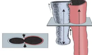
objects with significant noise and overlap at different levels. As shown in the figure the two cylinders are having gradually reducing radii which are going parallel. One cylinder is significantly larger than the other. The process starts with marking the seed followed by fuzzy segmentation which is done iteratively to produce better result. But only with the segmentation we can't separate the two cylinders as it can't find a threshold to separate the cylinder from top to bottom. Hence no straight forward morphological opening operator can solve the problem[10].
In Fuzzy connectivity paths, consider 2 cylinders with iso intensity objects with significant mutual overlap as shown in figure2. The intensity variation is fused regions are taken in consideration for separation of the fusible regions. Separating of fused cylinders is done using a suitable morphological operator. This morphological operator is used to combine locally separated regions and to determine the local size of the operators[11]. Selection of two seeds for each object is the initial step which is followed by segmentation and done iteratively to produce connectivity to the separated regions.
[image:2.595.91.248.384.479.2]The two cylinders are partially separated by using threshold that indicates the radius of the optimal erosion operator. The difficulty for separation at lower scale is that the scale of the annular remainder of the morphological erosion is at least equal to that of the regions not yet separated. This proposed morphological reconstruction operation is the main solution for this problem that fills the annular remainder while maintaining the separate identities of the two objects.
Fig 2: Morphologic erosion
Proceeding with separations at a lower scale and the method progresses iteratively separating fused structures at finer and finer scales starting with the larger one[12]. The entire process is summarized by the following three steps:
Step 1: Compute fuzzy segmentation of the assembly of
two mutually fused structures.
Step 2: Find the optimum morphological opening structure scale separating the two objects specified by two sets of seeds.
Step 3: Block the annular remainder around currently separated regions enabling morphological separations at lower scales (see Figure 2).
Step 4: Repeat Steps 2 & 3 until convergence.
Step 5:Segment artery region & compare normal image with Patient image.
The rest of this paper is organized as follows: Section B deals with the fuzzy segmentation, Section C deals with fuzzy
connectedness, Section D deals with local update with user interface and Section E deals with arteriosclerosis disease. Section III shows Results and Discussions.
2.2 Fuzzy Segmentation
Fuzzy connectedness principles have been efficient in segmenting a variety objects for large objects for image segmentation techniques. A new method has been introduced to segment tumors in 3 dimensional space using CT images using fuzzy rule based system. Initially the user selects a region which is called Region of Interest(ROI) within the tumor area. Depending on ROI's spatial and intensity properties, fuzzy inputs are generated for use in the fuzzy rules inference system. With a set of predefined fuzzy rules, the system generates a threshold value to segmenting the region. This creates a propagation of information from the previous slices, used to segment the current slice. The sub functions used during the fuzzification process are adaptive to the changes in the size and pixel intensities of the current ROI[13].
2.3 Fuzzy Connectedness
The most challenging job in image processing is defining the object in an image. Fuzzy connectedness framework aims at capturing this idea via a fuzzy topological notion called fuzzy connectedness which defines how the image elements hang together spatially in spite of their gradation of intensities. The author presented with a threshold on the strength of connectedness in his previous framework. Relative fuzzy connectedness provides a framework in which objects compete among each other and an image element is grabbed by the object within which the element has the largest fuzzy connectedness strength. It introduces the notion of iterative relative fuzzy connectedness that leads to more effective segmentations using relative connectedness[14].
A path π p, q
P(p, q) is one of the strongest morphological paths from p to q. The fuzzy morpho-connectivity between two points p ε𝑍
3 and q ε𝑍
3, denoted by𝛾
0(p, q), is the strength of one of the strongest morphological paths between p and q.
𝛾
0 (p,q) = πε P(p,q)
O(π)
𝑚𝑎𝑥
(1)
Fuzzy morpho-connectivity between two points p, q ε
𝑍
3 indicates the size of the morphological erosion operator that barely disconnects the two points[15].2.4 Local Update with an Effective User
Interface
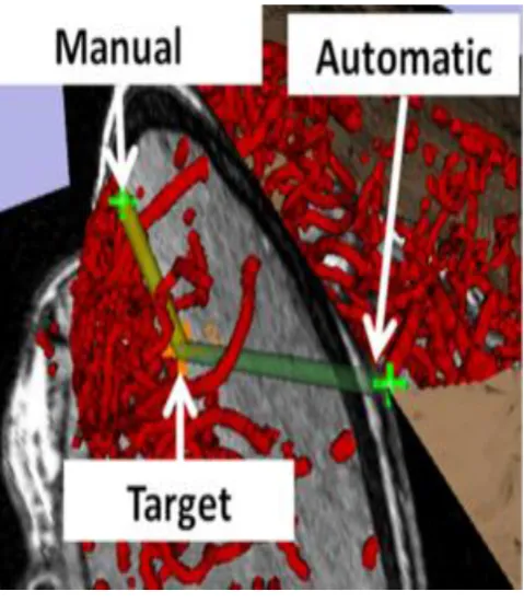
with a target[19]. And manual method deals with medical field that select seeds manually by a doctor.
Fig 3: 2D/3D graphical interface
(1)The local window inherits some A/V separation results from prior selected seeds possibly outside the local space.
(2) A/V separation inside the box needs to be partially or fully overruled with selection or deletion of seeds and separators[8].
Fig 4(a): Vessel structure Fig 4(b): Separation failure
in twoseeds
Fig 4(c): Selecting additional seed Fig 4(d): After global
update
The idea of local update is pictured in figure 4. The first one shows that it need to add some seeds in local space based on the results of A/v separation. The algorithm fails to eliminate faulty
segmentation if all voxels in arteries/veins are considered as seeds. So our main aim will be to use minimum set of seeds to receive global A/V separation in the local space. Any branch of an Vein or Artery should cross the outer region first before entering the inside and therefore regions of intersection between global separated veins/arteries. The outer surface of the local space is used to inherit seeds for arteries/veins during local updates, the seeds are called inherited seeds[20][21]. User specified seeds are also used simultaneously with inherited seeds to produce better result. Figure 4(a) shows the original vasculature with 2 inherited seed for artery and vein respectively. The result of initial seed can be seen in Figure 4(b) where we can seen an A/V separation is formed. We can see that a miscalculation is formed which resulted in formation of wrong venous branch, which requires a user interaction. An User specifies a seed manually in this region as shown in Figure 4(c) to rectify the error. As soon as the seed is added the process is invoked with new added seed as center. As a result correctly separated A/v region is formed as shown in Figure 4(d) but the connected arterial/venous structure in the whole image are not updated immediately, as shown in Figure 4(c). In order to get the new separated arterial/venous trees, a global update pre-process is needed, which takes more times[22].
2.5
Arterial Thrombosis
A blood clot forms within arteries is called arterial thrombosis. If artery has a hole or is damaged, thrombosis is helpful in stopping the bleeding and healing the artery[23]. Otherwise, the thrombosis can reduce or even block the blood supply, causing a stroke, a heart attack or peripheral vascular disease, when the arteries are not damaged. Main symptoms of arterial thrombosis include pain in the area of the clot, weakness, paralysis and paleness. The risk factors of the diseases include a poor diet, high cholesterol, diabetes, high blood pressure and a family history of the disease[24].
Fig 5(a): Artery with normal blood flow Fig 5(b): An artery with blood clot
[image:3.595.56.296.102.378.2] [image:3.595.54.292.377.694.2] [image:3.595.313.548.455.625.2]3. RESULTS AND DISCUSSIONS
Experiments are conducted on patient images collected from hospital. After pre-processing, the features mentioned in section I are connectedness from segmented images.
3.1
Patient Images
In 3D pulmonary CT imaging, consider about 50 medical data set for the 3D output images. For an example, just take 10 data set to reduce the time requirement.
Fig 6: A/V fused images of one patient
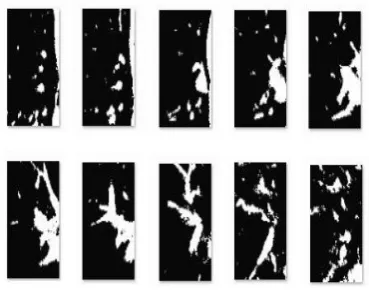
3.2 Fuzzy Segmented Output
[image:4.595.307.507.135.318.2]Image slices of the medical data set are loaded in order along with two blank template images having pixel values of the foreground and background data (denoted as white and black). The template images are progressively subtracted from the input image and the difference between the pixel values are noted. The pixel values of the template images are modified corresponding to the image data in the input image. Continued subtraction and classification of the input image into foreground and background gives a better segmentation result that a global threshold application. The output image can be optionally normalize and shifted to local scale.
Fig 7: Fuzzy segmented images
3.3 Fuzzy Connectedness Output
Two seed points are selected in the segmented image corresponding to the positions of artery and vein respectively in a single image. With this seed point a connection tree
[image:4.595.89.260.191.328.2]computation is run twice on the single image one each for both seeds. The output is an arterial or venal phase-wise segmentation of the input image. These images can be be saved separately and viewed in a 3d viewer by combining both sets and giving appropriate coloring and shading.
Fig 8: Fuzzy connectedness image
3.4 3D A/V Separated Output
The arterial and venal phases of the output image are given specific pixel values for later classification and passed on to the visualizer. Here the 2d images are stacked consecutively on top of each other to form the 3d volume. Isosurfaces connecting the voxels of similar value (here artery or vein) are drawn and appropriate colours are given which are then plotted on the 3d axis. Isosurfaces involve a connected 3d object made from a volume of connected pixels with the same pixel value.
Fig 9: 3D A/V separated images
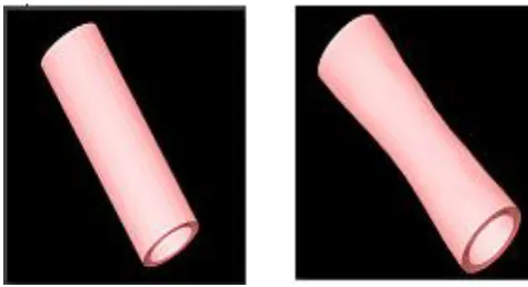
3.5 Arterial Thrombosis Image
[image:4.595.316.534.454.614.2] [image:4.595.90.275.532.680.2]average aperture data at specific vascular points in a normal human.
Fig 10: Comparison of normal image with arteriosclerosis image
4.
CONCLUSION AND FUTURE WORK
This work mainly concentrated on separation of artery /vein separation. To separate pulmonary arterial and venous trees through non contrast in vivo CT imaging, project presented a comprehensive system using the theory of arecently developed multi-scale fuzzy enhanced topo-morphologic opening algorithm and it embedded into a 2D-3D inter-connected graphical user interface[25].
The steps related to optimal erosion and constrained dilation using morpho connectivity and fuzzy segmentation within the multi-scale fuzzy enhanced topo-morphologic opening algorithm have been described. Also, we have introduced a new theory and methods for handling local update and separators essential for a practical A/V separation system with an effective user interface.
The same algorithm is used in modification. Here only artery region is focused to diagnose arterial thrombosis diseases with its radius calculation.
5 ACKNOWLEDGEMENT
Authors would like to thank all the researchers who have contributed in this field of research. The comments of anonymous reviewers to improve the quality of this paper are also acknowledged.
6 REFERENCES
[1] Saha P. K, Gao Z, Alford S. K, Sonka M and Hoffman E. A, “Topomorphologic separation of fused isointensity objects via multiscale opening: separating arteries and veins in 3-D pulmonary CT,” IEEE Trans Med Imag,. 840– 851, vol. 29, pp, 2010.
[2] Zhiyun Gao, Randall W. Grout, Colin Holtze, Eric A. Hoffman, and Punam K. Saha_, “A new paradigram of interactive Artery/Vein separation in non-contrast pulmonary CT imaging using multi-Scale topomorphologic opening,” IEEE Journ Med Imag 2012.
[3] G. Y. El-Khoury, M. H. Kathol, and W. W. Daniel, “Imaging of acute injuries of the cervical spine: Value of
plain radiography, CT, and MR imaging,” AJR Amer. J. Roentgenol., vol. 164, pp. 43–50, 1995.
[4] H. Shikata, E. A. Hoffman, and M. Sonka, “Automated segmentationof pulmonary vascular tree from 3D CT images,” in Proc. SPIE Int.Symp. Med. Imag., San Diego, CA, 2004, pp. 107–116.
[5] Z. H. Cho, J. P. Jones, and M. Singh, Foundation of Medical Imaging. New York: Wiley, 1993.
[6] X. Liu, D. Z. Chen, M. Tawhai, E. Hoffman, and M. Sonka, “Measurement,evaluation and analysis of wall thickness of 3D airway treesacross bifurcations,” in Proc. 2nd Int.Workshop Pulm. Image Process., London, UK, 2009, pp. 161–171.
[7] J. K. Udupa and G. T. Herman, “3D Imaging in Medicine”. Boca Raton,FL: CRC Press, 1991.
[8] K. C. Ciesielski, J. K. Udupa, P. K. Saha, and Y. Zhuge, “Iterative relative fuzzy connectedness for multiple objects with multiple seeds,” Comput. Vis. Image Understand., vol. 107, pp. 160–182, 2007.
[9] P. K. Saha and J. K. Udupa, “Relative fuzzy, P. K. Saha, and D. Odhner, “Artery-vein separation via MRA - an image processing approach,” IEEE Trans Med Imag, vol. 20, pp. 689–703, 2001.
[10] J. K. Udupa and P. K. Saha, “Fuzzy connectedness in image segmentation,” in Proc. IEEE, Emerging Med. Imag. Technol.,, 2003, vol. 91,pp. 1649–1669.
[11] T. Yonekura, “Classification algorithm of pulmonary vein and artery based on multi-slice CT image,” in Proc. SPIE: Med. Imag., San Diego, CA, vol. 6514, pp. 65 142E1–65 142E8 2007.
[12] Saha P. K and Udupa J. K, “Iterative relative fuzzy connectedness and object definition: theory, algorithms and applications in image segmentation,” in Proc of IEEE MMBIA 2000.
[13] T. Lei, J. K. Udupa segmentation,” in Proc. IEEE Workshop Math. Methods Biomed. Image Anal., Hilton Head, , pp. 28–35, SC, 2000.
[14] P. K. Saha and B. B. Chaudhuri, “3D digital topology under binary transformation with applications,” Comput. Vis. Image Understand.,vol. 63, pp. 418–429, 1996.
[15] C. M. van Bemmel, L. J. Spreeuwers, M. A. Viergever, and W. J. Niessen, “Level-set-based artery-vein separation in blood pool agent CE-MR angiograms,” IEEE Trans. Med. Imag., vol. 22, no. 10, pp. 1224–1234, Oct. 2003.
[16] A. L. D. Beckers and A. W. M. Smeulders, “Optimization of length measurements for isotropic distance transformations in three dimension,” CVGIP: Image Understand., vol. 55, pp. 296–306, 1991.
[17] P. K. Saha and J. K. Udupa, “Iterative relative fuzzy connectedness and object definition: Theory, algorithms, and applications in image connectedness among multiple objects: Theory, algorithms, and applications in image segmentation,” Comput. Vis. Image Understand., vol. 82, pp. 42–56, 2001.
[18] J. K. Udupa, P. K. Saha, and R. A. Lotufo, “Relative fuzzy connectedness and object definition: Theory, algorithms, and applications in image segmentation,” IEEE Trans. Pattern Anal. Mach. Intell., vol. 24, no. 11, pp. 1485–1500, Nov. 2002.
[image:5.595.54.292.102.231.2][20] LaMuraglia, G.M. ; Harvard Med. Sch., Massachusetts Gen. Hospital, Boston, MA, USA ; Prince, M.R. ; Nishioka, N.S. ; Obremski, S.,Optical properties of human arterial thrombus, vascular grafts, and sutures: implications for selective laser thrombus ablation,Quantum Electronics, IEEE Journal of (Volume:26 ,Issue: 12 ).
[21] Chun Xu ; Dept. of Mech. Eng. & Mech., Drexel Univ., Philadelphia, PA, USA ; Wootton, D.,“A model of platelet concentration sampling in arterial flow” (Volume:2, 2007).
[22] D. Pate, D. Resnick, M. Andre, D. J. Sartoris, S.Kursunoglu, D. Bielecki, P. Dev, and A. Vassiliadis, “Perspective: Three-dimensional imaging of the
musculoskeletal system,” AJR Amer. J. Roentgenol., vol.147, pp. 545–551, 1986.
[23] A. K. Jain,”Fundamentals of Digital Image Processing”. Upper Saddle River, NJ: Prentice Hall, 1989.
[24] T. Buelow, R. Wiemker, T. Blaffert, C. Lorenz, and S. Renisch, “Automatic extraction of the pulmonary artery tree from multi-slice CT data,” vol. 5746, pp, in Proc. SPIE: Med. Imag., San Diego, CA, 2005.