N A N O E X P R E S S
Open Access
In Vitro Study of Influence of Au
Nanoparticles on HT29 and SPEV Cell Lines
Elena Pavlovich
1, Nataliia Volkova
1*, Elena Yakymchuk
2, Olena Perepelitsyna
2, Michail Sydorenko
2and Anatoliy Goltsev
1Abstract
Cell culture models are excellent tools for potential toxicity of nanoparticles and fundamental investigations in cancer research. Thus, information about AuNP potential toxicity and effects on human health is necessary for the use of nanomaterials in clinical settings. The aim of our research is to examine the effects of AuNPs on the
epithelial origin cell lines: continuous and oncogenic. Embryonic porcine kidney epithelial inoculated (SPEV) cell line and colorectal carcinoma cell line (HT29) were used. In the test cultures, the cell proliferation, necrosis/apoptosis, and multicellular spheroids generation were evaluated. We demonstrated that AuNP concentrations of 6–12μg/ml reduced the proliferation of SPEV and HT29 cells and increased the cell number at early and late stages of
apoptosis and necrosis. It was shown that small concentrations of AuNPs (1–3μg/ml) stimulate multicellular spheroid formation by HT29 and SPEV cells. However, higher AuNP concentrations (6–12μg/ml) had both cytotoxic and anti-cohesive effects on cell in suspension. The large sensitiveness to the action of AuNPs was shown by the line of HT29 (6μg/ml) as compared to the SPEV cells (12μg/ml). This experimental study of the effect of AuNPs on SPEV and HT29 cell lines will justify their further application in AuNP-mediated anticancer treatment.
PACS:61.46+w, 61.48+c, 61.48De, 87.15−v, 87.64−t
Background
Production and investigation of the gold nanoparticles (AuNPs) have not only high potential for wide thera-peutic application of gold [1, 2] but also made them suit-able for specific biomedical applications such as target therapies [3, 4]. Recent reports have demonstrated that the use of AuNPs provides an opportunity for novel an-titumor therapies with a reduced risk for development of resistance. Thus, several studies have proven nanoparti-cle antitumor activity against breast, liver, gastric, colon, lung cancer [5, 6].
It is known that nanoparticles (NPs) can modulate cell fate, induce or prevent mutations, initiate cell–cell com-munication, and modulate cell structure [7, 8]. In addition, AuNPs have advantages over other metal NPs due to their biocompatibility and antitumor activity [8–12]. The cyto-toxic and genocyto-toxic effects of AuNPs are associated with their shape, size, charge, concentration, interaction time,
etc. [12–14]. Thus, information about their potential tox-icity and effects on human health is necessary for the use of nanomaterials in clinical settings.
Currently, despite the great success in the targeted therapy, the problem of selective delivery of AuNPs in the target tissue remains unsolved. Some studies have noted different rates of uptake of NPs by epithelial cells of different origin [15, 16]. Yet, investigations to explain this phenomenon are lacking even though they may help to achieve tissue-selective targeting of AuNPs. Anatom-ical or physiologAnatom-ical differences between different epithe-lia could explain differences in AuNPs uptake and transport rates. In particular, the rate of uptake may be influenced by the plasma membrane properties of the cells and the binding of nanoparticles to cell surface glycoproteins and proteoglycans, as well as the cells’ capacity for vesicular transport [17]. Thus, taking into account the impossibility of exclusively selective inter-action of nanoparticles with target cells, the comparative study of the features of their interaction with normal and oncogenic cells in order to avoid undesirable conse-quences of cancer therapy is topical [8–10].
* Correspondence:[email protected]
1Institute for Problems of Cryobiology and Cryomedicine, National Academy
of Science of Ukraine, Pereyaslavskaya str., 23, Kharkiv 61015, Ukraine Full list of author information is available at the end of the article
Although in vivo models are valuable for evaluating biological toxicity of nanoparticles, cell culture models are highly useful for preclinical physiological and toxico-logical studies. Currently, cell cultures are widely used in various fields of biology, medicine, veterinary medicine, and biotechnology. The use of cell cultures allows to explore biological processes that are difficult and some-times impossible, to study at the level of organisms. An important role of cell cultures is played in biotechnology in the production of many vaccines, test systems, and biologically active substances. Cell cultures are used to diagnose diseases of various etiologies, as test objects when testing new pharmacological, therapeutic, and cosmetic agents, as well as food additives [18].
In this work on cell culture models, we tried to examine the features of effects of AuNPs of the epithelial cells of continuous and oncogenic cell line origin. Monolayer cul-ture of epithelial cells SPEV (embryonic porcine kidney epithelial inoculated line) and HT-29 (colon carcinoma cell line) cells can be considered as a model of normal and cancer epithelial tissues when anti-tumor therapy with AuNPs is applied. Several traditional cytotoxicity assays, including the adhesion, proliferation, necrosis/apoptosis, and multicellular spheroids were employed to validate the cell cytotoxicity of AuNPs.
Methods
Culture of SPEV Cells
SPEV cells were cultured in plastic flasks in DMEM (Sigma, USA) with 5% FCS (v/v) (HyClone, USA) sup-plemented with penicillin/streptomycin (PAA, Austria) and amphotericin B (5 μg/ml) (5% CO2, 95% humidity)
as reported by [19]. Seeding concentration was 0.5– 2 × 10⁴cells/cm2. Culture medium was replaced every 2 days. Cells were passaged at 100% confluence [20]. SPEV cell line grew and preserved initial morphological structure of monolayer during serial passages without evidence of cell degeneration in culture.
Culture of HT29 Cells
HT29 cells were cultured in plastic flasks (Nunc, Denmark) in RPMI-1640 medium (Sigma, USA) with 10% FCS (v/v) (HyClone, USA) supplemented with 2 mM L-glutamine (Sigma, USA) and 40 mg/ml genta-mycin (Sigma, USA) under standard conditions (5% CO2, 95% humidity) [21]. The optimal cell density was
0.5–4.0 × 10⁴cells /cm2. The cells were kindly provided to us by the Bank of Cell Lines from Human and Animal Tissue of RE Kavetsky Institute of Experimental Path-ology, Oncology and Radiobiology NAS of Ukraine.
Manipulations with AuNPs
AuNPs were kindly provided by the Institute of Bio-chemistry and Physiology of Plants and Microorganisms, Russian Academy of Sciences. AuNPs were synthesized by citrate method [22]. The average size of nanoparticles was 15 nm. The initial concentration of gold was 57μg/ ml. The results of dark field electron microscopy, image of 15 nm AuNPs, and extinction spectra of 15 nm AuNPs (b) are shown in Fig. 1 (Fig. 1a; note––diagram of the size distribution) [23]. AuNPs were introduced in cells by passive diffusion at 37 °C. The investigated con-centrations were 1, 3, 6, and 12 μg/ml. Cells without AuNPs under the same conditions were taken as control ones.
Adhesion and Proliferation Cells
Morphofunctional state of cell cultures was judged by adhesive properties and proliferative activity. The adhe-sive properties of SPEV and HT29 cells were visually evaluated using an inverted microscope; the numbers of adhered and flattened cells were counted 30, 60, 120, 180, and 1440 min after culturing beginning.
The proliferation dynamics of SPEV and HT29 cells was studied for 1–4 days. To examine the increment of cell number in the studied cultures to the investigation terms, they were enzymatically (1:1 (0.25% trypsin
[image:2.595.59.539.560.702.2]solution: EDTA, PAA)) detached from plastic and the number of cells was counted. The total number of cultured cells was counted by the traditional method in Goryaev’s chamber.
Apoptotic/Necrotic Processes
Apoptotic and necrotic processes in SPEV and HT29 cells exposed to AuNPs were investigated in 4 days with Annexin-V (BD, USA), 7-Amino-Actinomycin (7AAD) (BD) dyes using a FACS Calibur Becton-Dickinson. The results were analyzed with WinMDI v.2.8 software.
Multicellular Spheroids
The multicellular spheroids (MSs) were generated to es-timate the in vitro impact of AuNPs on migration and aggregation potential of investigated cells. Spheroid (3-D) model system of SPEV and HT29 cells was cultured by the conventional method, which was reported by [24] and modified in our laboratory [25]. Briefly, cell suspen-sion were counted using trypan blue and equal numbers of cells (5 × 10⁴ cells/cm2) were planted in full supple-mented culture medium. MS generation was achieved by handling cell culture with 0.24% carboxy-methyl-cellulose (CMC) in 24-well plates coated with 1% agar with rotation (80 rpm) for 24 h. After that, 3-D cell cul-ture was maintained in standard conditions. To investi-gate dependence of the size and number of MSs on the AuNP concentration, MSs were generated in the pres-ence of AuNPs. Further cultivation was conducted for 48 h with constant rotation of plates. At the next stage, micro photo images were made by dark field method with a Carl Zeiss Stemi 2000 microscope. MS morph-ology was studied with the help of Axio Vision Release 4.7 program (Zeiss). This program allows measuring the geometric dimensions of cell aggregates. Then, the vol-ume of all MSs, which were in the files, was calculated. It was used with the following formula:V= 0.4 ×a×b2, where a and b––geometric diameters of MSs [24]. For statistical analysis, all cell aggregates were grouped by size from 1 × 10⁻⁴mm3to 1 × 10⁻²mm3with increment of 1 × 10⁻³ mm3. The MS number and median of MS volumes were estimated for each group.
Statistical Analysis
A single-factor analysis of variance and Student’s t test were used for statistical processing of the data with the software package Statistica 8. The significance threshold was 0.05. The results are presented as means and stand-ard errors (M ± SE).
Results
Effect of AuNPs on Adhesion of SPEV and HT-29 Cells
Cell adhesion is an indicator of functional state of cells, and it is necessary for further growth of culture. When
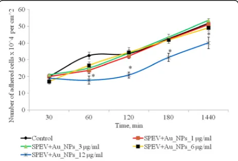
adhesion terminated, cells became flattened and gained appropriate morphology. Adhesive properties of SPEV cells are presented in Fig. 2.
After 1 h cultivation of SPEV cells with AuNPs at 1, 3, and 6μg/ml, the number of adhered cells was lower ver-sus the control value. The percentage of flattened cells in samples with AuNPs for these concentrations did not significantly differ from the control. Adhesion was slo-wed down after 1 h incubation with AuNPs at 12μg/ml. The number of adhered cells per squared centimeter was reduced by 1.8 times versus the control. This ten-dency in adhesion persisted for all the test periods. After 24 h of observation, the number of adhered cells was lower versus the control by 1.3 times. At the same time, incubation of AuNPs at small concentrations (1 and 3 μg/ml with tumor cells (HT29) had no significant ef-fect on the amount of adhesive cells. Increasing of AuNP concentration to 6 and 12μg/ml lead to decreasing the number of tumor cells in the adhesive fraction in 1.16 and 1.28 times, respectively, (Fig. 3). The obtained data can be influenced by several processes. The one is the cytostatic/cytotoxic effect of AuNPs on the adhesion fraction of both tumor and embryonic cell lines, which leads to cell death, transition to apoptosis, or necrosis. The other process is the reduction of cell adhesion, under the influence of AuNPs and transfer of cells into the suspension fraction. Notably, both processes can be realized simultaneously, and each one can contribute to the decrease in the number of living cells in the adhe-sion fraction.
Effect of AuNPs on Proliferation of SPEV and HT-29 Cells
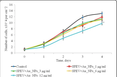
The effect of AuNPs within the concentration range of 1–12 μg/ml on proliferative processes in SPEV cell culture was studied (Fig. 4). On days 2–4 of culturing with AuNPs at 1, 3, and 6 μg/ml, the cell number did not significantly differ from the control. On day 4 of
[image:3.595.306.538.548.705.2]culturing with AuNPs at 3 and 6 μg/ml, this index decreased by 1.15 and 1.23 times, respectively, as com-pared with the control. Reduction in the cell number by 1.5 times (days 2 and 3) and by 1.15 times on day 4 of culturing with AuNPs at 12μg/ml was observed in SPEV culture versus the control. Thus, the AuNP concentra-tion, 12μg/ml, slowed down cell growth within the ob-served time period.
The effect of AuNPs at concentrations from 1 to 12μg/ml on the number of HT 29 cells in a monolayer culture is shown in Fig. 5. During the first 3 days of in-cubation, the number of cells in the control and in the presence of AuNPs was not statistically different. On the 4th day of cultivation, it was noted a dose-dependent de-creasing of the number of cells in 2D culture. So, after 4 days of cultivation, for low concentrations of AuNPs (1 and 3 μg/ml), the number of HT 29 cells is not sig-nificantly different in comparison with control. But at higher AuNP concentrations (6–12 μg/ml), HT29 cell
number was lower than the control in 1.33 and 1.44 times, respectively.
Effect of AuNPs on Apoptotic/Necrotic Processes in SPEV and HT-29 Cells
SPEV and HT-29 cells in the presence of AuNPs were cultured for 4 days under the standard conditions. The culturing of SPEV and HT29 cells with AuNPs at 1 and 3 μg/ml and the indexes of apoptotic/necrotic processes did not significantly differ from the control (Tables 1 and 2).
Culturing with AuNPs at 6–12 μg/ml increased the percentage of Annexin V+/7AAD+, Annexin V−/7AAD +, and Annexin V+/7AAD-cells as well as reduced the percentage of alive cells. The number of Annexin V
+
/7AAD+ SPEV cells was higher than the control value by 7.8 ± 0.7% (p ≤ 0.05) with 12 μg/ml of AuNPs. The number of Annexin V+/7AAD+ HT 29 cells was higher than the control value by 3.2 ± 0.4% (p ≤ 0.05) with 6 μg/ml of AuNPs and by 4.8 ± 0.6% (p ≤ 0.05) with 12μg/ml of AuNPs.
Effect of AuNPs on Generation of Multicellular Spheroids from SPEV and HT29 Cells
To determine the dependence of size and number of multicellular spheroids (MSs) on AuNP concentration, MSs were generated at various concentrations of AuNPs during 48 h. Our data demonstrated the variety ability of HT29 and SPEV cells to form multicellular spheroids under the same conditions of the microenvironment (Figs. 6 and 7).
So, if the control samples of HT29 cells for 48 h formed spheroids in average volume 5.19 × 10⁻³ mm3, the average volume spheroid of SPEV cells was 0.79 × 10
−5
mm3. At the same time, the influence of AuNPs on the HT29 and SPEV cells had the same trend. The pres-ence of AuNPs in the cell microenvironment stimulated Fig. 3Dynamics of adhesion of HT29 cells after exposure of AuNPs,
*p≤0.05 is significant versus with the control
Fig. 4Proliferation of SPEV cells after exposure of AuNPs, *p≤0.05 is significant versus with the control
[image:4.595.306.539.87.245.2] [image:4.595.58.292.87.251.2] [image:4.595.57.291.548.702.2]the formation of multicellular spheroids in both cultures. Thus, when the concentration of AuNPs was 1 and 3 μg/ml volume of MSs for SPEV increased by 9.7 and 7.4 times, respectively, compared with the control (Fig. 6), the same AuNP concentrations also stimulated an in-creasing volume of MSs for HT29 by 1.4 and 1.2 times, respectively (Fig. 7).
Further increasing of AuNP concentration leaded to decreasing average volume of MSs in both cultures. The elevation in AuNP concentration from 1 to 12μg/ml de-creased the volume of HT29 MSs from 7.18 × 10⁻³mm3 to 4.24 × 10⁻³, in 1.69 times, according to the control. As for SPEV, when the concentration of AuNPs was increased from 1 to 12 μg/ml, the volume of MSs decreased from 7.69 to 4.58 × 10⁻⁵mm3, in 1.68 times, according to control. However, increasing AuNP con-centration coincidences with reduction in volume of MSs and correlated with increases in the number of spheroids in culture of HT29 cells (Figs. 6 and 7). The number of HT29 MSs increased from 3 to 10 per field of view at AuNP concentration from 1 to 12 μg/ml. At the same time, the number of SPEV MSs decreased from 32 to 19, respectively.
The obtained data (Figs. 6 and 7) demonstrate that AuNPs are capable of influencing cohesive interactions in the cell-to-cell system. Our data show that small con-centrations of AuNPs (1–3μg/ml) stimulated the forma-tion of multicellular spheroids of both embryonic and tumor cells. However, higher AuNP concentrations (6– 12 μg/ml) had both cytotoxic and anti-cohesive effects on cell in suspension. This process contributed to for-mation of a larger number of HT29 MSs with the de-creased average volume. As for SPEV, the high concentration of AuNPs may have a cytostatic effect
which reduced cell numbers in the adhesive fraction and the number of MSs in suspension. Previously, the authors reported that carbon nanoparticles reduce the adhesion of cells to the substrate, stimulate cell transfer into the sus-pension, and leaded to formation of multicellular spher-oids [25, 26]. In the literature, there are data about AuNP ability to break the structure of actin/myosin microfila-ments and decrease cell proliferation, adhesion, and differ-entiation [27]. Our data confirmed this assumption.
Discussion
We evaluated the effects of AuNPs on proliferation, ne-crosis/apoptosis, and formation of multicellular spher-oids of the epithelial cells continuous and oncogenic cell line origin. It was shown that the AuNPs at 6–12μg/ml reduced the number of SPEV and HT29 cells and in-creased the cell number at early and late stages of apop-tosis and necrosis. The small concentrations of AuNPs stimulate formation of multicellular spheroids by HT29 and SPEV cells. However, higher AuNP concentrations had both cytotoxic and anti-cohesive effects on cell in suspension. The large sensitiveness to the action of AuNPs was shown by the line of HT29 (6 μg/ml) as compared to the SPEV cells (12μg/ml.)
[image:5.595.56.539.100.183.2]The effects of AuNPs on cellular morphology and cytoskeleton have only recently received more attention, and the underlying mechanism and forthcoming conse-quences have not been investigated in depth [28–30]. In this regard, it is important for all novel AuNP types to evaluate their endocytic uptake pathway and intracellular localization as a function of time. For different types of AuNPs, the effects have been described to be dependent on intracellular AuNP concentration and to be transient, where after recurrent cell divisions, the intracellular Table 1Cytofluorimetric analysis of SPEV cells after 4 days culturing with AuNPs, staining with Annexin V and 7AAD
Sample/region AnnexinV+/7AAD− Annexin V+/7AAD+ AnnexinV−/7AAD− AnnexinV−/7AAD+
Control 3.4 ± 0.4 4.5 ± 0.7 91.6 ± 1.0 0.5 ± 0.1
SPEV + Au_NPs_1μg/ml 3.5 ± 0.6 4.4 ± 0.8 91.4 ± 1.2 0.7 ± 0.2
SPEV + Au_NPs_3μg/ml 3.7 ± 0.5 3.6 ± 0.8 91.0 ± 1.1 1.7 ± 0.1
SPEV + Au_NPs_6μg/ml 3.9 ± 0.6 10.1 ± 0.5* 84.6 ± 1.2* 1.4 ± 0.2*
SPEV + Au_NPs_12μg/ml 5.1 ± 0.8* 12.5 ± 0.7* 81.1 ± 1.2* 1.3 ± 0.1*
Note*p≤0.05 is significant versus with the control
Table 2Cytofluorimetric analysis of HT29 cells after 4 days culturing with AuNPs, staining with Annexin V and 7AAD
Sample/region Annexin V+/7AAD− Annexin V+/7AAD+ Annexin V−/7AAD− Annexin V−/7AAD+
Control 3.1 ± 0.5 4.6 ± 0.6 91.8 ± 0.9 0.5 ± 0.2
HT29 + Au_NPs_1μg/ml 2.9 ± 0.7 3.8 ± 0.5 92.8 ± 1.1 0.5 ± 0.3
HT29 + Au_NPs_3μg/ml 3.0 ± 0.6 3.9 ± 0.7 92.5 ± 1.2 0.6 ± 0.2
HT29 + Au_NPs_6μg/ml 4.1 ± 0.5* 7.7 ± 0.4* 87.3 ± 1.5* 0.9 ± 0.3*
HT29 + Au_NPs_12μg/ml 5.3 ± 0.9* 9.2 ± 0.8* 84.0 ± 1.3* 1.2 ± 0.2*
[image:5.595.56.540.638.723.2]AuNP concentrations decrease exponentially and the ef-fects are no longer observed. Also, possible endosomal escape of the AuNPs must be assessed. As cytoskeleton defects have been described to be clearly dependent on AuNP concentrations, a wide concentration range of particles should be tested in order to try and assess the maximal cellular loading capacity without any effects. Furthermore, as the cytoskeleton is also involved in many intracellular signaling pathways, it remains to be investigated whether the AuNPs induced cytoskeletal disruption leads to secondary effects [31].
As NPs have certain physical dimensions, the intracel-lular volume they occupy can lead to alterations in cellu-lar morphology or affect the structure of the cellucellu-lar cytoskeleton network [28, 29, 31]. The later effects can also be due to the high demands of the NP pose on the cellular endocytic manner. AuNPs have been described to have a profound effect on the morphology of several
cell types, such as A549 human carcinoma lung cells [32]. AuNPs have also been described to have a concentration-dependent effect on the actin fibrils of human dermal fibroblasts [33, 34]. Mironava et al. [35, 36] further showed the cytoskeleton filaments to be dis-rupted as a function of AuNP exposure time, concentra-tion, and size of the NPs although actin or β-tubulin protein expression levels were not affected.
The cell type used is also of great importance as differ-ent cell types, even when closely related, can react quite differently for the same type of nanomaterials [37, 38]. Preferably, those cell types which are most involved in the (future) biomedical applications of the NPs should be tested (e.g., epithelial, endothelial cells), or multiple cells which are derived from the different germ layers. When investigating cytotoxic effects, the use of cancer cell types should be minimized, as these can lead to ab-errant results [39]. Cancer cells have several specific Fig. 6Number and volume of MS cells SPEV after incubation with AuNPs, #p≤0.01 (for number MSs); **p≤0.01 (for volume of MSs)
[image:6.595.59.540.88.270.2] [image:6.595.59.540.532.713.2]characteristics and altered intracellular signaling path-ways which are destined to upregulate proliferation and maintain cell viability, which will make them less prone to some NP-mediated cytotoxic effects.
In our opinion, binding of AuNPs to surface functional groups (e.g., transmembrane proteins) of cells can be re-versible or irrere-versible, resulting in temporary or perman-ent structural injuries [40, 41]. Potperman-ential implications of changes in biomechanical properties (e.g., hardness and elasticity), adhesiveness, and surface electrical properties of cells are perceivable. Thus, changes in hardness or elas-ticity are likely to influence the surface structural flexibil-ity, production of mechanical energy for cell division, and cell motility. As for adhesiveness, the cell microenviron-ment is normally composed of extracellular matrix with specific molecules that allow cells to adhere to their sur-roundings [42]. Surface charge undoubtedly plays an im-portant role in interactions between cells and their surroundings.
The other authors also reported of the NPs are prefer-entially localized in mitochondria and cause oxidative stress as well as potentiate structural damage [40]. A re-cent article by Pan et al. describes that 1.4 nm AuNPs induce necrosis via oxidative stress and mitochondrial damage in Hela cells [43]. Accumulation of nanoparti-cles in cell medium upon biodegradation is unsafe be-cause it may disrupt organelles and even be-cause genetic mutations.
Changes occurring in cells during apoptosis are similar for most of cell types. In apoptotic cells, there are changes of lipid composition of plasma membrane: phosphatidyl serine transfers from cytoplasmic part of bilayer to outer side, causing caspase cascade activation, chromatin condensation, and disorder of electron trans-port chain in mitochondria and eventually arresting ATP synthesis. Programmed cell death can be triggered by receptor-mediated physiological stimuli resulted from genetic disorders, exposure to chemical or physical fac-tors as well as by other changes in cells. We observed this effect is with 6–12μg/ml of AuNPs.
Multicellular aggregates (spheroids, embryoid body) represent an intermittent level between monolayer grow-ing cells and tissue culture. Spheroids are objective model of the cell three-dimensional growth and organization, the cell-to-cell interactions and influence of microenviron-mental conditions, for example, AuNP concentration, on intensiveness of proliferation as well as on cell adhesive-ness and formation of microaggregates. MS formation is a well-established culture method as for tumor as for em-bryonic cell lines [24, 44, 45]. In our work, formation and growth of spheroids is achieved by adding CMC as part of artificial extracellular matrix and surface coating by 1% agar which inhibited cell adhesion to surface and stimu-lated cell aggregation. At these conditions, MS culture can
be realized until the aggregates forming central necrosis, due to limited cell mass growth or spontaneous differenti-ation of embryonic cells.
In literature, there is information about interaction AuNPs with colon cancer cell line and embryonic cell lines [46, 47]. According these data, exposure to even very low concentrations of AuNPs may have a damaging effect on the Human Embryonic Neural Precursor Cells and HT29 by stressing cell proliferation, differentiation, and apoptotic cell death.
There are published data that the effect of AuNPs is based on G0/G1 phase accumulation, S and G2/M phase depletion, as well as on reduced ATP levels in human oral squamous carcinoma cells (HSC-3) [48]. Cell cycle regulation may be addressed by violation of focal con-tacts of cells with substrate and cell transfer to sus-pended fraction in 2D culture and inhibition cell-to-cell contacts in gap junction in 3D culture [48–50]. Due to nanosize of AuNPs (near 15 nm), it cannot be centers of cohesion for cells. At the same time, intercalation of AuNPs into cell membrane [51], influence on cell mem-brane AuNP zeta potential [32], and influence on forma-tion cell-to-cell/cell-to-surface contacts obviously can trigger mechanism for necrosis/apoptosis, cytotoxic ef-fect, and cell cycle arrest. Violation of focal contacts of cells with substrate and cell transfer to suspended frac-tion is a way of cell cycle regulafrac-tion [48, 49]. Small con-centrations of AuNPs exerted no statistically significant cytotoxic effect on cells. However, higher AuNP concen-trations had both cytotoxic and anti-cohesive effects on cell in suspension. This process contributed to formation of a larger number of MSs with the decreased average volume. We suppose that AuNP wedge into cohesive contacts of cells and compromise them. Thus, our ex-periments on the effects of AuNPs on SPEV and HT29 cell lines support their further application in develop-ment of AuNP-mediated cancer therapies.
Although future studies will be necessary to confirm anti-cancer effects on the in vivo animal studies. Never-theless, our deep conviction is that if we know the na-ture of substance and its possible negative influence, we are able to avoid the detrimental effects of AuNP and use their positive biotechnological potential. Our investi-gation could be applied quite reliably in effective mate-rials for anti-cancer treatment context with maximum advantage for medicine.
Conclusions
necrosis. Also, it was shown that small concentrations of AuNPs (1–3μg/ml) stimulate formation of multicellular spheroids. However, higher AuNP concentrations had both cytotoxic and anti-cohesive effects on cell in sus-pension. The large sensitiveness to the action of AuNPs was shown by the line of HT29 (6 μg/ml) as compared to the SPEV cells (12μg/ml.)
Acknowledgements
We are deeply grateful to Dr. L.A. Dykman, who kindly provided us with gold nanoparticles and their characterization.
Authors’Contributions
MS and OP provided the study design. AG and NV contributed to the study design. NV and EP carried out experiments, analyzed data, and wrote the manuscript. EP performed the cytofluorimetric analysis. EY and OP took part in experiments, data analyzing, and writing the manuscript. EY ensured the cytotoxic assay. OP carried out the 3D culture experiments and analyzed the data. All authors read and approved the final manuscript.
Competing Interests
The authors declare that they have no competing interests.
Publisher’s Note
Springer Nature remains neutral with regard to jurisdictional claims in published maps and institutional affiliations.
Author details
1
Institute for Problems of Cryobiology and Cryomedicine, National Academy of Science of Ukraine, Pereyaslavskaya str., 23, Kharkiv 61015, Ukraine.
2
Department for Biotechnical Problems of Diagnostic, Institute for Problems of Cryobiology and Cryomedicine, National Academy of Science of Ukraine, 42/1Nauky av., Kiev 03028, Ukraine.
Received: 1 December 2016 Accepted: 4 August 2017
References
1. Leu JG, Chen SA, Chen HM, Wu WM, Hung CF, Yao YD, Tu CS, Liang YJ (2012) The effects of gold nanoparticles in wound healing with antioxidant epigallocatechin gallate andα-lipoic acid. Nanomedicine. doi:10.1016/j.nano. 2011.08.013
2. Volkova N, Yukhta M, Pavlovich O, Goltsev A (2016) Application of cryopreserved fibroblast culture with Au nanoparticles to treat burns. Nanoscale Res Lett 11:22. doi:10.1186/s11671-016-1242-y
3. Alam F, Naim M, Aziz M, Yadav N (2015) Unique roles of nanotechnology in medicine and cancer-II. Indian J Cancer 52(1):1–9. doi:10.4103/0019-509X. 175591
4. Oh MH, Yu JH, Kim I, Nam YS (2015) Genetically programmed clusters of gold nanoparticles for cancer cell-targeted photothermal therapy. ACS Appl Mater Interfaces 7(40):22578–22586. doi:10.1021/acsami.5b07029 Epub 2015 Oct 1
5. Egusquiaguirre SP, Igartua M, Hernández RM, Pedraz JL (2012) Nanoparticle delivery systems for cancer therapy: advances in clinical and preclinical research. Clin and Transll Oncol 14(2):83–93. doi:10.1007/s12094-012-0766-6. 6. Loutfy SA, Al-Ansary NA, Abdel-Ghani NT, Hamed AR, Mohamed MB, Craik
JD, Eldin TA, Abdellah AM, Hussein Y, Hasanin MT, Elbehairi SE (2015) Anti-proliferative activities of metallic nanoparticles in an in vitro breast cancer model. Asian Pac J Cancer Prev 16(14):6039–6046
7. Behzadi S, Serpooshan V, Tao W, Hamaly MA, Alkawareek MY, Dreaden EC, Brown D, Alkilany AM, Farokhzad OC, Mahmoudi M (2017) Cellular uptake of nanoparticles: journey inside the cell. Chem Soc Rev. doi:10.1039/ c6cs00636a
8. Bao H, Zhang Q, Xu H, Yan Z (2016) Effects of nanoparticle size on antitumor activity of 10-hydroxycamptothecin-conjugated gold nanoparticles: in vitro and in vivo studies. Int J Nanomedicine 11:929–940. doi:10.2147/IJN.S96422
9. Xiang-Yu S, Liu P-D, Wu H, Ning G (2014) Enhancement of radiosensitization by metal-based nanoparticles in cancer radiation therapy. Cancer Biol Med 11(2):86–91
10. Priya KMR, Lyer PR (2015) Anticancer studies of the synthesized gold nanoparticles against MCF 7 breast cancer cell lines. Appl Nanosci 5:443–448 11. Zhao P, Li N, Astruc D (2013) State of the art in gold nanoparticle synthesis.
Coord Chem Rev 257:638–665
12. Jana NR, Gearheart L, Murphy CJ (2001) Wet chemical synthesis of high aspect ratio cylindrical gold nanorods. J Phys Chem B 105:4065–4067 13. Brown KR, Natan MJ (1998) Hydroxylamine seeding of colloidal Au
nanoparticles insolution and on surfaces. Langmuir 14:726–728 14. Nikoobakht B, El-Sayed MA (2003) Preparation and growth mechanism of
gold nanorods (NRs) using seed-mediated growth method. Chem Mater 15: 1957–1962
15. Freese C, Uboldi C, Gibson MI, Unger RE, Weksler BB, Romero IA, Couraud PO, Kirkpatrick CJ (2012) Uptake and cytotoxicity of citrate-coated gold nanospheres: comparative studies on human endothelial and epithelial cells. Part Fibre Toxicol 9:23. doi:10.1186/1743-8977-9-23
16. Jia YP, Ma BY, Wei XW, Qian ZY (2017) The in vitro and in vivo toxicity of gold nanoparticles. Chin Chem Lett 28(4):691–702
17. Gromnicova R, Kaya M, Romero IA, Williams P, Satchell S, Sharrack B, Male D (2016) Transport of gold nanoparticles by vascular endothelium from different human tissues. PLoS One 11(8):e0161610. doi:10.1371/journal.pone. 0161610
18. Kunzmann A, Andersson B, Thurnherr T (2010) Toxicology of engineered nanomaterials: focus on biocompatibility, biodistribution and biodegradation. Biochim Biophys Acta 1810(3):361–373 19. Freshni R (1989) Culture of animal cells. Methods. Mir, Moscow 20. Birger MO (1982) Manual for microbiological and virological research
methods. Medistyna, Moscow
21. Perepelitsyna OM, Garmanchouk LV, Sydorenko MV, Ostapchenko LI (2010) Formation of multicellular aggregates under different conditions of microenvironment. Cytol Genet 44(1):19–22
22. McFarland AD, Haynes CL, Mirkin CA, Van Duyne RP, Godwin HA (2004) Color my Nanoworld. J Chem Educ 81:544A
23. Khlebtsov NG, Melnikov AG, Dykman LA, Bogatyrev VA (2004) Optical properties and biomedical applications of nanostructures based on gold and silver bioconjugates. Photopolarimetry in remote sensing, pp 265–308 24. Bjerkvig R (1992) Spheroid culture in cancer research, CRC Press, vol 335 25. Perepelytsina O, Sydorenko M, Yakymchuk O, Dobrydnev A (2015) Effect of
single-walled carbon nanotubes on tumor cells viability and formation of multicellular tumor spheroids. Nanoscale Res Lett 10(1):150. doi:10.1186/ s11671-015-0858-7
26. Perepelytsina O, Yakymchuk O, Rud A, Kirian I, Sydorenko M (2014) Impact of carbon nanomaterials on the formation of multicellular spheroids by tumor cells. Phys Status Solidi A:1–7. doi:10.1002/pssa.201431358 27. Soenen SJ, Parak WJ, De Smedt S et al (2012) Cytotoxic effects of gold
Nanoparticles: a multiparametric study. ACS Nano 6(7):5767–5783. doi:10. 1021/nn301714n
28. Soenen SJ, Illyes E, Vercauteren D, Braeckmans K, Majer Z, De Smedt SC De Cuyper M (2009) The role of nanoparticle concentration-dependent induction of cellular stress in the internalization of non-toxic cationic magnetoliposomes. Biomaterials 30: 6803–6813.
29. Soenen SJ, Nuytten N, De Meyer SF, De Smedt SC, De Cuyper M (2010) High intracellular iron oxide nanoparticle concentrations affect cellular cytoskeleton and focal adhesion kinase-mediated signaling. Small 6(7):832– 42 832–42. doi:10.1002/smll.200902084
30. Singh A, Sahoo SK (2015) Stem Cell Nanoengineering, pp 87–95 31. Soenen SJ, Rivera-Gil P, Montenegro JM, Montenegro JM, Parak WJ, De
Smedt SC, Braeckmans K (2011) Cellular toxicity of inorganic nanoparticles: common aspects and guidelines for improved nanotoxicity evaluation. Nano Today 6:446–465. doi:10.1016/j.nantod.2011.08.001\30
32. Soenen SJ, Manshian B, Montenegro JM, Amin F, Meermann BR, Thiron T (2012) Cytotoxic effects of gold nanoparticles: a multiparametric study. ACS Nano 6(7):5767–5783
33. Pernodet N, Fang X, Sun Y, Bakhtina A, Ramakrishnan A, Sokolov J, Ulman A, Rafailovich M (2006) Adverse effects of citrate/gold nanoparticles on human dermal fibroblasts. Small 2(6):766–773
35. Mironava T, Hadjiargyrou M, Simon M, Jurukovski V, Rafailovich MH (2010) Gold nanoparticles cellular toxicity and recovery: effect of size, concentration, and exposure time. Nanotoxicology 4(1):120–137 36. Xia Q, Li H, Liu Y, Zhang S, Feng Q, Xiao K (2017) The effect of particle size
on the genotoxicity of gold nanoparticles. J Biomed Mater Res A 105(3): 710–719
37. Barua S, Rege K (2009) Cancer cell phenotype dependent differential intracellular trafficking of unconjugated quantum dots. Small 5(3):370–376 38. Joris F, Manshian BB, Peynshaert K, De Smedt SC, Braeckmans K, Soenen SJ
(2013) Assessing nanoparticle toxicity in cell-based assays: influence of cell culture parameters and optimized models for bridging the in vitro–––in vivo gap. Chem Soc Rev 42(21):8339–8359
39. Almeida JPM, Chen AL, Foster A, Drezek R (2011) In vivo biodistribution of nanoparticles. Nanomedicine 6(5):815–835
40. Nel AE, Mädler L, Velegol D, Xia T, Hoek EM, Somasundaran P, Klaessig F, Castranova V, Thompson M (2009) Understanding biophysicochemical interactions at the nano-bio interface. Nat Mater 8:543–557
41. Volle CB, Ferguson MA, Aidala KE, Spain EM, Núñez ME (2008) Quantitative changes in the elasticity and adhesive properties ofEscherichia coliZK1056 prey cells during predation by Bdellovibrio bacteriovorus 109 J. Langmuir 24:8102–8110
42. Derfus AM, Chan WCW, Bhatia SN (2004) Intracellular delivery of quantum dots for live cell labeling and organelle tracking. Adv Mater 16(12):961–966 43. Pan Y, Leifert A, Ruau D, Neuss S, Bornemann J, Schmid G, Brandau W,
Simon U, Jahnen-Dechent W (2009) Gold nanoparticles of diameter 1.4 nm trigger necrosis by oxidative stress and mitochondrial damage. Small 5: 2067–2076
44. Zujur D, Kanke K, Lichtler AC et al (2017) Three-dimensional system enabling the maintenance and direct differentiation of pluripotent stem cells under defined conditions. Sci Adv 3:e1602875
45. Petrenko Y, Sykova E, Kubionova S (2017) The therapeutic potential of three-dimentional multupotent mesenchymal stromal cell spheroids. Stem Cell Res Ther 8:94. doi:10.1186/s13287-017-0558-6
46. Huang R-FS, Wei Y-J, Inbaraj BS, Chen B-H (2015) Inhibition of colon cancer cell growth by nanoemulsion caring gold nanoparticles and lycopene. Int J Nanomedicine 10:2823–2846
47. Sodestjerna E, Johansson F, Klefbohm B et al (2013) Gold and silver nanoparticles affect the growth characteristics of human embryonic neural precursor cells. PLoS One 8(3):e58211
48. Mackey MA, Saira F, Mahmoud MA, El-Sayed MA (2013) Inducing cancer cell death by targeting its nucleus: solid gold nanospheres versus hollow gold nanocages. Bioconjug Chem 24(6):897–906
49. Gérard C, Goldbeter A (2014) The balance between cell cycle arrest and cell proliferation: control by the extracellular matrix and by contact inhibition. Interface Focus. doi:10.1098/rsfs.2013.0075
50. Schkie S, Mazur K, Bintig W, Ngezahayo A (2010) Cell cycle dependent regulation of gap junction coupling and apoptosis in GFSHR-17 granulosa cells. JBiomedical Science Endineering 3:884–891