S T U D Y P R O T O C O L
Open Access
The effects of deep neck muscle-specific
training versus general exercises on deep
neck muscle thickness, pain and disability
in patients with chronic non-specific neck
pain: protocol for a randomized clinical trial
(RCT)
Pegah Kashfi
1, Noureddin Karimi
1, Anneli Peolsson
2and Leila Rahnama
1,3*Abstract
Background:Altered thickness, cross-sectional area and activity of deep neck muscles have frequently been reported in patients with chronic non-specific neck pain (CNNP). It is claimed that these muscles do not recover spontaneously. These muscles provide a considerable amount of cervical stability. Therefore, various therapeutic exercises have been recommended to recover from resulting complications. However, most exercise protocols do not target deep neck muscles directly. Thus, this might be a reason for long-lasting complications. Accordingly, the purpose of the present study is to discuss a randomized controlled trial (RCT) protocol in which we aim to
investigate and compare the effects of neck-specific exercise programmes versus general exercise programmes in patients with CNNP.
Methods:A 2*2 factorial RCT with before-after design. Sixty-four participants with CNNP will be recruited into the study. They will be randomly divided into two groups, including specific neck exercise and general exercise. Each exercise programme will be carried out three times a week and will last for 8 weeks. Primarily, dorsal and ventral neck muscle thickness, pain and disability and secondarily, muscle strength, quality of life, sleep quality, fear avoidance and neck range of motion will be assessed at the baseline and immediately at the end of the exercise protocol.
Discussion:The results of this study will inform clinicians on which type of exercise is more beneficial for patients with CNNP.
Trial registration:IRCT2017091620787N2, Sep 16 2017.
Keywords:Neck pain, Exercise, Deep neck muscles, Muscle thickness
© The Author(s). 2019Open AccessThis article is distributed under the terms of the Creative Commons Attribution 4.0 International License (http://creativecommons.org/licenses/by/4.0/), which permits unrestricted use, distribution, and reproduction in any medium, provided you give appropriate credit to the original author(s) and the source, provide a link to the Creative Commons license, and indicate if changes were made. The Creative Commons Public Domain Dedication waiver (http://creativecommons.org/publicdomain/zero/1.0/) applies to the data made available in this article, unless otherwise stated.
* Correspondence:[email protected];[email protected]
1
Department of Physiotherapy, University of Social Welfare and Rehabilitation Sciences, Tehran, Iran
3Department of Physical Therapy, University of North Georgia, Dahlonega,
USA
Background
Two thirds of the adult population suffer from chronic non-specific neck pain (CNNP) [1], which is associated with disability, activities of daily living (ADL) difficulties, work dissatisfaction, and economic and social costs [2– 4]. In addition, altered muscle cross-sectional area, thickness, size, and activity of deep neck muscles have been frequently reported in previous research studies [4–7]. Falla et al. [5] and Kim et al. [6] showed deep neck flexors atrophy and altered electromyography activ-ity (EMG) in patients with neck pain. Rahnama et al. [7] and Fernández-de-las-Peñas et al. [8] demonstrated deep neck extensors muscle atrophy and altered EMG activity following chronic neck pain. These structural and activ-ity changes in deep neck muscles are claimed to be rea-sons for chronicity and recurrences of the neck pain [9,
10]. To reduce and compensate for negative impacts of such changes, therapeutic exercise is one of the most common and acceptable treatments. Various exercises including neck muscle strengthening, stretching and sta-bilizing exercises were recommended to overcome these complications in patients with CNNP [11–20]. However, the answer to the question of which exercises are the most effective remained controversial.
Janda suggested that in the presence of pain, neck and back superficial muscles are prone to guarding while the deep muscles are vulnerable to weakening [21]. muscle guarding could be induced by pain. When the pain remains the vicious cycle may lead more muscle guarding [22]. In this regard, stretching exercises may be recommended to reduce neck muscle guard in patients with CNNP [23, 24]. Add-itionally, general neck exercises are believed to im-prove general fitness and physiological interactions [17]. On the other hand, deep neck flexor training re-gimes have increased deep neck flexor thickness and strength in patients with CNNP. Landén Ludvigsson et al. [25] and Peolsson et al. [26] studied different exercise regimes in individuals with chronic whiplash associated disorder (WAD) and found more psycho-logic and clinical benefits from specific neck exercise (SNE) compared to general neck exercise (GNE). Deep cervical muscle thickness and the effects of ex-ercise on their atrophy were not assessed [25, 26]. However, considering the efficacy of both mentioned exercise regimes, no study has yet compared the ef-fects of these two exercise approaches on CNNP. Fur-thermore, despite the important role of deep neck extensor muscles in providing neck stability and healthy function [27], their training has been missed in many studies. Therefore, prescribing exercises tar-geting deep neck extensor muscles is essential as it is claimed that their recovery following pain inhibition would not happen automatically [6, 7].
General neck exercises are commonly prescribed by clinicians in routine management of CNNP based in order to improve general muscle activity and function and reduce muscle guarding. However, to the best of our knowledge, there are not yet any studies comparing the effects of general neck exercises and specific exer-cises targeting deep neck muscles (both flexor and ex-tensor muscles) in CNNP patients. Therefore, the aim of the present clinical trial is to evaluate and compare the effects of two exercise programmes including GNE and SNE on muscle morphology, pain, disability and func-tional measures in CNNP patients.
Study hypotheses
Primary hypothesis: There would be significant improve-ments in pain, disability and deep cervical muscle thick-nesses in both groups, but changes would be more prominent in the SNE group compared to the GNE group in people with CNNP.
Secondary hypothesis: There would be significant im-provements in cervical active range of motions, max-imum voluntary isometric contraction, sleep quality, quality of life and fear avoidance in both groups, but changes would be more prominent in the SNE group compared to the GNE group in people with CNNP.
Methods Trial design
The trial utilizes a single blinded design and conforms to the SPIRIT guidelines (Additional file1).
Study setting
This study will be conducted at the University of Social Welfare and Rehabilitation Sciences’ physiotherapy Re-search Lab and has been registered in the Iranian Regis-try of Clinical Trials (WHO subgroup) with the clinical trial registry number IRCT2017091620787N2. The pro-ject is in accordance with the ethical principles and na-tional norms and standards approved by the University of Social Welfare and Rehabilitation Sciences, Tehran, Iran (IR.USWR.REC.1396.194).
Participants
of pain anywhere in posterior of cervical spine, from su-perior nuchal line to the first thoracic spinous process) [30] of at least 3 months’duration in the past year [31], 3) pain intensity greater than 30 mm on the visual analogue scale (VAS) [5], and 5) diagnosed with CNNP. Neck pain was defined as having pain on the posterior aspect of the cervical spine anywhere from nuchal line to the first thoracic spine [30]. Volunteers will be ex-cluded if they report acute neck pain, history of any spinal surgery and disc disease, cervical fracture or tumour, radicular pain into their shoulders or any posi-tive neurologic signs, history of cervical trauma or crash injury, congenital abnormality of the spine, inflammatory diseases, vertigo or vestibular disorders [18,31,32].
Procedure
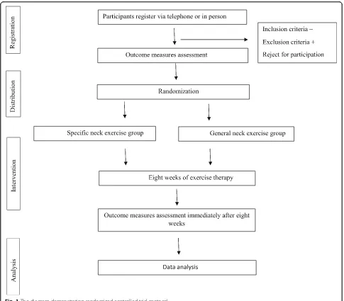
All necessary information about the trial including the study purpose and procedure will be given to the partici-pants both orally and in writing. The participartici-pants will then be allocated randomly to two exercise groups, the SNE and GNE groups. Randomization will be performed using sealed envelopes. None of the participants will be aware of the other training group. Both exercise pro-grammes will continue for 8 weeks (3 days per week with three sets on each day and five repetitions in each set). One set will be supervised by a physiotherapist at the university physiotherapy clinic and two other sets will be carried out at home by the participant himself/ herself [26, 33]. Before beginning the study, after base-line measurements and randomization, each participant will be familiarized with his or her own exercise programme. They will be taught how to perform their exercises and will be monitored to ensure that they carry out them correctly. Each participant will receive a pamphlet explaining all exercises (SNE or GNE) using schematic pictures. The primary and secondary outcome measures will be assessed before and after 8 weeks of intervention, except for pain which will be assessed daily (Fig.1).
The participants will be advised not to use other forms of treatments during the trial. However, they will be asked to notify the researcher at every session if they use analgesic medication in an unavoidable situation.
Reliability
In order to assess the repeatability level of measure-ments, a primary study will be conducted on 10 partici-pants with measuring dorsal and ventral muscles thicknesses, neck active range of motions and maximum voluntary isometric contraction (MVIC). After 3–7 days, the participants will be asked to return and the measure-ment process will be carried out again. After the second assessment, participants will start training as previously
described. Interclass coefficient correlation (ICC) and standard error of measurements (SEM) will be reported.
Assessor
The study will be conducted by a physiotherapist with 2 years of clinical practice who has been trained for ultra-sound imaging for 6 months. The physiotherapist will as-sess and record outcome measures and will supervise one of the three sets of exercises to make sure that par-ticipants perform the exercises correctly (three times per week). The other two sets of exercises will be performed by the participants without the physiotherapist’s supervi-sion. The participants will be asked to notify the trial physiotherapist if they feel any discomfort when carrying out the exercises. The test-retest reliability of the trial physiotherapist in measuring the outcome measures will be assessed before beginning the study procedures on two separate days, 3 days apart.
Intervention programmes
The exercise period will be performed for 8 weeks, 3 days per week, three sets each day with five repetitions in each set. The final goal is to increase the exercise difficulty to 20 repetitions in each set [26]. The exercise difficulty will be increased by two repetitions per week considering par-ticipants’ tolerance. If increasing the exercise repetition causes participants to feel pain, the exercise repetition is therefore beyond their tolerance and the repetition num-ber will not change for the next week [26]. There will be no specific exercise order in either group.
Specific neck exercise group (SNE)
The participant will lie on the experimental bed in a su-pine position with bent knees and relaxed hands laid be-side him or her on the bed. A thin layer of a towel will be placed under the participant’s head to keep his or her head and neck in the neutral position (forehead and chin should be parallel to the ceiling) [26,33] (Table1).
General neck exercises
All exercises will be performed in the standing position (the participant will stand relaxed while looking forward with the head and neck in a neutral position), except for press-up exercises which will be performed in a sitting position (feet on the ground, hands on the armrest of a chair). A 1 kg weight will be added to shoulder shrug ex-ercise from week 4 to the end of the programme [13,15,
26] (Table2).
Outcome measures
Primary outcome measures
0.98 according to a previous study [34]. The number 0 on this scale means no pain and 100 means the worst imaginable pain. The participants will be asked to show their painful areas on the posterior region of their necks using their hands. They will be instructed to show us if their pain is on their left and right upper cervical, lower cervical, and the trapezius. The participants’current pain
will be measured before and after 8 weeks of interven-tion. In addition, pain intensity will be measured at each intervention session before and after performing exer-cises [34].
[image:4.595.56.541.86.511.2]Disability The Iranian version of neck disability index (NDI) questionnaire (ICC = 0.90–0.97) [35] will be used Fig. 1The diagram demonstrating randomized controlled trial protocol
Table 1Details of specific neck exercises
I. Participants move their eyes upward and backward without any movement of the head and neck and hold for 5 s. II. Participants move their eyes downward and forward without any movement of the head and neck and hold for 5 s. III. Participants do chin talk by bringing their chin closer to their sternum, and hold for 5 s.
IV. Participants perform a light isometric nodding accompanied by downward movement of their eyes. They will be asked to apply moderate resistance to their chin with their own hands in the opposite direction of the chin movement.
[image:4.595.53.540.655.731.2]to determine the participants’disability. This index con-tains ten items including questions about activities of daily living (seven items), pain (two items), and concen-tration (one item). Each question is scored from zero to five. The NDI scores will be presented as a percentage of the maximum score, in which 0% indicates no disability and 100% indicates maximum disability [36].
Cervical muscle thickness The thicknesses of dorsal neck muscles including trapezius, splenius capitis, semispinalis capitis, semispinalis cervicis, and multifi-dus and ventral neck muscles including longus colli and sternocleidomastoid will be measured on the pain-ful side using an ultrasound device (Ultrasonix ES 500) with linear array, 45 mm, and 6.6 MHz probe for dorsal muscles and 12 MHz probe for ventral muscles. Ac-cording to established ultrasonographic studies, Ultra-sound is a valid and reliable device to measure dorsal (ICC = 0.98–0.99) [37] and ventral (ICC = 0.98–0.99) neck muscles thickness [38]. Muscle thickness will be recorded at rest and during maximum voluntary iso-metric contraction (MVIC).
Dorsal neck muscle imaging
Participants will be asked to sit on the experimental chair with their head and neck in a neutral position, with their hands at rest on their legs and their feet on the ground [39]. The assessor then palpates the neck to find the fourth cervical vertebral (C4) spinous process [39]. The probe will be placed on C4 transversely, and will then be moved slightly towards the painful side to see the echogenic vertebral lamina clearly [39]. At this level the measurement will be taken from the muscle’s sur-rounding fascia which are the superior and inferior fascia at the widest distance. at rest and during a 10 s MVIC. While participants keep the pressure constant, the ultra-sound image will be frozen for thickness measurement. The procedure will be repeated three times and the mean thicknesses will be used for data analyses to re-duce measurement errors [39–41].
Ventral neck muscle imaging
Longus colli and sternocleidomastoid muscles thick-nesses will be measured while participants lie supine with bent knees and their hands resting on the bed. It is essential that participants’heads and necks are in a neu-tral position. To achieve this, a thin layer of a towel will be put under participants’ occiputs in order to ensure that their foreheads are parallel to the ceiling [42]. The assessor will then place the probe 2 cm below the Ad-am’s apple and move it about 1 cm laterally towards the painful side to observe the muscle, carotid artery and thyroid cartilage [42]. The muscle thicknesses will be measured at rest and during a 10 s MVIC. To record the flexor muscle thicknesses during contraction, a pressure biofeedback will be placed under participants’ occiputs [12]. Participants will be asked to nod, holding the nod until the pressure unit shows 30 mmHg and then hold it for 10 s. While participants keep the pressure constant, the ultrasound image will be frozen for thickness meas-urement. The procedure will be repeated three times to reduce measurement errors [42].
Secondary outcome measures
Neck active range of motion (AROM)
[image:5.595.53.540.101.212.2]Cervical AROM in flexion, extension, rotation to the right and the left and lateral flexion to the right and the left will be measured with a universal goniometer. Go-niometric assessment of neck AROM is a reliable tech-nique with ICC ranged from 0.83 to 0.98 [43]. To measure flexion and extension AROM, the centre of the goniometer will be placed over the external auditory me-atus, the stationary arm will be perpendicular to the ground and the moving arm will be aligned parallel to the longitudinal axis of the nose. To measure the lateral flexion AROM, the centre of the goniometer will be placed over the spinous process of the seventh cervical vertebra, the stationary arm will be perpendicular to the ground (in the direction of the thoracic vertebral spinous process) and the moving arm will be aligned to the dor-sal head midline (the line passing the occipital protuber-ance). To assess rotational AROM, the centre of the goniometer will be placed over the centre of the cranial Table 2Details of general neck exercises
I. Participants move their heads slowly up and down without holding at the end ranges.
II. Participants rotate their heads to the right and left slowly (to see their shoulders), without holding at the end ranges.
III. Participants bend their heads to the side, bringing their ears close to their right and left shoulders. No holding at the end ranges. IV. Participants abduct their shoulders by bringing their arms into the frontal plane.
V. Participants flex their shoulders by bringing their arms into the sagittal plane.
VI. Participants hold their elbows in 90° of flexion and their forearms in pronation. They then move their hands towards and away from their trunks. VII. Participants try to bring their shoulders up as close to their ears as possible. They then lower their shoulders without holding at the end ranges.
(Shoulder shrugs.)
aspect of head, the stationary arm will be parallel to an imaginary line passing between the two acromial pro-cesses, and the moving arm will be aligned with the tip of the nose. First, the assessor will show the movements to participants and instruct them to perform them cor-rectly so that they do not use their thoracic vertebra. The participants will then be asked to move their heads in three anatomical planes in six directions so that the assessor can measure their neck AROM [44].
Neck muscle maximum voluntary isometric contraction (MVIC)
A tensiometer will be used to record neck extension and flexion MVICs. Measuring MVIC using dynamometry is a reliable technique with ICC = 0.94 [45]. The tensiome-ter has two bands, one fixed to a wall and the other one placed around the participants’ heads [45]. Participants will be asked to sit on a chair with their feet on ground and their arms resting on their thighs. To record neck extension MVIC, they will be instructed to push their head backwards without any movements in their heads and trunks [46]. To record neck flexor MVIC, partici-pants will turn back to the tensiometer and the tensiom-eter band will be placed on the participants’ foreheads. Participants will then be instructed to push their fore-heads towards the band. All MVIC measurements will be repeated three times and the maximum MVIC will be recorded for further analyses. There is a 30 s rest be-tween each MVIC performance and the physiotherapist will lead the patient orally by pushing during the task [47,48].
Sleep quality
The Iranian version of the Pittsburgh Sleep Quality Index (PSQI) questionnaire with the reported ICC equal to 0.77 [49], will be used to assess participants’ sleep quality. The PSQI is a self-rating questionnaire with 19 questions in seven categories: sleep quality, sleep latency, sleep duration, habitual sleep efficiency, sleep distur-bances, use of sleeping medication, and daytime dys-function. Each component is rated from zero to three. Higher scores indicate poorer sleep quality [49].
Quality of life
The Iranian cultural comparable version of the short-form 36 (SF-36) questionnaire with ICC equal to 0.70 [50] will be used to assess participants’ quality of life. This questionnaire contains 36 questions in eight dimen-sions of quality of life, including physical functioning (ten questions), role limitations due to physical health problems (four questions), social functioning (two ques-tions), bodily pain (two quesques-tions), general mental health (five questions), vitality (four questions), role limitations due to emotional health (three questions), general health
perceptions (five questions) and reported health transi-tion (one questransi-tion) [51].
Fear avoidance
The Iranian version of the Tampa scale questionnaire with ICC larger than 0.80 [52] will be used to investigate participants’ fear of movement. This questionnaire con-tains 17 questions, each of which are scored from 1 to 4. Total scores range from 17 to 68, with the higher scores indicating stronger fear avoidance beliefs [52].
Randomization and allocation concealment
The participants will be allocated randomly to one of the two training groups: SNE and GNE. Simple randomization with sealed envelopes in which one of the letters A or B is written will be used for group allocation. Each participant will choose one of the sealed envelopes to be allocated to one of the exercise groups. The enve-lope will then be returned to the enveenve-lope box The randomization will be carried by a physiotherapist who is independent of the study. The allocation concealment will be revealed after the final measurement.
Sample size
The sample size estimates are based on relevant studies (SD1= 0.32, SD2= 0.56) and the mean difference of 1.1
cm for deep cervical muscle thickness changes [53].We accept the significance level of 5% and the power equal to 80%. Accordingly, 32 participants have been calcu-lated to be recruited in each group [53].
Trial status
Among 64 recruited participants of this study, 56 partic-ipants have completed their exercise programs and eight participants is still performing their exercise programs. The estimated final day of the trial is 18 the March 2019.
Statistical analysis
any effect of each intervention program on the strength of the evaluated correlations. In order to make the re-sults comparable, the effect size, mean differences, and their confidence intervals will be reported.
Discussion
Neck pain is a common musculoskeletal problem, and has episodic and periodic types which cause ADL and work difficulties, disability and economic and social costs for both patients and society [2–4]. Therefore, introdu-cing the most effective treatment protocol would seem to be essential in order to decrease not only the pain but also the complications which are not spontaneously re-versible. This trial seeks to be more precise about the types of training and investigates whether a specific training that targets deep muscles has any superiority to general exercises for the neck. Based on present evi-dence, specific exercise training is effective and general training has positive effects on clinical symptoms. How-ever, to the best of our knowledge, no study has yet ex-amined the effects of specific exercises targeting the deep neck muscles, including deep neck extensor and flexor muscles, in patients with chronic neck pain. The results of this trial are expected to increase the efficacy of prescriptive exercise training and also to bring about improvements in individuals with CNNP conditions.
Supplementary information
Supplementary informationaccompanies this paper athttps://doi.org/10. 1186/s12891-019-2880-x.
Additional file 1:SPIRIT 2013 Checklist: Recommended items to address in a clinical trial protocol and related documents*.
Abbreviations
AROM:Active range of motion; CNNP: Chronic non-specific neck pain; MVIC: Maximum voluntary isometric contraction; SD: Standard deviation; SPIRIT: Standard Protocol Items: Recommendations for Interventional Trials; SPSS: Statistical Package for the Social Sciences; VAS: Visual analogue scale
Acknowledgements
The authors would like to thank all the participants.
Authors’contributions
Study design: LR, AP, NK, Data collection: PK, Data analyses: LR, NK, PK, AP, Data interpretation: LR, AP, NK, Writing the first draft of the manuscript: PK, Intellectual revision of the first draft: LR, AP, NK, Final approval of the manuscript: PK, NK, AP, LR.
Funding
University of Social Welfare and Rehabilitation Sciences will fund this study by providing our free access to study setups including the ultrasound and tensiometer devices.
Availability of data and materials
The data set will be available on reasonable request to the corresponding author.
Ethics approval and consent to participate
This study was approved by“Research Ethics Committee”, University of Social Welfare and Rehabilitation Sciences, Tehran, Iran. Participants will be included if they give their signed, written informed consent.
Consent for publication
Not applicable.
Competing interests
The authors declare that they have no competing interests.
Author details
1Department of Physiotherapy, University of Social Welfare and Rehabilitation
Sciences, Tehran, Iran.2Department of Medical and Health Sciences, Division of Physiotherapy, Faculty of Health Sciences, Linköping University, Linköping, Sweden, Linköping University, Linköping, Sweden.3Department of Physical
Therapy, University of North Georgia, Dahlonega, USA.
Received: 23 December 2018 Accepted: 3 October 2019
References
1. Chiu TT, Leung AS. Neck pain in Hong Kong: a telephone survey on prevalence, consequences, and risk groups. Spine. 2006;31(16):E540–4. 2. Tsakitzidis G, Remmen R, Dankaerts W, Van Royen P. Non-specific neck pain
and evidence-based practice. Eur Sci J. 2013;9(3):1–19.
3. Cagnie B, Danneels L, Van Tiggelen D, De Loose V, Cambier D. Individual and work related risk factors for neck pain among office workers: a cross sectional study. Eur Spine J. 2007;16(5):679–86.
4. Wermeling M, Scherer M, Himmel W. GPs’experiences of managing non-specific neck pain--a qualitative study. Fam Pract. 2011;28(3):300–6. 5. Falla DL, Jull GA, Hodges PW. Patients with neck pain demonstrate reduced
electromyographic activity of the deep cervical flexor muscles during performance of the craniocervical flexion test. Spine. 2004;29(19):2108–14. 6. Kim JY, Kwag KI. Clinical effects of deep cervical flexor muscle activation in
patients with chronic neck pain. J Phys Ther Sci. 2016;28(1):269–73. 7. Karimi N, Rezasoltani A, Rahnama L, Noori-Kochi F, Jaberzadeh S.
Ultrasonographic analysis of dorsal neck muscles thickness changes induced by isometric contraction of shoulder muscles: a comparison between patients with chronic neck pain and healthy controls. Man Ther. 2016;22: 174–8.
8. Fernández-De-Las-Peñas C, Albert-Sanchís JC, Buil M, Benitez JC,
Alburquerque-Sendín F. Cross-sectional area of cervical multifidus muscle in females with chronic bilateral neck pain compared to controls. J Orthop Sports Phys Ther. 2008;38(4):175–80.
9. O’Leary S, Falla D, Elliott JM, Jull G. Muscle dysfunction in cervical spine pain: implications for assessment and management. J Orthop Sports Phys Ther. 2009;39(5):324–33.
10. Panjabi MM, Cholewicki J, Nibu K, Grauer J, Babat LB, Dvorak J. Critical load of the human cervical spine: an in vitro experimental study. Clin Biomech. 1998;13(1):11–7.
11. Durall CJ. Therapeutic exercise for athletes with nonspecific neck pain: a current concepts review. Sports Health. 2012;4(4):293–301.
12. Chiu TT, Lam T-H, Hedley AJ. A randomized controlled trial on the efficacy of exercise for patients with chronic neck pain. Spine. 2005;30(1):E1–7. 13. Rezasoltani A, Mohammad M, KhalkhaliZavieh M, Tabatabaee SM. A study
on the effectiveness of shoulder strengthening exercises on thickness of the neck extensor muscles. Rehabil Med. 2013;2(1):7–13.
14. Khan M, Soomro RR, Ali SS. The effectiveness of isometric exercises as compared to general exercises in the management of chronic non-specificneck pain. Pak J Pharm Sci. 2014;27(5):1719–22.
15. Akhter S, Khan M, Ali SS, Soomro RR. Role of manual therapy with exercise regime versus exercise regime alone in the management of non-specific chronic neck pain. Pak J Pharm Sci. 2014;27(6 Suppl):2125–8.
16. Jull G, Falla D, Vicenzino B, Hodges P. The effect of therapeutic exercise on activation of the deep cervical flexor muscles in people with chronic neck pain. Man Ther. 2009;14(6):696–701.
18. Rudolfsson T, Djupsjobacka M, Hager C, Bjorklund M. Effects of neck coordination exercise on sensorimotor function in chronic neck pain: a randomized controlled trial. J Rehabil Med. 2014;46(9):908–14. 19. Gallego Izquierdo T, Pecos-Martin D, Lluch Girbes E, Plaza-Manzano G,
Rodriguez Caldentey R, Mayor Melus R, Blanco Mariscal D, Falla D. Comparison of cranio-cervical flexion training versus cervical proprioception training in patients with chronic neck pain: a randomized controlled clinical trial. J Rehabil Med. 2016;48(1):48–55.
20. Borisut S, Vongsirinavarat M, Vachalathiti R, Sakulsriprasert P. Effects of strength and endurance training of superficial and deep neck muscles on muscle activities and pain levels of females with chronic neck pain. J Phys Ther Sci. 2013;25(9):1157–62.
21. Page P, Frank C, Lardner R. Assessment and treatment of muscle imbalance: the Janda approach: human kinetics; 2010.
22. Mense S. Muscle pain: mechanisms and clinical significance. Dtsch Arztebl Int. 2008;105(12):214–9.
23. Tunwattanapong P, Kongkasuwan R, Kuptniratsaikul V. The effectiveness of a neck and shoulder stretching exercise program among office workers with neck pain: a randomized controlled trial. Clin Rehabil. 2016;30(1):64–72. 24. Ylinen J, Kautiainen H, Wiren K, Hakkinen A. Stretching exercises vs manual
therapy in treatment of chronic neck pain: a randomized, controlled cross-over trial. J Rehabil Med. 2007;39(2):126–32.
25. Landen Ludvigsson M, Peterson G, Peolsson A. The effect of three exercise approaches on health-related quality of life, and factors associated with its improvement in chronic whiplash-associated disorders: analysis of a randomized controlled trial. Qual Life Res. 2018;357–368.
26. Peolsson A, Ludvigsson ML, Tigerfors A-M, Peterson G. Effects of neck-specific exercises compared to waiting list for individuals with chronic whiplash-associated disorders: a prospective, randomized controlled study. Arch Phys Med Rehabil. 2016;97(2):189–95.
27. O’Leary S, Cagnie B, Reeve A, Jull G, Elliott JM. Is there altered activity of the extensor muscles in chronic mechanical neck pain? A functional magnetic resonance imaging study. Arch Phys Med Rehabil. 2011;92(6):929–34. 28. Elliott J, Jull G, Noteboom J, Durbridge G, Gibbon W. Magnetic resonance
imaging study of cross-sectional area of the cervical extensor musculature in an asymptomatic cohort. Clin Anat. 2007;20(1):35–40.
29. Maigne JY, Cornelis P, Chatellier G. Lower back pain and neck pain: is it possible to identify the painful side by palpation only? Ann Phys Rehabil Med. 2012;55(2):103–11.
30. Misailidou V, Malliou P, Beneka A, Karagiannidis A, Godolias G. Assessment of patients with neck pain: a review of definitions, selection criteria, and measurement tools. J Chiropr Med. 2010;9(2):49–59.
31. Rahnama L, Rezasoltani A, Zavieh MK, NooriKochi F, Baghban AA. Differences in cervical multifidus muscle thickness during isometric contraction of shoulder muscles: a comparison between patients with chronic neck pain and healthy controls. J Manip Physiol Ther. 2015; 38(3):210–7.
32. Rezasoltani A, Ali-Reza A, Khosro KK, Abbass R. Preliminary study of neck muscle size and strength measurements in females with chronic non-specific neck pain and healthy control subjects. Man Ther. 2010;15(4):400–3. 33. Lieber RL. Skeletal muscle structure, function, and plasticity: the
physiological basis of rehabilitation; 2010.
34. Bijur PE, Silver W, Gallagher EJ. Reliability of the visual analog scale for measurement of acute pain. Acad Emerg Med Off J Soc Acad Emerg Med. 2001;8(12):1153–7.
35. Mousavi SJ, Parnianpour M, Montazeri A, Mehdian H, Karimi A, Abedi M, Ashtiani AA, Mobini B, Hadian MR. Translation and validation study of the Iranian versions of the neck disability index and the neck pain and disability scale. Spine. 2007;32(26):E825–31.
36. Hassan Azarsa M, Shadmehr A, Jalaei S. The effect of the loading on dynamic stability and scapular asymmetry. J Rehabil Sci Res. 2014;1(1):12–6. 37. Rankin G, Stokes M, Newham DJ. Size and shape of the posterior neck
muscles measured by ultrasound imaging: normal values in males and females of different ages. Man Ther. 2005;10(2):108–15.
38. Jeong BL, Ha SM, Jeon IC, Hong KH. Reliability of ultrasonography measurement for the longus colli according to inward probe pressure. J Phys Ther Sci. 2015;27(11):3579–81.
39. Rahnama L, Rezasoltani A, Khalkhali Zavieh M, Noori Kochi F, Akbarzadeh Baghban A. The effects of isometric contraction of shoulder muscles on cervical multifidus muscle dimensions in healthy office workers. J Bodyw Mov Ther. 2014;18(3):383–9.
40. Lee J-P, Tseng W-YI, Shau Y-W, Wang C-L, Wang H-K, Wang S-F. Measurement of segmental cervical multifidus contraction by ultrasonography in asymptomatic adults. Man Ther. 2007;12(3):286–94. 41. Kristjansson E. Reliability of ultrasonography for the cervical multifidus
muscle in asymptomatic and symptomatic subjects. Man Ther. 2004;9(2): 83–8.
42. Cagnie B, Derese E, Vandamme L, Verstraete K, Cambier D, Danneels L. Validity and reliability of ultrasonography for the longus colli in asymptomatic subjects. Man Ther. 2009;14(4):421–6.
43. Farooq MN, Mohseni Bandpei MA, Ali M, Khan GA. Reliability of the universal goniometer for assessing active cervical range of motion in asymptomatic healthy persons. Pak J Med Sci. 2016;32(2):457–61. 44. Norkin CC, White DJ. Measurement of joint motion : a guid to goniometry;
2004. p. 16.
45. Goodarzi F, Rahnama L, Karimi N, Baghi R, Jaberzadeh S. The effects of forward head posture on neck extensor muscle thickness: an ultrasonographic study. J Manip Physiol Ther. 2018;41(1):34–41. 46. Rezasoltani A, Nasiri R, Faizei AM, Zaafari G, Mirshahvelayati AS,
Bakhshidarabad L. The variation of the strength of neck extensor muscles and semispinalis capitis muscle size with head and neck position. J Bodyw Mov Ther. 2013;17(2):200–3.
47. Meldrum D, Cahalane E, Conroy R, Fitzgerald D, Hardiman O. Maximum voluntary isometric contraction: reference values and clinical application. Amyotroph Lateral Scler. 2007;8(1):47–55.
48. Schomacher J, Erlenwein J, Dieterich A, Petzke F, Falla D. Can neck exercises enhance the activation of the semispinalis cervicis relative to the splenius capitis at specific spinal levels? Man Ther. 2015;20(5):694–702.
49. Moghaddam JF, Nakhaee N, Sheibani V, Garrusi B, Amirkafi A. Reliability and validity of the Persian version of the Pittsburgh Sleep Quality Index (PSQI-P). Sleep Breath. 2012;16(1):79–82.
50. Montazeri A, Goshtasebi A, Vahdaninia M, Gandek B. The short form health survey (SF-36): translation and validation study of the Iranian version. Qual Life Res. 2005;14(3):875–82.
51. Jenkinson C, Coulter A, Wright L. Short form 36 (SF36) health survey questionnaire: normative data for adults of working age. BMJ. 1993; 306(6890):1437–40.
52. Askary-Ashtiani A, Ebrahimi-Takamejani I, Torkaman G, Amiri M, Mousavi SJ. Reliability and validity of the Persian versions of the fear avoidance beliefs questionnaire and Tampa scale of Kinesiophobia in patients with neck pain. Spine. 2014;39(18):E1095–102.
53. Javanshir K, Amiri M, Mohseni Bandpei MA, De las Penas CF, Rezasoltani A. The effect of different exercise programs on cervical flexor muscles dimensions in patients with chronic neck pain. J Back Musculoskelet Rehabil. 2015;28(4):833–40.
Publisher’s Note