0095-1137/07/$08.00⫹0 doi:10.1128/JCM.01870-06
Copyright © 2007, American Society for Microbiology. All Rights Reserved.
Using a Resequencing Microarray as a Multiple Respiratory Pathogen
Detection Assay
䌤
†
Baochuan Lin,
1* Kate M. Blaney,
2Anthony P. Malanoski,
1Adam G. Ligler,
2Joel M. Schnur,
1David Metzgar,
3Kevin L. Russell,
3and David A. Stenger
1Center for Bio/Molecular Science and Engineering, Code 6900, Naval Research Laboratory, Washington, DC 203751;
NOVA Research Incorporated, Alexandria, Virginia 223082; and Department of Defense Center for
Deployment Health Research, Naval Health Research Center, San Diego, California 921863
Received 8 September 2006/Returned for modification 27 October 2006/Accepted 15 November 2006
Simultaneous testing for detection of infectious pathogens that cause similar symptoms (e.g., acute respiratory infections) is invaluable for patient treatment, outbreak prevention, and efficient use of antibiotic and antiviral agents. In addition, such testing may provide information regarding possible coinfections or induced secondary infections, such as virally induced bacterial infections. Furthermore, in many cases, detection of a pathogen requires more than genus/species-level resolution, since harmful agents (e.g., avian influenza virus) are grouped with other, relatively benign common agents, and for every pathogen, finer resolution is useful to allow tracking of the location and nature of mutations leading to strain variations. In this study, a previously developed resequencing microarray that has been demon-strated to have these capabilities was further developed to provide individual detection sensitivity ranging
from 101
to 103
genomic copies for more than 26 respiratory pathogens while still retaining the ability to detect and differentiate between close genetic neighbors. In addition, the study demonstrated that this system allows unambiguous and reproducible sequence-based strain identification of the mixed pathogens. Successful proof-of-concept experiments using clinical specimens show that this approach is potentially very useful for both diagnostics and epidemic surveillance.
Accurate and rapid identification of infectious pathogens that cause similar symptoms, such as acute respiratory infec-tions (ARIs), can be a critical factor in the successful treatment of the illness, outbreak control measures, and the efficient use of precious antibiotics and antiviral drugs (30, 40). Simulta-neous testing for all possible pathogens is an efficient means to obtain a conclusive result. In addition, assaying for all potential pathogens may yield information regarding possible coinfec-tions or induced secondary infeccoinfec-tions (e.g., virally induced bac-terial infections). Currently, many promising approaches using reverse transcription-PCR (RT-PCR)/PCR amplification strat-egies as multiplexed approaches for testing several organisms are being developed (1, 2, 4–10, 14–16, 22, 23, 25, 28, 36, 37, 41, 42). While these approaches are versatile, additional strategies must be implemented to ensure good specificity for the assay. In cases where closely related organisms can have very differ-ent clinical consequences and epidemiological patterns (e.g.,
Bordetella pertussisversusBordetella parapertussis),
discrimina-tion to the species, serotype/subtype, or even strain level is required to reduce the incidence of false positives (22). Influ-enza virus detection is a case where serotype/subtype discrim-ination, at a minimum, is required. In addition, strain-level information can be used to quickly discern the effectiveness of vaccines and make proper recommendations for appropriate
outbreak control measures. Furthermore, the sequence infor-mation of new circulating human isolates and possible zoonotic strains [e.g., avian influenza virus (H5N1)] that are detected will be of immediate and obvious value for potential vaccine candidate prediction.
A resequencing microarray approach is advantageous in ad-dressing these issues. This platform can maintain specificity, provide information on mutation hot spots and strain variants, and monitor all these aspects in a few long sections of DNA/ RNA sequence. Our previous work has demonstrated the poten-tial of short-oligonucleotide resequencing arrays to simulta-neously provide both species-level and strain-level identification of amplicons from respiratory pathogens (19, 39). This ap-proach has been used to efficiently and simultaneously detect, type, and genetically characterize geographically diverse influ-enza viruses (39). The sequences produced by the resequenc-ing arrays were identical to the results from conventional se-quencing methods with the exception of ambiguous base calls (N⬘s) (19, 39). The microarray results were analyzed using a new approach that compared the sequence of bases deter-mined to all previously sequenced results. Thus, it is possible both to correctly identify the organism and to determine the difference in sequence between the organism detected and previously sequenced organisms (21). In the initial studies, random amplification methods were used as the enrichment method to provide the sensitivity necessary for this potential diagnostic assay. This broadly targeted enrichment method was initially selected for its potential benefits in reducing biased amplification of one organism over another and in amplifying mutated organisms that a more specific amplification method might miss.
* Corresponding author. Mailing address: Center for Bio/Molecular Science and Engineering, Code 6900, Naval Research Laboratory, Washington, DC 20375. Phone: (202) 767-0289. Fax: (202) 767-9594. E-mail: [email protected].
† Supplemental material for this article may be found at http://jcm .asm.org/.
䌤Published ahead of print on 29 November 2006.
443
on May 16, 2020 by guest
http://jcm.asm.org/
While the initial results using random amplification and a resequencing microarray are very promising, several issues must be addressed before this technology can be considered ready for use in a surveillance or diagnostic application. Spe-cifically, the generic amplification methods, which performed well with analytic test samples, did not consistently detect or-ganisms in complex clinical samples with lower titers. In addi-tion, the analysis method was complex and time-consuming and required the expertise of highly trained individuals. In this study, in order to overcome the sensitivity issue related to random target amplification, we developed an alternate ampli-fication strategy, optimized multiplex PCR, which provides greater sensitivity for clinical samples while still being capable of identifying the presence of close genetic neighbors (e.g., various adenovirus serotypes). Furthermore, to demonstrate that this approach is effective for the simultaneous detection of multiple pathogens, we tested the system’s capability for de-tecting mixed multiple pathogens in analytic control samples and complex backgrounds (mimicking real-world situations). The results demonstrate that this approach allows unambigu-ous and reproducible sequence-based strain identification of the mixed pathogens. Further testing, using clinical specimens (throat swabs) obtained from patients presenting febrile ill-ness, showed that we can achieve high species-level concor-dance with standard reference assays while at the same time producing correct species and strain-level identification via direct sequence reads in an assay time of around 8.5 h. These results show that this approach is amenable to the develop-ment of a robust microarray-based platform that offers com-prehensive coverage of the significant respiratory pathogens for both diagnostic and surveillance purposes.
MATERIALS AND METHODS
RPM v.1 chip design.The design of respiratory pathogen microarray, version 1 (RPM v.1), chips includes 57 target genes comprising partial sequences from the genes containing the diagnostic regions of each pathogen (i.e.,E1A,hexon, andfiberfor human adenoviruses [HAdV’s]; thehemagglutinin(HA), neuramin-idase(NA), andmatrixgenes for influenza A viruses). This allows resequencing of 29.7 kb of sequences from 26 respiratory pathogens and biowarfare agents (Table 1) known to cause “flu-like” symptoms at early stages of infection, as described in detail in a previous report (19). Briefly, RPM v.1 arrays consist of prototype sequences (ProSeqs), which are a collection of probes chosen to represent portions of the genomic DNA/RNA of a targeted organism. ProSeqs consist of sequential 25-mer perfect-match probes, each in its own probe set, representing each base of the prototype sequence chosen from the genome of the target organism. For each perfect-match probe, the remainder of the probe set consists of three mismatch probes representing the three possible single-nucle-otide polymorphisms of the center position. Thus, hybridization to a series of probe sets provides redundant presence/absence information for the organism while also revealing strain-specific single-nucleotide polymorphisms relative to the sequence chosen.
Prototype strains.All control and field strains used to test the sensitivity and specificity of RPM v.1 and their sources are listed in Table 1.
Clinical samples.Archived throat swabs were collected from patients with symptoms of ARI at various military recruit training centers, at U.S.-Mexico border sites, and on deployed naval ships from 1999 to 2005. These were imme-diately placed in 2-ml cryogenic vials containing 1.5 ml of viral transport medium (MICROTEST, Multi-Microbe Media; Remel Inc., Lenexa, KS), frozen, and stored at or below⫺80°C to maintain the viral particles during transport. Sam-ples were then shipped to the Naval Health Research Center (NHRC; San Diego, CA), a molecular diagnostics laboratory certified by the College of Amer-ican Pathologists (CAP), where they were thawed, aliquoted, and tested for human adenoviruses and influenza virus using CAP-approved diagnostic RT-PCR/PCR and culture tests. Frozen aliquots were then submitted for microarray-based detection in a blinded fashion. Informed consent was obtained from all
participants after the nature and possible consequences of the studies were explained.
These samples were collected, and this research has been conducted, in compli-ance with all applicable federal and international regulations governing the protec-tion of human subjects in research, under Naval Health Research Center (NHRC) work unit number 60501, Department of Defense protocols NHRC.1999.0002.31271 and NHRC.2003.0002.
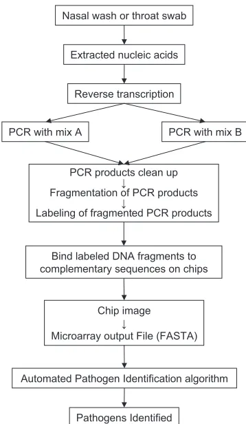
Chip processing protocol.Figure 1 is a schematic diagram of the processing protocol. The details of each processing step are described below.
Nucleic acid extraction.Nucleic acids were extracted from clinical samples using either the MasterPure DNA purification kit (Epicentre Technologies, Madison, WI), omitting RNase digestion, or the MagNA Pure Compact nucleic acid isolation kit I (Roche Applied Science, Indianapolis, IN) according to the manufacturer’s recommended protocols.
Internal controls.TwoArabidopsis thaliana plant genes, corresponding to NAC1 and triosphosphate isomerase (TIM), were chosen as internal controls for RT and PCRs, since they would be unlikely to occur naturally in clinical samples. Two plasmids, pSP64poly(A)-NAC1 and pSP64poly(A)-TIM, containing⬃500 bp of the two genes, were kindly provided by Norman H. Lee of The Institute for Genome Research (Rockville, MD) (38). NAC1 was amplified by PCR with SP6 and M13R primers, and the PCR products were purified using a QIAquick PCR purification kit (QIAGEN, Valencia, CA). To generate RNA from pSP64poly(A)-TIM, the plasmids were linearized with EcoRI and in vitro transcribed from the SP6 promoter using the MEGAscript high-yield transciption kit (Ambion, Austin, TX). Sixty femtograms each of NAC1 and TIM were used as internal controls for check-ing the amplification efficiency and the presence of inhibitors in the specimens.
Primer design and multiplex RT-PCR amplification.The gene-specific primer pairs for all targets on the RPM v.1 chip (listed in Table S1 in the supplemental material) were designed to meet the minimum amplification efficiency require-ment (to provide a detection sensitivity of 101to 103genome copies per target)
for multiplex PCR. All primers were designed to have similar annealing temperatures and were checked to ensure uniqueness using a full search of the GenBank database with the BLAST program for known sequences. All primers were checked for potential hybridization to other primers in order to reduce the potential of primer-dimer formation. In addition, we adapted a method developed by Shuber et al. and Brownie et al. (3, 32) to further suppress primer-dimer formation by adding a linker sequence of 22 bp (primer L) to the 5⬘ends of primers used in this study. To further minimize the possibility of intraprimer interactions, the primers were divided into two independent reac-tions to simplify primer design and optimization. Fine-tuning adjustments to both mixtures (replacing primers that amplified poorly with new ones) were carried out to ensure that all target genes from the 26 targeted pathogens (West Nile virus is included on the array but not in this amplification scheme) would amplify sufficiently to generate detectable hybridization.
RT reactions were performed in 20-l volumes containing 50 mM Tris-HCl (pH 8.3), 75 mM KCl, 3 mM MgCl2, 500M each dATP, dCTP, dGTP, and
dTTP, 40 U of RNaseOUT, 10 mM dithiothreitol, 2M primer LN, 200 U of Superscript III (Invitrogen Life Technologies, Carlsbad, CA), 60 fg of each of the two internal controls (NAC1 and TIM), and 5 to 8l of the extracted clinical specimen or laboratory control. Reactions were carried out in a Peltier thermal cycler-PTC240 DNA Engine Tetrad 2 (MJ Research Inc., Reno, NV) using the manufacturer’s recommended protocol.
The RT reaction products were split up into two 10-l volumes to be subjected to two different multiplex PCRs. Primer mix A contains 19 primer pairs and amplifies 18 gene targets from three different influenza A viruses, one influenza B virus, three HAdV serotypes, and one internal control (TIM). Primer mix B contains 38 primer pairs and amplifies the remaining 37 gene targets and the other internal control (NAC1). PCRs were performed in 50-l volumes contain-ing 20 mM Tris-HCl (pH 8.4), 50 mM KCl, 2 mM MgCl2, 400M each dATP,
dCTP, dGTP, and dUTP, 1 U of uracil-DNA glycosylase, heat-labile (USB Corporation, Cleveland, OH), 2M primer L, 40 nM each primer from mix A or 50 nM each primer from mix B, 10 U of PlatinumTaqDNA polymerase (Invitrogen Life Technologies, Carlsbad, CA), and 10l of the RT product. The amplification reaction was carried out in a Peltier thermal cycler-PTC240 DNA Engine Tetrad 2 (MJ Research Inc., Reno, NV) with initial incubation at 25°C for 10 min; preliminary denaturation at 94°C for 3 min, followed by 5 cycles of 94°C for 30 s, 50°C for 90 s, and 72°C for 120 s; 35 cycles of 94°C for 30 s and 64°C for 120 s; and a final extension at 72°C for 5 min. The amplified products from the two PCRs were combined into a single volume and subjected to purification and processing prior to hybridization to the RPM v.1 chips (see below).
Microarray hybridization and processing.Microarray hybridization and pro-cessing were carried out according to the manufacturer’s recommended protocol (Affymetrix Inc., Santa Clara, CA) using a GeneChip resequencing assay kit
on May 16, 2020 by guest
http://jcm.asm.org/
(Affymetrix Inc.) with the following modification. Purified PCR products were fragmented for 5 min and then labeled for 30 min. Hybridization was carried out at 45°C for 2 h. The images were scanned and processed as previously described to produce FASTA output files (19).
Automatic pathogen identification algorithm.A new software program, the computer-implemented biological sequence-based identifier system, version 2 (CIBSI 2.0), was used to automate the pathogen identification process for the RPM v.1 array. CIBSI 2.0 incorporates the general concept of the resequencing pathogen identification (REPI) program (19) and in addition analyzes the result and makes decisions, steps that were previously carried out manually. A broader discussion of this protocol, including an improved REPI algorithm, is described in detail elsewhere (21).
Quantification of pathogens.For sensitivity assessments, real-time PCR assays were conducted on an iCycler or MyiQ instrument (Bio-Rad Laboratories, Her-cules, CA) to determine the number of pathogen genomes in each sample. The findings for the samples were compared to those for 10-fold serial dilutions of prototype genomic DNA templates with known copy numbers (101to 106copies)
by using specific primers and RT-PCR/PCR conditions as previously described in the literature (see Table S2 in the supplemental material) (11, 17, 24, 34, 35). The
genomic copy number of the pathogen was calculated by measuring the DNA/ RNA concentration from purified genomic DNA/RNA and using conversion factors as follows. Molecular mass (y) is calculated as the number of base pairs⫻
660 Da (average molecular mass for 1 bp at 330 Da for each nucleotide). Grams of DNA per genome copy is calculated asydaltons⫻1.67⫻10⫺24g (e.g., a
single adenovirus genome of⬃35 kb has a molecular mass of 2.31⫻107
Da, which is equivalent to 3.86⫻10⫺17g; 1 ng of purified adenovirus genomic DNA
is equivalent to 2.6⫻107
genome copies) (31). Real-time PCRs were carried out in 25-l reaction volumes containing 2.5l FastStart reaction mix SYBR Green I (Roche Applied Science, Indianapolis, IN), 20 mM Tris-HCl (pH 8.4), 50 mM KCl, 3 mM MgCl2, 200M each dATP, dTTP, dGTP, and dCTP, 200 nM
primers, and genomic DNA (1 to 4l of clinical specimen or DNA extracts).
RESULTS
Analytical sensitivity and specificity of the RPM v.1 assay.A
[image:3.585.49.540.80.470.2]split multiplex PCR protocol that divides the sample to amplify the 57 gene targets on the RPM v.1 chip was developed to
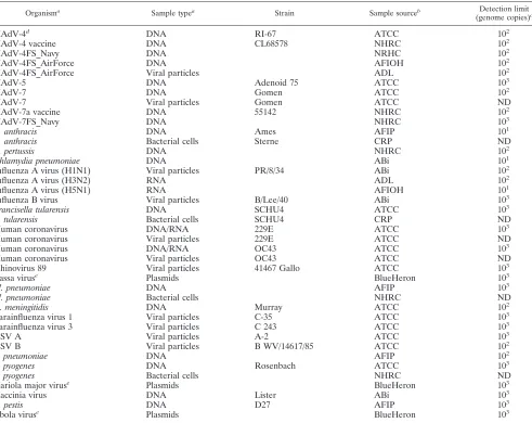
TABLE 1. Analytic sensitivity of microarray-based detection for prototype control strains
Organisma Sample typea Strain Sample sourceb Detection limit
(genome copies)c
HAdV-4d DNA RI-67 ATCC 102
HAdV-4 vaccine DNA CL68578 NHRC 102
HAdV-4FS_Navy DNA NRHC 102
HAdV-4FS_AirForce DNA AFIOH 102
HAdV-4FS_AirForce Viral particles ADL 102
HAdV-5 DNA Adenoid 75 ATCC 103
HAdV-7 DNA Gomen ATCC 102
HAdV-7 Viral particles Gomen ATCC ND
HAdV-7a vaccine DNA 55142 NHRC 102
HAdV-7FS_Navy DNA NHRC 103
B. anthracis DNA Ames AFIP 101
B. anthracis Bacterial cells Sterne CRP ND
B. pertussis DNA NHRC 102
Chlamydia pneumoniae DNA ABi 101
Influenza A virus (H1N1) Viral particles PR/8/34 ABi 102
Influenza A virus (H3N2) RNA ADL 102
Influenza A virus (H5N1) RNA AFIOH 101
Influenza B virus Viral particles B/Lee/40 ABi 103
Francisella tularensis DNA SCHU4 ATCC 103
F. tularensis Bacterial cells SCHU4 CRP ND
Human coronavirus DNA/RNA 229E ATCC 103
Human coronavirus Viral particles 229E ATCC ND
Human coronavirus DNA/RNA OC43 ATCC 103
Human coronavirus Viral particles OC43 ATCC ND
Rhinovirus 89 Viral particles 41467 Gallo ATCC 103
Lassa viruse Plasmids BlueHeron 103
M. pneumoniae DNA AFIP 103
M. pneumoniae Bacterial cells NHRC ND
N. meningitidis DNA Murray ATCC 102
Parainfluenza virus 1 Viral particles C-35 ATCC 103
Parainfluenza virus 3 Viral particles C 243 ATCC 103
RSV A Viral particles A-2 ATCC 103
RSV B Viral particles B WV/14617/85 ATCC 102
S. pneumoniae DNA AFIP 102
S. pyogenes DNA Rosenbach ATCC 103
S. pyogenes Bacterial cells NHRC ND
Variola major viruse Plasmids BlueHeron 103
Vaccinia virus DNA Lister ABi 103
Y. pestis DNA D27 AFIP 103
Ebola viruse Plasmids BlueHeron 103
aSamples were generated by mixing purified nucleic acid templates in Tris-EDTA buffer to create 106genome copies/l of stock solution. Starting from this
concentration, 10-fold serial dilutions in Tris-EDTA buffer were prepared. NA, not applicable.
bATCC, American Type Culture Collection; AFIOH, Air Force Institute for Operational Health; ADL, Advanced Diagnostics Laboratory; AFIP, Armed Forces
Institute of Pathology; CRP, Critical Reagents Program; ABi, Advanced Biotechnologies Incorporated (Columbia, MD).
cND, not determined. The lowest detection limit tested was 101. dPlaque purified.
eTarget genes were constructed and cloned into pUC119 by BlueHeron Biotechnology (Bothell, WA).
on May 16, 2020 by guest
http://jcm.asm.org/
overcome the sensitivity issues related to random target am-plification. The specificity of this assay was confirmed using various prototype strains and clinical samples. The results showed only noninterfering hybridization, by which we mean hybridization of nontarget amplicons, resulting in base calls occurring for only a few target probe sets, which was insuffi-cient to cause an incorrect identification. No significant cross-hybridization occurred between any targets. The largest num-ber of base calls generated from noninterfering hybridization was observed on HAdVhexongenes when the different sero-types were used as the template. But the sequences generated still did not cause misidentification. This is due to the fact that sequence information produced on the microarray can distin-guish noninterfering hybridization from hybridization of the intended target.
The analytical sensitivity of the RPM v.1 assay was then evaluated using serial 10-fold dilutions of the nucleic acid tem-plates of the prototype strains. Table 1 shows the lowest de-tectable dilution of each pathogen. The results revealed an individual sensitivity that ranged from 101to 103genomic
cop-ies per reaction for the prototype strains, which is comparable to the sensitivity of standard multiplex RT-PCR/PCR methods. The genome copy number should not be equated to the CFU or PFU but was used so that comparisons could be made between pathogens from different sources. The number of genome copies represented by the CFU or PFU of a patient sample can vary from one to several orders of magnitude greater for various respiratory pathogens.
The capability of RPM v.1 to identify and discriminate between near genetic neighbors is dependent not only on the capabilities of the microarray but also on the amplification strategy. The capability of RPM v.1 was first demonstrated with random am-plification protocols and has been reproduced with this multi-plexed amplification protocol. This assay distinguished between 11 different serotypes of ARI-associated HAdV’s and also differ-entiated four different strains of HAdV-4, three strains of HAdV-7, and two strains of HAdV-3. These results demonstrated that the newly developed multiplexed amplification would detect a range of variants comparably to random amplification methods (Table 2).
Simultaneous detection and differentiation of respiratory
pathogens.In this study, we further assess the ability of the
RPM v.1 assay to identify multiple pathogens simultaneously, i.e., coinfections, by the preparation of various combinations of pathogen templates (see Tables S3 and S4 in the supplemental material). Serial dilutions of nucleic acid templates were used to evaluate the detection sensitivity and specificity for multiple pathogens. Initially, samples containing 103 to 106 genome copies per reaction of four pathogens—HAdV-4,Streptococcus
pyogenes, Mycoplasma pneumoniae, and Yersinia pestis—were
[image:4.585.72.251.66.374.2]prepared. Tests using the RPM v.1 assay demonstrated repro-ducible sequence-based identification of all four pathogens, even at the lowest concentration of 103 genomic copies per target per reaction (see Table S3 in the supplemental mate-rial). In addition to tests in which targets are present at the same concentration, experiments were carried out diluting only one of two pathogens, representing a situation possible for many clinical specimens, especially those identified as coinfec-tions. Using the combination of HAdV-4 (105genome copies)
TABLE 2. Differentiation by RPM v.1 of various HAdVs causing febrile respiratory infections
Samplea Strain Strain identification by RPM v.1
(GenBank accession no.)
HAdV-4 RI-67 HAdV-4 (AY594253)
HAdV-4 vaccine CL68578 HAdV-4 vaccine (AY594254)
HAdV-4FS_Navy HAdV-4FS_Navy (AY599835)
HAdV-4FS_AirForce HAdV-4FS_AirForce (AY599837)
HAdV-5 Adenoid 75 HAdV-5 (AY339865)
HAdV-1 Adenoid 71 HAdV-1 (AY490817)
HAdV-2 Adenoid 6 HAdV-2 (J01917)
HAdV-6 Tonsil 99 HAdV-6 (DQ149613)
HAdV-7 Gomen HAdV-7 (AY594255)
HAdV-7a vaccine 55142 HAdV-7 (AY 594256)
HAdV-7FS_Navy HAdV-7FS_Navy (AY601634)
HAdV-3 GB HAdV-3 (DQ086466)
HAdV-3FS_Navy HAdV-3 U.S. Navy field strain (AY 599836)
HAdV-16 Ch. 79 HAdV-16 (AY601636)
HAdV-21 AV-1645 [128] HAdV-21 (AF492353) HAdV-11 Slobitski HAdV-11 (AY598970)
HAdV-14 De Wit HAdV-14 (AY803294)
[image:4.585.301.541.541.715.2]aFS, field strain.
FIG. 1. Schematic of the processing protocol. Clinical samples (na-sal swab or na(na-sal wash specimens) were collected from patients pre-senting ARI symptoms. Nucleic acids were extracted from these sam-ples, followed by reverse transcription. The products of reverse transcription were split into two multiplex PCR mixtures for amplifi-cation. PCR products from the two mixtures were combined after amplification, cleaned up, fragmented, and labeled. The labeled prod-ucts were then hybridized to RPM v.1 chips. After the chips were washed and stained, the sequences generated from RPM v.1 were exported as FASTA-formatted files and further analyzed for pathogen identification using the automatic pathogen identification algorithm.
on May 16, 2020 by guest
http://jcm.asm.org/
andStreptococcus pneumoniae(105to 101genome copies), the results showed that RPM v.1 can detect both pathogens to a dilution of 100 genome copies of S. pneumoniae (data not shown). In similar experiments, the combinations of influenza A virus (H1N1) withS. pneumoniaeand ofS. pyogeneswithS.
pneumoniae also demonstrated that both pathogens are
de-tected to a dilution of 100 genome copies of S. pneumoniae
(data not shown).
The effectiveness of simultaneous multiple pathogen detec-tion was also tested with more-complex mixtures. Three to seven available cultured organisms were spiked at different titers (102 to 105 CFU or PFU/ml) into pooled nasal wash samples collected from volunteers, and 150l of the prepared samples was used for testing. Initial results revealed that this
approach allowed unambiguous detection of seven patho-gens—HAdV-4, HAdV-7, influenza A virus (H1N1), parain-fluenza virus 1, respiratory syncytial virus A (RSV-A),M.
pneu-moniae, and S. pyogenes—simultaneously at the lowest titer,
[image:5.585.50.539.81.531.2]100 CFU (or PFU)/ml (see Table S4A in the supplemental material). Further assessment with a set of six pathogens showed that HAdV-4, Bacillus anthracis, influenza A virus (H1N1), RSV-A, and M. pneumoniae were detected at the lowest titer tested, 100 CFU (PFU)/ml, andS. pyogenes was detected at 1,000 CFU/ml (see Table S4B in the supplemental material). For further confirmation, RPM v.1 was tested using eight cultured organisms combined to form three different sets of three pathogens (see Table S4C in the supplemental mate-rial). In all sets tested, the assay could reproducibly detect
TABLE 3. Quantitative real-time PCR results for 40 clinical samples
Sample ID RPM resulta NHRC
result
PCR result for the following organism (genome copies/l):
HAdV-4 Influenza A
virus S.pneumoniae M.pneumoniae
31844 H3N2,S. pneumoniae H3N2 —b 381 198 —
43536 HAdV-4,S. pneumoniae, N. meningitidis
Negative 45,000 91.6 1,110 —
6738 HAdV-4,S. pneumoniae Negative 6,000 — 1,190 —
12358 HAdV-4 Negative 17,000 — — —
70793 H3N2,S. pneumoniae, N. meningitidis
H3N2 — 4,400 17,600 —
48934 H3N2,S. pneumoniae, N. meningitidis
H3N2 — 1,320 5,710 —
49376 H3N2,S. pneumoniae, N. meningitidis
H3N2 — 32.5 19,400 —
1388 HAdV-4,S. pneumoniae Negative 14,600 — 2,620 —
12007 S. pneumoniae,N. meningitidis H3N2 — — 7,920 —
41394 H3N2 H3N2 — 672 — —
21474 HAdV-4 Negative 55,000 198 4,630 —
10499 H3N2,S. pneumoniae H3N2 — 1,270 19,000 —
10552 H3N2 H3N2 — 3,330 222 —
32329 H3N2 H3N2 — 6,250 — —
61596 H3N2 H3N2 — 1,800 — —
9464 Negative Negative — — — —
62416 H3N2,S. pneumoniae H3N2 — 2,150 806 200
51672 H3N2 H3N2 — 612 — —
49424 S. pneumoniae H3N2 — — 18,100 —
62687 H3N2,M. pneumoniae, S. pneumoniae
Negative — — 461 24,000
32349 H3N2,S. pneumoniae H3N2 — — 354 —
48930 H3N2 H3N2 — 83.4 — —
39002 H1N1,N. meningitidis H1N1 — 418 108 —
90909 S. pneumoniae H3N2 — — 55,000 —
62686 HAdV-4,S. pneumoniae Negative — — 5,840 —
90781 H3N2,S. pneumoniae H3N2 — 189 521 —
12364 HAdV-4 Negative 14,000 — — —
49369 H3N2 H3N2 — 108 — —
566771484 HAdV-4 Negative 205 — 1,140 —
43269 H3N2,S. pneumoniae H3N2 — 126 377 —
70246 H1N1,S. pneumoniae H1N1 — 1,080 35,600 —
50833 H3N2 H3N2 — 134 — —
3600 HAdV-4 Negative 8,000 — — —
90900 Negative Negative — — — —
21475 HAdV-4 Negative 190,000 — — —
30491 H3N2,S. pneumoniae H3N2 — 332 10,600 —
12359 HAdV-4 Negative 3,400 — 154 —
20694 H3N2,N. meningitidis H3N2 — 612 — —
51673 H3N2 H3N2 — 502 2,210 —
38076 S. pneumoniae,N. meningitidis Negative — — 121 —
aAbbreviations: H3N2, influenza A virus (H3N2); H1N1, influenza A virus (H1N1). b—, not detected.
on May 16, 2020 by guest
http://jcm.asm.org/
HAdV-4, M. pneumoniae, B. anthracis, influenza A virus (H1N1), human coronavirus 229E, or RSV-A at titers as low as 100 CFU (PFU)/ml and S. pyogenes to only 1,000 CFU/ml. These results indicate that the resequencing array-based ap-proach is an effective means of detecting and typing various pathogens directly from nasal wash samples with the benefit of high sensitivity and specificity, even when as many as seven pathogens are present.
Assessment of clinical specimens. After the capability of
the RPM v.1 assay for pathogen detection was successfully demonstrated, it was used for prospective and retrospective diagnoses of infections causing ARI. Clinical specimens, collected primarily from military recruits presenting with ARI, were used to compare the utility of the microarray-based diagnostic to more-established methods of respiratory pathogen detection. The samples (n ⫽ 101) consisted of throat swabs, in viral transport medium, from patients with clinically documented respiratory illness. Samples were cho-sen randomly from sets that had tested positive for HAdV or influenza virus by assays performed at NHRC, a CAP-cer-tified molecular diagnostic laboratory (cell culture and/or PCR), but that were not tested for any other pathogen. As controls, samples that had tested negative for HAdV or influenza virus were also included for testing. These were blinded (randomly renumbered and separated from the as-sociated clinical records) and sent to the Naval Research Laboratory for RPM v.1 testing, and the sample identities were revealed only after the results had been finalized. For pathogens detected in at least two samples each (HAdV, influenza A virus,S. pneumoniae, and M. pneumoniae) by RPM v.1, published species-specific (11, 24) or selected in-house specific PCR primers (see Table S2 in the supplemen-tal material) were used to perform quantitative real-time PCR for a subset of the clinical samples (limited to only 40 due to the availability of the samples) (Table 3). The lack of samples makes it difficult to properly estimate the clinical sensitivity and specificity for some of the pathogens detected by RPM v.1, and so data for these pathogens, such as
Neis-seria meningitidis, are not reported.
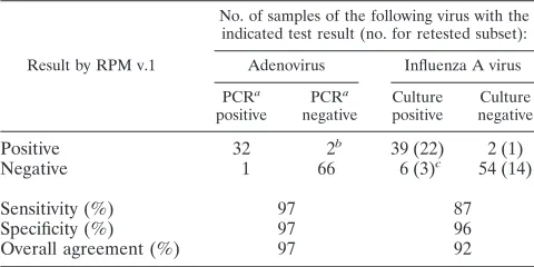
For influenza A virus, the RPM v.1 method showed a de-tection sensitivity of 87% and a specificity of 96% with respect to the initial diagnostic result and an overall agreement of 92% (Table 4). For adenovirus, the RPM v.1 detection sensitivity was 97% with 97% specificity, for an overall agreement of 97% (Table 4). For influenza A virus, the RPM v.1 result had better agreement with culture than PCR did, which suggests that the assay has better sensitivity and specificity than PCR (Table 5). It should be noted that three of the six false-negative and one of two false-positive clinical samples by RPM v.1 were samples that were retested by quantitative real-time PCR. The three false-negative samples also were not detected by PCR, suggest-ing that the samples no longer contained any detectable viral material due to possible degradation of the RNA during stor-age or transport. The false positive, however, was confirmed, since real-time PCR did not detect influenza A virus. In addi-tion to detecting single pathogens in the clinical samples, the assay also detected several multiply infected samples, primarily possible coinfections. The presence of multiple pathogens was verified in a subset of 40 clinical samples by using quantitative real-time PCR. The differences in the titers of the pathogens confirmed the assay’s ability to detect patho-gens at similar titers and at very different titers in the same sample. It is well known that S. pneumoniae(26% of sam-ples) andN. meningitidis (16% of samples) are commensal bacteria in the mouth and upper respiratory system, so it is not surprising that these were commonly found in clinical samples. However, quantitative real-time PCR data showed that 32% of the influenza virus-positive samples harbored titers higher than expected forS. pneumoniae (7/25) orN.
meningitidis(1/25) (ⱖ104genome copies/l) (Table 3). The
high titers of bacteria present in these clinical samples were possibly due to virally induced bacterial superinfection, con-sistent with the findings of Madhi and Klugman (20) and Peltola and McCullers (26).
[image:6.585.43.283.90.210.2]This study further demonstrated the capability of this assay to identify the subtypes of the influenza viruses and track genetic changes of the influenza virus strains in clinical sam-ples. This is especially critical for influenza epidemiology, since antigenic drift is the mechanism by which influenza viruses escape from immunological pressure induced by previous nat-ural exposures and vaccination. Analysis of the HA and NA sequences generated from RPM v.1 for the influenza A virus-positive clinical samples recapitulated the known lineage
TABLE 4. Evaluation of RPM v.1 for adenovirus, influenza A virus, and negative-control detection in clinical samples
Result by RPM v.1
No. of samples of the following virus with the indicated test result (no. for retested subset):
Adenovirus Influenza A virus
PCRa
positive
PCRa
negative
Culture positive
Culture negative
Positive 32 2b 39 (22) 2 (1)
Negative 1 66 6 (3)c 54 (14)
Sensitivity (%) 97 87
Specificity (%) 97 96
Overall agreement (%) 97 92
aCAP-approved PCR.
bOne of the samples testing negative by CAP-approved PCR was cultured for
influenza A virus, and RPM v.1 showed HAdV-4 and influenza A coinfection. The other negative sample was confirmed to have low-titer HAdV-4.
cThree samples that were culture positive for influenza A virus could not be
[image:6.585.301.542.98.197.2]detected by quantitative real-time PCR as well, suggesting that the templates were degraded.
TABLE 5. Comparison of the culture method with a real-time PCR assay for detection of influenza A virus-positive and -negative
controls in 40 clinical samples
Result by PCR
No. of samples testing as follows for influenza A virus by culture:
Positive Negative
Positive 21 2
Negative 4a 13
Sensitivity (%) 84
Specificity (%) 87
Overall agreement (%) 85
a
Three of four samples positive for influenza A virus by culture could not be detected by RPM v.1 as well.
on May 16, 2020 by guest
http://jcm.asm.org/
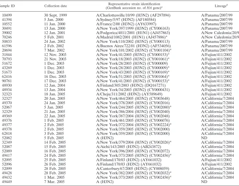
changes occurring from 1999 to 2005 through antigenic drifting (Table 6). Seven influenza A virus (H3N2) specimens collected prior to the 2003-to-2004 influenza season were identified as belonging to the A/Panama/2007/99-like lineage, while nine influenza A virus (H3N2) samples collected during the 2003-to-2004 influenza season were clearly carrying signature A/ Fujian/411/2002-like lineage nucleotide substitutions in the HA gene. The shift from an A/Fujian/411/2002-like strain to an A/California/7/2004-like strain is evident for the 18 influenza A virus (H3N2) samples collected during the 2004-to-2005 influ-enza season. Three samples were identified as A/Fujian/411/ 2002-like strains, while the rest showed signature California-like nucleic acid substitutions in the HA gene (12, 33). Two samples collected during the same period could be identified only as influenza A virus (H3N2). This was due to poor am-plification and/or hybridization of targets, resulting in insuffi-cient sequence information for strain-level identification. Two influenza A virus (H1N1) samples collected in 2000 to 2001 were identified as closely related to A/New Caledonia/20/99.
DISCUSSION
Clinical syndromes are seldom specific to single pathogens, so assays that allow testing for, and discrimination among, a large number of candidate pathogens will undoubtedly be ben-eficial to public health efforts (2). The data presented here indicate that the RPM v.1 system is potentially such an assay for direct (uncultured) clinical specimens, obtaining results that will correlate well with those of conventional detection methods while providing sequence information. While earlier studies (13, 18, 39) demonstrated the usefulness of the RPM v.1 array for discriminating among 20 common respiratory and 6 biothreat pathogens, failure to meet requirements for detect-ing low-titer pathogens in complex clinical samples led to al-teration of the amplification strategy. The use of a properly designed multiplex PCR amplification method with a rese-quencing array avoids some of the trade-off between specificity and sensitivity often seen in other diagnostic assays. The sen-sitivity of the new protocol for all pathogens, by using control
TABLE 6. Influenza virus strain and lineage identification using RPM v.1
Sample ID Collection date Representative strain identification
(GenBank accession no. ofHAgene)a Lineageb
10499 30 Sept. 1999 A/Charlottesville/10/99 (H3N2) (AF297094) A/Panama/2007/99
41394 5 Jan. 2000 A/Sydney/5/97 (H3N2) (AF180584) A/Panama/2007/99
10552 11 Jan. 2000 A/France/2/00 (H3N2) (AY633997) A/Panama/2007/99
30491 13 Jan. 2000 A/New York/397/1999 (H3N2) (CY006163) A/Panama/2007/99
39002 12 Jan. 2001 A/Podgorica/4011/2001 (H1N1) (AJ457863) A/New Caledonia/20/99
70246 5 Feb. 2001 A/Madrid/1082/2001 (H1N1) (AJ457886)* A/New Caledonia/20/99
50833 24 Jan. 2002 A/New York/110/2002 (H3N2) (CY000113) A/Panama/2007/99
61596 2 Feb. 2002 A/Buenos Aires/722/01 (H3N2) (AF534056) A/Panama/2007/99
20694 7 Mar. 2002 A/New York/101/2002 (H3N2) (CY001104)* A/Panama/2007/99
31844 12 Nov. 2003 A/New York/41/2003 (H3N2) (CY000153)* A/Fujian/411/2002
70793 21 Nov. 2003 A/New York/18/2003 (H3N2) (CY001061)* A/Fujian/411/2002
51672 1 Dec. 2003 A/New York/28/2003 (H3N2) (CY000009) A/Fujian/411/2002
43269 1 Dec. 2003 A/New York/28/2003 (H3N2) (CY000009)* A/Fujian/411/2002
51673 1 Dec. 2003 A/New York/43/2003 (H3N2) (CY000169)* A/Fujian/411/2002
62416 16 Dec. 2003 A/New York/51/2003 (H3N2) (CY001064)* A/Fujian/411/2002
90781 17 Dec. 2003 A/New York/41/2003 (H3N2) (CY000153)* A/Fujian/411/2002
48930 12 Jan. 2004 A/Finland/305/2003 (H3N2) (DQ167271) A/Fujian/411/2002
48934 13 Jan. 2004 A/New York/34/2003 (H3N2) (CY0000431) A/Fujian/411/2002
32323 18 Jan. 2005 A/Cheju/311/2002 (H3N2) (AY589649) A/Fujian/411/2002
32327 20 Jan. 2005 A/New York/464/2005 (H3N2) (CY003648) A/California/7/2004
49370 24 Jan. 2005 A/New York/378/2005 (H3N2) (CY002016) A/California/7/2004
52067 5 Jan. 2005 A/New York/244/2005 (H3N2) (CY002080)* A/California/7/2004
32329 21 Jan. 2005 A/New York/386/2004 (H3N2) (CY002040) A/California/7/2004
49369 22 Jan. 2005 A/New York/387/2004 (H3N2) (CY002048) A/California/7/2004
49376 1 Feb. 2005 A/New York/461/2005 (H3N2) (CY006076) A/California/7/2004
49377 2 Feb. 2005 A/New York/372/2004 (H3N2) (CY002224)* A/California/7/2004
49378 2 Feb. 2005 A/New York/359/2005 (H3N2) (CY002000) A/California/7/2004
62818 3 Feb. 2005 A/New York/359/2005 (H3N2) (CY002000) A/California/7/2004
52082 5 Feb. 2005 A (H3N2) ND
32349 14 Feb. 2005 A/New York/379/2004 (H3N2) (CY002026)* A/California/7/2004
52087 15 Feb. 2005 A/Aichi/143/2005 (H3N2) (AB243872) A/California/7/2004
52089 16 Feb. 2005 A/New York/396/2005 (H3N2) (CY002072) A/California/7/2004
49417 24 Feb. 2005 A/New York/373/2005 (H3N2) (CY002456)* A/California/7/2004
52095 25 Feb. 2005 A/Finland/170/03 (H3N2) (AY661032) A/Fujian/411/2002
52096 26 Feb. 2005 A/Finland/170/03 (H3N2) (AY661032) A/Fujian/411/2002
52099 28 Feb. 2005 A/Canterbury/67/2005 (H3N2) (CY008059) A/California/7/2004
49428 28 Feb. 2005 A/New York/382/2005 (H3N2) (CY002032)* A/California/7/2004
49432 1 Mar. 2005 A/New York/373/2005 (H3N2) (CY002456)* A/California/7/2004
49449 7 Mar. 2005 A (H3N2) ND
a
*, only a single strain was identified.
b
ND, not detected.
on May 16, 2020 by guest
http://jcm.asm.org/
[image:7.585.46.536.80.462.2]samples either in extraction buffer or together as complex mixtures spiked into healthy-patient clinical samples, was on a par with that of alternate detection methods and met diagnos-tic requirements. Furthermore, genediagnos-tically close neighbors could still be detected, as demonstrated for HAdV’s, indicating that the multiplex PCR method maintained an ability for broad target enrichment. This study shows specifically that this assay exhibits the ability to resolve complex coinfections of as many as seven pathogens without a loss of sensitivity in simulated samples. The data show for clinical samples that the assay offers a sensitivity equivalent to those of accepted RT-PCR/ PCR- and culture-based methods for both HAdV and influ-enza A virus, using 101 throat-swab samples from patients with influenza-like illnesses. This group of samples also contained examples of coinfections that were successfully detected. While the RPM v.1 has provided confirmation of many concepts, fully establishing the clinical sensitivity and specificity of every pathogen is not worthwhile, since this particular microarray represents an incomplete coverage of respiratory pathogens and would not be ideal for real-world diagnostics or surveil-lance. Our ongoing efforts have focused on a new microarray that has more-complete coverage of the respiratory pathogens and for which a complete clinical validation would be worth-while.
Commensal microflora has been viewed as a source of spec-imen contamination and an occasional opportunistic pathogen, but it may play a more important role in health and disease than was once thought (29). Madhi and Klugman (20) and Peltola and McCullers (26) have reported that⬃30% of respi-ratory viral infections predispose patients, both adults and children, to bacterial superinfection. These superinfections are thought to be the source of many influenza-related pneumo-nias and subsequent deaths (27). It is not surprising that RPM v.1 methods detected S. pneumoniae and N. meningitidis in several clinical samples, since it is well known that both are commensal bacteria present in the upper respiratory tract. However, it is noteworthy that while most of these bacteria appeared to be present at low titers in the samples, high titers of these pathogens were regularly seen in influenza A virus-positive samples. Although the RPM v.1 assay does not deter-mine the titers of the pathogens detected, the assay can effec-tively detect coinfection. This type of assay would be highly effective in studies to establish more firmly whether a clinical correlation of disease exists in cases of coinfection. If situations where this occurs are established, a diagnostic test based on a resequencing array will be valuable for the effective treatment of the primary agent and secondary coinfecting organisms, with prompt treatment using appropriate antibiotics.
Unlike traditional methods, the optimized RPM v.1 assay not only identifies pathogens but also provides sequence infor-mation, allowing a large number of pathogens to be detected and phylogenetically categorized for genetic variation analysis in the same assay (19, 39). This utility was clearly demonstrated for the influenza A virus-positive clinical samples, where the array using the HA gene tracked the lineage changes from A/Panama/2007/99-like strains (prior to the 2003 influenza sea-son) to A/Fujian/411/2002-like strains in the 2003-to-2004 in-fluenza season and then to A/California/7/2004-like strains in the 2004-to-2005 influenza season. While the specific HA genes allowed detailed tracking of changes in a specific
sub-type, one M gene (H1N1) sequence that is relatively conserved among influenza A viruses tiled on the RPM v.1 was still able to detect homologous regions of disparate subtypes, allowing correct differentiation (Table 6). This M gene ProSeq would theoretically allow detection of any other type of influenza virus for which specific antigenic HA and NA sequences were not tiled on the array. In the future, this method of mixing specific and conserved targeting will be useful for enhancing clinical management and epidemic outbreak responses by per-mitting accurate fingerprinting, antibiotic resistance profiling, genetic drift/shift analysis, forensics, and many other param-eters of important pathogens while maintaining coverage of a large number of pathogens. This capability will be invaluable for rapid detection of emerging diseases, such as avian influ-enza virus (H5N1), and biological terrorism events. In addi-tion, with the capability for simultaneous resequencing of doz-ens of gene targets from multiple pathogdoz-ens in a single assay, the technology is an excellent tool for identifying minor vari-ants within a population that may emerge and become domi-nant when selection pressure changes, without the need to isolate before proceeding with the sequencing reaction.
While the RPM v.1 chips with multiplex amplification dem-onstrated remarkable diagnostic and surveillance capabilities, the following factors must be taken into account before intro-ducing this technique in the diagnostic laboratory. One major hurdle that limits the use of a resequencing array to broad-spectrum pathogen diagnostics has been the selection of the primers for amplification of chosen target species and near neighbors prior to microarray hybridization. In this study, we showed that multiplex PCR can reach the desired clinical sen-sitivity and detect the presence of close genetic neighbors; however, the current system remains somewhat vulnerable to the rapid mutation of the RNA virus, and each new resequenc-ing array design would require recalibratresequenc-ing the mix of multi-plex primers. Future work will attempt to simplify the redesign method and also try to find alternative amplification methods that provide the necessary sensitivity with more-comprehen-sive coverage.
Another issue, in the current array format, is that a limited number of probe sets can be placed on the microarray, and so very detailed information cannot be obtained for every patho-gen. Currently the targets must be carefully chosen, or the microarray will not provide the coverage or detailed informa-tion desired. As the number of probe sets that can be placed on the microarray continues to increase, the trade-off between specific and conserved targets in the effort to provide suffi-cient detailed information while maintaining coverage will become less of an issue. Indeed, our newer chip design has improved upon the content of the detectable pathogens, which will further broaden the detection capability of the resequencing array.
In its current format, this assay can be performed at central-ized laboratories in 8.5 h with a completely automated patho-gen identification process. However, this is not suitable for a point-of-care diagnostic tool. The simplicity of the assay should allow it to be adapted to a fully automated process, increasing throughput, further decreasing the assay time, and reducing the error rate caused by human handling. With these alter-ations, it should be possible to implement this assay as a point-of-care analysis system. The cost of this assay compared to that
on May 16, 2020 by guest
http://jcm.asm.org/
of rapid single assays is high, although factoring the cost of all the rapid assays required to give the same information indi-cates that the relative cost difference for the two methods is not as significant. Considering costs on a per-test basis, the ad-vancements occurring in microarray production technology should provide even higher density microarrays with a reduced chip cost, and steps taken to produce a point-of-care system would also result in a reduction in the overall assay cost, which leads us to believe that newer-generation chips providing more-comprehensive coverage will be cost-effective in the near future. The benefits of this approach, with clear paths to over-come or reduce its limitations, make this an attractive assay method to develop for detecting respiratory pathogens. Future development will include addressing the most important limi-tations: designing a new resequencing array with more-compre-hensive coverage of respiratory pathogens, improving primer se-lection, and integrating microarrays and microfluidics into a portable device, allowing the possibility of transferring this refer-ence lab method into the point-of-care field.
ACKNOWLEDGMENTS
Funding support for this research was obtained from the Office of Naval Research via the Naval Research Laboratory Base program and is very much appreciated. We also gratefully acknowledge the previous support provided by the Defense Threat Reduction Agency, the U.S. Army Medical Research and Materiel Command, and the Joint Pro-gram Executive Office. Partial support from the Air Force Medical Service (Office of HQ, USAF Surgeon General), which helped make this research possible, is also gratefully appreciated.
We thank Margaret Ryan and Christopher Barrozo at NHRC, Linda Canas and Luke Daum at AFIOH, and Ted Hadfield at AFIP for kindly providing samples used in this study. We also thank Carolyn E. Meador for providing technical support and advice. Constructive ad-vice from Frances Ligler, Gary Vora, Zheng Wang, and Chris Myers is gratefully appreciated.
The opinions and assertions contained herein are those of the au-thors and are not to be construed as official or as reflecting the views of the Department of Defense or the U.S. Government.
REFERENCES
1.Bellau-Pujol, S., A. Vabret, L. Legrand, J. Dina, S. Gouarin, J. Petitjean-Lecherbonnier, B. Pozzetto, C. Ginevra, and F. Freymuth.2005. Develop-ment of three multiplex RT-PCR assays for the detection of 12 respiratory RNA viruses. J. Virol. Methods126:53–63.
2.Briese, T., G. Palacios, M. Kokoris, O. Jabado, Z. Liu, N. Renwick, V. Kapoor, I. Casas, F. Pozo, R. Limberger, P. Perez-Brena, J. Ju, and W. I. Lipkin.2005. Diagnostic system for rapid and sensitive differential detection of pathogens. Emerg. Infect. Dis.11:310–313.
3.Brownie, J., S. Shawcross, J. Theaker, D. Whitcombe, R. Ferrie, C. Newton, and S. Little.1997. The elimination of primer-dimer accumulation in PCR. Nucleic Acids Res.25:3235–3241.
4.Call, D. R., M. K. Bakko, M. J. Krug, and M. C. Roberts.2003. Identifying antimicrobial resistance genes with DNA microarrays. Antimicrob. Agents Chemother.47:3290–3295.
5.Call, D. R., M. K. Borucki, and F. J. Loge.2003. Detection of bacterial pathogens in environmental samples using DNA microarrays. J. Microbiol. Methods53:235–243.
6.Chizhikov, V., A. Rasooly, K. Chumakov, and D. D. Levy.2001. Microarray analysis of microbial virulence factors. Appl. Environ. Microbiol.67:3258– 3263.
7.Chizhikov, V., M. Wagner, A. Ivshina, Y. Hoshino, A. Z. Kapikian, and K. Chumakov.2002. Detection and genotyping of human group A rotaviruses by oligonucleotide microarray hybridization. J. Clin. Microbiol.40:2398– 2407.
8.Coiras, M. T., J. C. Aguilar, M. L. Garcia, I. Casas, and P. Perez-Brena.
2004. Simultaneous detection of fourteen respiratory viruses in clinical spec-imens by two multiplex reverse transcription nested-PCR assays. J. Med. Virol.72:484–495.
9.Coiras, M. T., M. R. Lopez-Huertas, G. Lopez-Campos, J. C. Aguilar, and P. Perez-Brena.2005. Oligonucleotide array for simultaneous detection of res-piratory viruses using a reverse-line blot hybridization assay. J. Med. Virol.
76:256–264.
10.Conejero-Goldberg, C., E. Wang, C. Yi, T. E. Goldberg, L. Jones-Brando, F. M. Marincola, M. J. Webster, and E. F. Torrey.2005. Infectious pathogen detection arrays: viral detection in cell lines and postmortem brain tissue. BioTechniques39:741–751.
11.Corless, C. E., M. Guiver, R. Borrow, V. Edwards-Jones, A. J. Fox, and E. B. Kaczmarski.2001. Simultaneous detection ofNeisseria meningitidis, Hae-mophilus influenzae, and Streptococcus pneumoniaein suspected cases of meningitis and septicemia using real-time PCR. J. Clin. Microbiol.39:1553– 1558.
12.Daum, L. T., M. W. Shaw, A. I. Klimov, L. C. Canas, E. A. Macias, D. Niemeyer, J. P. Chambers, R. Renthal, S. K. Shrestha, R. P. Acharya, S. P. Huzdar, N. Rimal, K. S. Myint, and P. Gould.2005. Influenza A (H3N2) outbreak, Nepal. Emerg. Infect. Dis.11:1186–1191.
13.Davignon, L., E. A. Walter, K. M. Mueller, C. P. Barrozo, D. A. Stenger, and B. Lin.2005. Use of resequencing oligonucleotide microarrays for identifi-cation ofStreptococcus pyogenesand associated antibiotic resistance deter-minants. J. Clin. Microbiol.43:5690–5695.
14.Dunbar, S. A.2006. Applications of Luminex xMAP technology for rapid, high-throughput multiplexed nucleic acid detection. Clin. Chim. Acta363:
71–82.
15.Grondahl, B., W. Puppe, A. Hoppe, I. Kuhne, J. A. Weigl, and H. J. Schmitt.
1999. Rapid identification of nine microorganisms causing acute respiratory tract infections by single-tube multiplex reverse transcription-PCR: feasibil-ity study. J. Clin. Microbiol.37:1–7.
16.Gruteke, P., A. S. Glas, M. Dierdorp, W. B. Vreede, J. W. Pilon, and S. M. Bruisten.2004. Practical implementation of a multiplex PCR for acute res-piratory tract infections in children. J. Clin. Microbiol.42:5596–5603. 17.Hardegger, D., D. Nadal, W. Bossart, M. Altwegg, and F. Dutly.2000. Rapid
detection ofMycoplasma pneumoniaein clinical samples by real-time PCR. J. Microbiol. Methods41:45–51.
18.Lin, B., K. M. Blaney, A. P. Malanoski, A. G. Ligler, J. M. Schnur, D. Metzgar, K. L. Russell, and D. A. Stenger.2006. Rapid testing for over 20 respiratory pathogens simultaneously in clinical samples using resequencing arrays. Naval Research Laboratory, Washington, DC.
19.Lin, B., Z. Wang, G. J. Vora, J. A. Thornton, J. M. Schnur, D. C. Thach, K. M. Blaney, A. G. Ligler, A. P. Malanoski, J. Santiago, E. A. Walter, B. K. Agan, D. Metzgar, D. Seto, L. T. Daum, R. Kruzelock, R. K. Rowley, E. H. Hanson, C. Tibbetts, and D. A. Stenger.2006. Broad-spectrum respiratory tract pathogen identification using resequencing DNA microarrays. Genome Res.16:527–535.
20.Madhi, S. A., and K. P. Klugman.2004. A role forStreptococcus pneumoniae in virus-associated pneumonia. Nat. Med.10:811–813.
21.Malanoski, A. P., B. Lin, Z. Wang, J. M. Schnur, and D. A. Stenger.2006. Automated identification of multiple micro-organisms from resequencing DNA microarrays. Nucleic Acids Res.34:5300–5311.
22.McDonough, E. A., C. P. Barrozo, K. L. Russell, and D. Metzgar.2005. A multiplex PCR for detection ofMycoplasma pneumoniae,Chlamydophila pneumoniae,Legionella pneumophila, andBordetella pertussisin clinical spec-imens. Mol. Cell. Probes19:314–322.
23.Miyashita, N., A. Saito, S. Kohno, K. Yamaguchi, A. Watanabe, H. Oda, Y. Kazuyama, and T. Matsushima.2004. Multiplex PCR for the simultaneous detection ofChlamydia pneumoniae,Mycoplasma pneumoniaeandLegionella pneumophilain community-acquired pneumonia. Respir. Med.98:542–550. 24.Molling, P., S. Jacobsson, A. Backman, and P. Olcen.2002. Direct and rapid identification and genogrouping of meningococci andporAamplification by LightCycler PCR. J. Clin. Microbiol.40:4531–4535.
25.Osiowy, C.1998. Direct detection of respiratory syncytial virus, parainfluenza virus, and adenovirus in clinical respiratory specimens by a multiplex reverse transcription-PCR assay. J. Clin. Microbiol.36:3149–3154.
26.Peltola, V. T., and J. A. McCullers.2004. Respiratory viruses predisposing to bacterial infections: role of neuraminidase. Pediatr. Infect. Dis. J.23:S87– S97.
27.Peltola, V. T., K. G. Murti, and J. A. McCullers.2005. Influenza virus neuraminidase contributes to secondary bacterial pneumonia. J. Infect. Dis.
192:249–257.
28.Puppe, W., J. A. Weigl, G. Aron, B. Grondahl, H. J. Schmitt, H. G. Niesters, and J. Groen.2004. Evaluation of a multiplex reverse transcriptase PCR ELISA for the detection of nine respiratory tract pathogens. J. Clin. Virol.
30:165–174.
29.Relman, D. A.1999. The search for unrecognized pathogens. Science284:
1308–1310.
30.Roth, S. B., J. Jalava, O. Ruuskanen, A. Ruohola, and S. Nikkari.2004. Use of an oligonucleotide array for laboratory diagnosis of bacteria responsible for acute upper respiratory infections. J. Clin. Microbiol.42:4268–4274. 31.Saitoh-Inagawa, W., A. Oshima, K. Aoki, N. Itoh, K. Isobe, E. Uchio, S.
Ohno, H. Nakajima, K. Hata, and H. Ishiko. 1996. Rapid diagnosis of adenoviral conjunctivitis by PCR and restriction fragment length polymor-phism analysis. J. Clin. Microbiol.34:2113–2116.
32.Shuber, A. P., V. J. Grondin, and K. W. Klinger.1995. A simplified proce-dure for developing multiplex PCRs. Genome Res.5:488–493.
33.Smith, D. J., A. S. Lapedes, J. C. de Jong, T. M. Bestebroer, G. F.
on May 16, 2020 by guest
http://jcm.asm.org/
Rimmelzwaan, A. D. Osterhaus, and R. A. Fouchier.2004. Mapping the antigenic and genetic evolution of influenza virus. Science305:371–376. 34.Stone, B., J. Burrows, S. Schepetiuk, G. Higgins, A. Hampson, R. Shaw, and
T. Kok.2004. Rapid detection and simultaneous subtype differentiation of influenza A viruses by real time PCR. J. Virol. Methods117:103–112. 35.Vabret, A., F. Mouthon, T. Mourez, S. Gouarin, J. Petitjean, and F.
Freymuth.2001. Direct diagnosis of human respiratory coronaviruses 229E and OC43 by the polymerase chain reaction. J. Virol. Methods97:59–66. 36.Wang, D., L. Coscoy, M. Zylberberg, P. C. Avila, H. A. Boushey, D. Ganem,
and J. L. DeRisi.2002. Microarray-based detection and genotyping of viral pathogens. Proc. Natl. Acad. Sci. USA99:15687–15692.
37.Wang, D., A. Urisman, Y. T. Liu, M. Springer, T. G. Ksiazek, D. D. Erdman, E. R. Mardis, M. Hickenbotham, V. Magrini, J. Eldred, J. P. Latreille, R. K. Wilson, D. Ganem, and J. L. DeRisi.2003. Viral discovery and sequence recovery using DNA microarrays. PLoS Biol.1:E2.
38.Wang, H. Y., R. L. Malek, A. E. Kwitek, A. S. Greene, T. V. Luu, B. Behbahani,
B. Frank, J. Quackenbush, and N. H. Lee.2003. Assessing unmodified 70-mer oligonucleotide probe performance on glass-slide microarrays. Genome Biol.
4:R5.
39.Wang, Z., L. T. Daum, G. J. Vora, D. Metzgar, E. A. Walter, L. C. Canas, A. P. Malanoski, B. Lin, and D. A. Stenger.2006. Identifying influenza viruses with resequencing microarrays. Emerg. Infect. Dis.12:638–646. 40.Whelen, A. C., and D. H. Persing.1996. The role of nucleic acid amplification
and detection in the clinical microbiology laboratory. Annu. Rev. Microbiol.
50:349–373.
41.Wilson, K. H., W. J. Wilson, J. L. Radosevich, T. Z. DeSantis, V. S. Viswanathan, T. A. Kuczmarski, and G. L. Andersen.2002. High-density microarray of small-subunit ribosomal DNA probes. Appl. Environ. Micro-biol.68:2535–2541.
42.Wilson, W. J., C. L. Strout, T. Z. DeSantis, J. L. Stilwell, A. V. Carrano, and G. L. Andersen.2002. Sequence-specific identification of 18 pathogenic mi-croorganisms using microarray technology. Mol. Cell. Probes16:119–127.