0095-1137/10/$12.00
doi:10.1128/JCM.02374-09
Copyright © 2010, American Society for Microbiology. All Rights Reserved.
Evaluation of a New Selective Medium, BD BBL CHROMagar MRSA
II, for Detection of Methicillin-Resistant
Staphylococcus aureus
in
Different Specimens
䌤
C. Wendt,
1* N. L. Havill,
2K. C. Chapin,
3J. M. Boyce,
2R. Dickenson,
3U. Eigner,
4S. Schu
¨tt,
1and A. M. Fahr
4University Hospital Heidelberg, Heidelberg, Germany
1; Hospital of St. Raphael, New Haven, Connecticut
2; Rhode Island Hospital and
Brown Albert Medical School, Providence, Rhode Island
3; and Labor Limbach, Heidelberg, Germany
4Received 4 December 2009/Returned for modification 27 January 2010/Accepted 1 April 2010
The sensitivity of screening for methicillin-resistant
Staphylococcus aureus
(MRSA) can be improved by
adding other specimen sites to nares. We describe an evaluation of a new selective medium, BBL CHROMagar
MRSA II (CMRSAII), for its ability to detect MRSA from different specimen types. CMRSAII is a chromogenic
medium which incorporates cefoxitin for the detection of MRSA. A study was performed at four clinical
laboratories with the following specimens: 1,446 respiratory, 694 stool, 1,275 skin, and 948 wound specimens
and 688 blood culture bottles containing Gram-positive cocci. The recovery of MRSA on traditional culture
media was compared to results with CMRSAII.
S. aureus
was tested by cefoxitin disk diffusion. CMRSAII was
interpreted as positive for MRSA at 24 h (range, 18 to 28 h) based solely on the visualization of mauve-colored
colonies and at 48 h (range, 36 to 52 h) based on detection of mauve colonies with subsequent confirmation as
S. aureus
(by coagulase or latex agglutination testing). MRSA was recovered more frequently on CMRSAII
(89.8% at 24 h and 95.6% at 48 h) than on traditional culture plates (83.1% at 24 h and 79.8% at 48 h) for all
specimen types combined (
P
< 0.001). The percent sensitivities of CMRSAII at 24- and 48-h reads, respectively,
were 85.5 and 92.4% for respiratory specimens, 87.9% and 98.3% for stool specimens, 88.4% and 96.1% for skin
specimens, 92.1% and 94.6% for wound specimens, and 100% and 100% for positive blood cultures. The
specificity was 99.8% for respiratory specimens and 100% for all others. In conclusion, CMRSAII is a reliable
screening medium for multiple specimen types.
Controlling the spread of multidrug-resistant
microorgan-isms and especially methicillin-resistant
Staphylococcus aureus
(MRSA) has become a major infection control objective in the
United States (4) and many European countries (3, 4, 21). A
part of most programs to control the spread of MRSA is
screening of patients (4, 8, 14), and screening has even become
mandatory in some countries (11, 31).
Traditionally, MRSA screening included mainly the
cultur-ing of naris swabs. However, it has been demonstrated that up
to 35% of MRSA carriers may be colonized only from sites
other than the nares, for example, the throat or the rectum (1,
2, 16).
Usage of chromogenic media can improve the sensitivity and
pace of MRSA detection (5, 6, 9, 10, 12, 13, 15, 17,19, 20,
22–24, 26–30); however, currently available media that have
been marketed at this time are recommended only for nasal
specimens.
This study was designed to compare the performance of
BBL CHROMagar MRSAII (CMRSAII), a chromogenic
dium which incorporates cefoxitin, with traditional culture
me-dia in the recovery and identification of MRSA isolates from
clinical specimens, including respiratory, lower
gastrointesti-nal, and skin specimens as well as wound cultures and blood
culture bottles with Gram-positive cocci. In addition, it was
designed to determine whether CMRSAII results may be
re-ported as presumptive or definitive with no (or one)
confirma-tory test at 24 and 48 h of incubation.
(These data were presented in part at the 48th Interscience
Conference on Antimicrobial Agents and Chemotherapy,
Washington, DC, 25 to 28 October 2008.)
MATERIALS AND METHODS
Participating centers.Four centers participated in the study, two in the United States (Hospital of St. Raphael, New Haven, CT [RAPH] and Rhode Island Hospital and Brown Albert Medical School, Providence, RI [RIH]) and two in Germany (University Hospital Heidelberg, Heidelberg, Germany [HYG] and Labor Limbach, Heidelberg, Germany [LIMB]). The study began in October 2007 and ended in March 2008.
The sponsor trained technologists performing testing at each site. Emphasis was placed on appropriate storage and examination of the medium, as well as proper performance of antimicrobial susceptibility tests. All sites and participat-ing technologists were required to pass a proficiency panel prior to participation in the study.
The study sites followed their own laboratory approved methods and proce-dures for collection and transport of all clinical specimens. The following col-lecting devices were used: uni-Ter Amies CLR, sterile tubes, Bactec media (Plus Aerobic, Plus Anaerobic, and PEDS Plus) (HYG); Amies agar with charcoal Copan 114, Amies flexible wire Copan 190C, Bactec media (Plus Aerobic, Plus Anaerobic, and PEDS Plus) (LIMB); culture swab with Stuarts, Copan 139CQ, sterile cups, Trek media (standard aerobic, 80 ml; standard anaerobic, 80 ml) (RIH, used double swabs); sterile culturette Starplex Scientific 2162, sterile cup, BacT/Alert media (standard aerobic, standard anaerobic) (RAPH).
Media and inoculation.The study sites followed their own laboratory ap-proved procedures and order of inoculation of specimens onto standard culture media. All sites inoculated specimens to a traditional reference blood agar plate in addition to the CMRSAII plate. Each site chose the placement of
* Corresponding author. Mailing address: Hygiene-Institut,
Uni-versity of Heidelberg, INF 324, 69 120 Heidelberg, Germany.
Phone: 49-6221-568202. Fax: 49-6221-565627. E-mail: Constanze
[email protected].
䌤
Published ahead of print on 14 April 2010.
2223
on May 16, 2020 by guest
http://jcm.asm.org/
the CMRSAII plate, but each specimen was plated first to the reference medium and then to the CMRSAII plate. The following reference media were used: HYG, Columbia agar with 5% sheep blood for all specimens; LIMB, Columbia agar with colistin, nalidixic acid, and 5% sheep blood for stool specimens and Columbia agar with 5% sheep blood for all other specimens; RIH and RAPH, Columbia agar with colistin, nalidixic acid, and 5% sheep blood for stool specimens and Trypticase soy agar (TSA) with 5% sheep blood for all other specimens.
All media were streaked for isolation and incubated at 35 to 37°C. All CMRSAII plates were required to be read at 18 to 28 h for the 24-h read range. If mauve colonies were observed during this reading range, no further incubation or reading was performed. If no mauve colonies were observed at the 24-h read range, plates were required to be reincubated and read again at 36 to 52 h for the 48-h read range.
Detection and identification ofS. aureusand MRSA.Recovery and identifi-cation of MRSA from traditional media was considered the reference method. Following incubation, the traditional media were examined for colonies sugges-tive ofS. aureus, identified using conventional laboratory tests, including coag-ulase testing (HYG, Pastorex Staph plus [Bio-Rad]; LIMB, SlidexStaph Plus [bioMe´rieux] and/or coagulase plasma [rabbit with EDTA]; RIH and RAPH, Staphaurex [Remel]), and Vitek (RIH), Vitek 2 (HYG and LIMB), or Micro-Scan (RAPH). Methicillin resistance was determined by the criteria specified in the standard test method documents (CLSI M2-A9 [7]) for cefoxitin disk diffu-sion.
Recovery and identification of MRSA on CMRSAII were considered the test method. Interpretive categories for the CMRSAII medium were the following. Growth of one or more mauve-colored colonies was considered positive for MRSA; a coagulase test was performed to confirm each CMRSAII mauve colony as positive forS. aureus during the study. Nonmauve colony growth or the absence of growth was considered negative for MRSA. MRSA was confirmed from the reference plate by cefoxitin disk diffusion testing. If the reference plate did not indicate anyS. aureuscolonies (e.g., due to no growth or overgrowth by other normal flora) and the CMRSAII plate recovered a presumptively positive MRSA (mauve colony, coagulase positive), then the cefoxitin disk diffusion testing was performed using the CMRSAII plate. Confirmation of MRSA using either a reference plate and/or CMRSAII was considered a true positive.
Data analysis.Sample size estimation was done separately for each specimen group (respiratory, stool, and skin specimens and wound and positive blood culture bottles containing Gram-positive cocci). Depending on the study site and the specimen type, an estimatedS. aureusisolation rate of 5 to 25% was ex-pected. Of theseS. aureusisolates, approximately 5 to 40% were expected to be MRSA depending on the site and specimen type. The goal was to recover approximately 350 to 475 total MRSA clinical isolates across specimen types and all participating sites with a target of approximately 50 to 75 MRSA isolates positive for each of respiratory, stool, and skin samples, as well as 100 to 125 MRSA-positive wounds and approximately 100 to 125 MRSA isolates from positive blood cultures containing Gram-positive cocci. Addressing the preva-lence of MRSA and the probable recovery of MRSA isolates from the various specimen sites was necessary to allow a statistically powered study for the com-parison of the new media.
Only those specimens that were compliant with the protocol were included in the data analysis. For study subjects with multiple positive MRSA specimens over the study period, only up to two MRSA-positive isolates from each speci-men type per subject were included. Subsequent multiple MRSA isolates from the same specimen type and subject (3 or more) were excluded.
Data entry was performed primarily through the use of automated case report forms (CRF) using the Cardiff TeleForm System. Data were stored in a Mi-crosoft SQL server database. Data verification was performed during data entry, using both visual inspection and programming.
Poolability analyses based on conditional logistic regression were conducted to determine if sites and specimen types had statistically significant effects on the overall agreement of the CMRSAII final result (24-h range without a confirma-tion test and 48-h range with a confirmaconfirma-tion test) with the cefoxitin disk test result for each specimen group (18). All the statistical analyses were performed in the SAS (Statistical Analysis System) 9.1 software program (SAS, Cary, NC). The performance of the CMRSAII plate was evaluated by three main criteria: the accuracy of the identification as MRSA with and without a confirmatory test (coagulase/latex agglutination), positive, negative, and overall percent agreement of MRSA recovery from CMRSAII versus traditional reference media for each specimen group, and sensitivity and specificity of the CMRSAII result compared to the cefoxitin disk diffusion method for each specimen group. Statistical sig-nificance of differences was tested using logistic regression.Pvalues of less than 0.05 were considered significant.
RESULTS
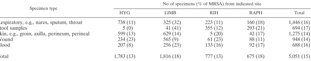
A total of 5,148 specimens were enrolled in the study, of
which 97 had to be excluded due to noncompliance with the
study protocol, e.g., specimen types that were not included in
the protocol or repeated positive samples from the same
pa-tient. From the evaluated 5,051 specimens, 1,186
S. aureus
isolates were recovered, of which 778 (65.6%) were MRSA.
The overall MRSA rate was 15% and differed between 13%
and 18% by study site and between 14 and 17% by type of
specimen (Table 1).
Poolability.
For respiratory and stool specimens and blood,
data were poolable across sites (
P
value
⫽
0.1092, 1.0000, and
1.0000, respectively) at the 0.05 significance level. For skin and
wound specimens, an outlier position was detected for one site
(RIH). However, this was probably attributable to the low
numbers of skin specimens and the perfect performance for
wound specimens at this clinical site. The 95% confidence
intervals of the overall agreement for this site demonstrate the
overlapping with the 95% confidence intervals of the overall
agreement for the other three sites. Therefore, the skin and
wound data were deemed poolable across the study sites, and
the data for all sites were included in the evaluation.
Accuracy of identification as MRSA with and without a
confirmatory test.
After 24 h (18 to 28 h) of incubation time,
99.7% (671/673) of mauve colonies on CMRSAII were
con-firmed as MRSA. Of 243 additional plates with mauve colonies
at 48 h (36 to 52 h) of read time, 170 were not
S. aureus
as
revealed by a recommended confirmatory test (coagulase or
Staphylococcus
latex agglutination test). Based on these data, it
was determined that mauve-colored colonies visible at 24 h
(read range, 18 to 28 h) on CMRSAII need not be confirmed
as
S. aureus
by a coagulase or
Staphylococcus
latex
[image:2.585.48.542.82.179.2]agglutina-tion test. Conversely, it was determined that mauve-colored
colonies visible at 48 h (read range, 36 to 52 h) should be
TABLE 1. Number of specimens and MRSA recovery by site and specimen type
Specimen type
No of specimens (% of MRSA) from indicated site
HYG LIMB RIH RAPH Total
Respiratory, e.g., nares, sputum, throat
738 (11)
325 (32)
223 (11)
160 (18)
1,446 (16)
Stool samples
5 (0)
41 (41)
355 (12)
293 (21)
694 (17)
Skin, e.g., groin, axilla, perineum, perineal
599 (13)
629 (14)
5 (20)
42 (17)
1,275 (14)
Wound
234 (23)
565 (9)
61 (23)
88 (11)
948 (14)
Blood
207 (8)
256 (23)
133 (16)
92 (17)
688 (16)
Total
1,783 (13)
1,816 (18)
777 (13)
675 (18)
5,051 (15)
on May 16, 2020 by guest
http://jcm.asm.org/
confirmed as
S. aureus
by a confirmatory coagulase or latex
agglutination test.
Positive and negative percent agreement of CMRSAII
ver-sus traditional reference media.
The positive percent
agree-ment between CMRSAII and traditional reference media was
87.8% (545/621) at the 24-h read time without a confirmatory
test (coagulase or
Staphylococcus
latex agglutination test) and
94.5% (587/621) at the 48-h read time with a confirmatory test.
Negative percent agreement was 97.1% (4,302/4,430) at the
24-h read time with no confirmatory test and 96.4% (4,271/
4,430) at the 48-h read time with the confirmatory test. Overall
agreement with the reference media was 96% (4,847/5,051)
when colony color (alone) was used to report MRSA at 24 h
and 96.2% (4,858/5,051) with a confirmatory
S. aureus
test at
48 h (coagulase or latex agglutination at 48 h).
Sensitivity and specificity of CMRSAII result compared to
cefoxitin disk diffusion.
The sensitivities of CMRSAII
com-pared to the cefoxitin disk test at the 24-h (without
confirma-tion test) and 48-h (with confirmaconfirma-tion test) reads were between
85.5% and 100%, with the lowest sensitivity for respiratory
specimens and the highest sensitivity for blood cultures
con-taining Gram-positive cocci. The specificity was 99.8% for
res-piratory specimens and 100% for all others.
Recovery of MRSA on CMRSAII versus traditional culture
reference plate and cefoxitin.
Overall recovery of MRSA from
5,051 compliant specimens for CMRSAII was 95.6% (744/
778), compared to recovery on reference blood plates of 79.8%
(621/778) (
P
⬍
0.001) (Table 2). For each specimen type
group, recovery of MRSA on CMRSAII was better than
re-covery of MRSA from the traditional culture method (or equal
in the case of positive blood cultures containing Gram-positive
cocci, where both were 100%). Only 2 false positives were
encountered on the CMRSAII plate at 24 h and none at 48 h
when a confirmatory test was used. The false-positive rates
were 0.05% at 24 h (2 of 4,340) and 48 h (2/4,273).
DISCUSSION
According to the manufacturer, CMRSAII is a selective
medium that is specifically designed for screening for MRSA
from various kinds of microbial specimens, including sputum,
wound swabs, and stool. This implies that the composition of
the medium allows suppression of a broad range of normal
flora. Otherwise, it is likely that high numbers of fast-growing
microorganisms overgrow the MRSA colonies. In this
multi-center study, we evaluated the medium and tested its ability to
detect MRSA even from high-flora samples with mixed flora,
such as stool and rectal samples.
Apart from positive blood cultures, the highest sensitivity
and recovery rate were found for stool samples, from which the
broadest range of microbial flora is expected. In this kind of
material,
S. aureus
colonies can be difficult to detect among
other microorganisms on the nonselective reference media.
The lowest sensitivity and recovery rates were found for
respi-ratory samples, although the difference was not significant. The
fraction of
S. aureus
in the normal flora of the respiratory tract
is certainly higher than that in other body sites. In addition, fast
growing Gram-negative rods are less often detected from
res-piratory samples, and thus,
S. aureus
or MRSA, respectively,
can be more easily detected on the nonselective reference
media even if they are present in small numbers.
The recovery of MRSA from different kinds of samples has
been compared in a few other studies (19, 22, 26, 29). For some
types of specimens, the recovery rate was as low as 50% (29),
but this does not seem to be equally distributed for different
brands of selective media. For example, Nahimana et al. (22)
compared three different chromogenic media for the detection
of MRSA and found the lowest recovery from throat
speci-mens for some media, which is comparable with our results,
but for another medium, the lowest level of recovery was
de-tected for perineal specimens. The differences between the
sensitivities of different chromogenic media may be caused by
additives in the media that inhibit growth of the concomitant
microorganisms but that also inhibit the growth of MRSA (25).
However, groups that compared different media usually did
not compare the recovery rates for different specimens on
selective media to those on nonselective media (19, 22, 26, 29).
Since there is a lack of a gold standard for MRSA screening,
most authors compared the recovery rate of a specific medium
with the combined result of all media or methods that have
been used (5, 6, 9, 10, 12, 13, 15, 19, 20, 21–24, 26–30). This
prevents the misclassification of true-positive results, i.e.,
de-tection of MRSA classified as false positive if the comparison
method failed to detect MRSA. However, this method is prone
to considerable bias, because it depends on the quality and
characteristics of the studied medium itself and all media and
methods that have been included. In addition, recovery rates
for a single test medium seem to be lower if the number of
included reference media is high (22, 26) or if a direct PCR is
included in the methods (28, 29). Nahimana et al. (22) and
Stoakes et al. (26) both compared 4 different selective media
and found recovery rates of approximately 80% after 48 h of
incubation. Van Hal et al. (28, 29), who used PCR detection of
MRSA for comparison in two studies, found recovery rates
of around 75% for the selective media. Groups that compared
only one selective medium with nonselective media reported
recovery rates above 90% (9, 12, 19, 24, 27). Hence,
compar-ison of results gained from different studies can only be
per-formed very cautiously.
CHROMagar MRSA is the predecessor of the medium that
has been studied here. Flayhart et al. (12) compared this
me-TABLE 2. MRSA recovery from traditional culture and CMRSAII
Specimen
category Read time (h)
% MRSA recovery (no. positive by indicated test/total no. positive)
Traditional
culture CMRSAII
Respiratory
24
79.8 (182/228)
85.5 (195/228)
48
76.8 (182/237)
92.4 (219/237)
Stool samples
24
86.9 (93/107)
87.9 (94/107)
48
77.5 (93/120)
98.3 (118/120)
Skin
24
68.6 (118/172)
88.4 (152/172)
48
66.3 (118/178)
96.1 (171/178)
Wound
24
90.6 (115/127)
92.1 (117/127)
48
88.5 (115/130)
94.6 (123/130)
Blood
24
100 (113/113)
100 (113/113)
48
100 (113/113)
100 (113/113)
Total
24
83.1 (621/747)
89.8 (671/747)
48
79.8 (621/778)
95.6 (744/778)
on May 16, 2020 by guest
http://jcm.asm.org/
[image:3.585.43.284.80.250.2]dium in a multicenter study to a nonselective medium and
found a recovery rate for CHROMagar MRSA of 95%,
com-pared to 86% on TSA blood agar for nasal specimens. The
recovery rate of CMRSAII for respiratory swabs, which
in-cluded nasal swabs, was 92%, compared to 77% for the
refer-ence media, and was thus in the same range.
Recovery rates of MRSA from positive automated blood
culture bottles with Gram-positive cocci were 100% for
CMRSAII, identical to those that have been previously
de-scribed for another chromogenic medium (9). Positive blood
cultures are mostly positive for only one species, and the
num-ber of microorganisms is high, making lack of sensitivity or
overgrowth with other microorganisms unlikely. On the other
side, false-positive results may occur due to growth of
sus-ceptible organisms that have been inoculated on the
me-dium in high numbers (9). This phenomenon was not
ob-served in this study for CMRSAII.
Similar to most chromogenic media containing cefoxitin, the
specificity of CMRSAII was very high. At the 24-h read time,
the total number of false positives was only 2 of 673 (0.02%),
allowing for the possibility of reporting mauve colonies as
MRSA after a 24-h read time without further confirmation.
Compernolle et al. (10) found 2% false-positive results on
chromogenic media (MRSA ID [bioMe
´rieux] and CMRSA
[BD Diagnostics]) after 24 h of incubation time in their
insti-tution and suggested performing confirmatory testing at the
24-h read time. They included a large number of samples for
surveillance cultures that were taken from patients in the
in-tensive care unit, where resistant organisms that may grow on
the selective media are more prevalent than in other
depart-ments. We did not collect data on the location of the patients;
however, a high number of specimens were taken from
univer-sity hospitals that often have higher rates of multiresistant
organisms without impairing the specificity.
Because 9.8% of MRSA isolates (73/744) were detected only
after 48 h of incubation, the extension of the incubation time is
valuable for every specimen except positive blood cultures.
After 48 h of incubation, 170 out of 243 specimens with mauve
colonies on CMRSAII did not grow MRSA on the reference
plate, but all of these mauve colonies could be easily proven to
be MRSA by using a confirmatory test, such as coagulase or
cefoxitin disk diffusion testing. When a confirmatory test was
included, the specificity of CMRSAII was 100% and was
com-parable to or better than those described for other
chromo-genic media (9, 10, 12, 19, 22, 23, 26). In conclusion, CMRSAII
is a reliable screening medium for multiple specimen types.
ACKNOWLEDGMENTS
The study was funded by BD Diagnostic Systems, Sparks, MD.
We thank the technicians for their dedicated work.
REFERENCES
1.Batra, R., A. C. Eziefula, D. Wyncoll, and J. Edgeworth.2008. Throat and rectal swabs may have an important role in MRSA screening of critically ill patients. Intensive Care Med.34:1703–1706.
2.Bignardi, G. E., and S. Lowes.2009. MRSA screening: throat swabs are better than nose swabs. J. Hosp. Infect.71:373–374.
3.Bundesministerium fu¨r Gesundheit. 2009. DART Deutsche Antibiotika-Resistenzstrategie. Bundesministerium fu¨r Gesundheit, Bonn, Germany. www.bmg.bund.de. Accessed 3 March 2009.
4.Calfee, D. P., C. D. Salgado, D. Classen, K. M. Arias, K. Podgorny, D. J. Anderson, H. Burstin, S. E. Coffin, E. R. Dubberke, V. Fraser, D. N. Gerding, F. A. Griffin, P. Gross, K. S. Kaye, M. Klompas, E. Lo, J. Marschall, L. A.
Mermel, L. Nicolle, D. A. Pegues, T. M. Perl, S. Saint, R. A. Weinstein, R. Wise, and D. S. Yokoe.2008. Strategies to prevent transmission of methicil-lin-resistantStaphylococcus aureusin acute care hospitals. Infect. Control Hosp. Epidemiol.29(Suppl. 1):S62–S80.
5.Carson, J., B. Lui, L. Rosmus, H. Rennick, and J. Fuller.2009. Interpreta-tion of MRSASelect screening agar at 24 hours of incubaInterpreta-tion. J. Clin. Microbiol.47:566–568.
6.Cherkaoui, A., G. Renzi, P. Franc¸ois, and J. Schrenzel.2007. Comparison of four chromogenic media for culture-based screening of meticillin-resistant Staphylococcus aureus. J. Med. Microbiol.56:500–503.
7.Clinical and Laboratory Standards Institute.2006. Approved standard M2-A9. Performance standard for antimicrobial disk susceptibility tests; ap-proved standard, 9th ed. CLSI, Wayne, PA.
8.Coia, J. E., G. J. Duckworth, D. I. Edwards, M. Farrington, C. Fry, H. Humphreys, C. Mallaghan, D. R. Tucker, Joint Working Party of the British Society of Antimicrobial Chemotherapy, Hospital Infection Society, and Infection Control Nurses Association.2006. Guidelines for the control and prevention of meticillin-resistantStaphylococcus aureus(MRSA) in health-care facilities. J. Hosp. Infect.63(Suppl. 1):S1–S44.
9.Colakoglu, S., H Aliskan, S. S. Senger, T. Turunc, Y. Z. Demiroglu, and H. Arslan.2007. Performance of MRSA ID chromogenic medium for detection of methicillin-resistantStaphylococcus aureusdirectly from blood cultures and clinical specimens. Diagn. Microbiol. Infect. Dis.59:319–323. 10.Compernolle, V., G. Verschraegen, and G. Claeys.2007. Combined use of
Pastorex Staph-Plus and either of two new chromogenic agars, MRSA ID and CHROMagar MRSA, for detection of methicillin-resistant Staphylococ-cus aureus. J. Clin. Microbiol.45:154–158.
11.Dancer, S.2008. Considering the introduction of universal MRSA screening. J. Hosp. Infect.69:315–320.
12.Flayhart, D., J. F. Hindler, D. A. Bruckner, G. Hall, R. K. Shrestha, S. A. Vogel, S. S. Richter, W. Howard, R. Walther, and K. C. Carroll.2005. Multicenter evaluation of BBL CHROMagar MRSA medium for direct detection of methicillin-resistantStaphylococcus aureus from surveillance cultures of the anterior nares. J. Clin. Microbiol.43:5536–5540.
13.Han, Z., E. Lautenbach, N. Fishman, and I. Nachamkin.2007. Evaluation of mannitol salt agar, CHROMagar Staph aureus and CHROMagar MRSA for detection of meticillin-resistantStaphylococcus aureusfrom nasal swab spec-imens. J. Med. Microbiol.56:43–46.
14.Kommission fu¨r Krankenhaushygiene und Infektionspra¨vention, Robert Koch-Institut.2008. Commentary on “Guidelines for prevention and control of MRSA in hospitals and other medical institutions” references on popu-lation with risk of MRSA colonization (August 2008). Epidemiol. Bull. 42:363–364.
15.Krishna, B. V., M. Smith, A. McIndeor, A. P. Gibb, and J. Dave.2008. Evaluation of chromogenic MRSA medium, MRSA select and oxacillin resistance screening agar for the detection of methicillin-resistant Staphylo-coccus aureus. J. Clin. Pathol.61:841–843.
16.Kunori, T., B. Cookson, J. A. Roberts, S. Stone, and C. Kibbler.2002. Cost-effectiveness of different MRSA screening methods. J. Hosp. Infect. 51:189–200.
17.Lagace´-Wiens, P. R., M. J. Alfa, K. Manickam, and G. K. Harding.2008. Reductions in workload and reporting time by use of methicillin-resistant Staphylococcus aureusscreening with MRSASelect medium compared to mannitol-salt medium supplemented with oxacillin. J. Clin. Microbiol.46: 1174–1177.
18.Littell, R. C., W. W. Stroup, and R. J. Freund.2002. Generalized linear models, p. 328–353.InSAS for linear models, 4th ed. SAS Institute Inc., Cary, NC.
19.Louie, L., D. Soares, H. Meaney, M. Vearncombe, and A. E. Simor.2006. Evaluation of a new chromogenic medium, MRSA Select, for detection of methicillin-resistantStaphylococcus aureus. J. Clin. Microbiol.44:4561–4563. 20.Malhotra-Kumar, S., K. Haccuria, M. Michiels, M. Ieven, C. Poyart, W. Hryniewicz, H. Goossens, and the MOSAR WP2 Study Team.2008. Current trends in rapid diagnostics for methicillin-resistant Staphylococcus aureus and glycopeptide-resistant enterococcus species. J. Clin. Microbiol.46:1577– 1587.
21.Molstad, S., O. Cars, J. Struwe, and A. Strama.2008. Swedish working model for containment of antibiotic resistance. Euro Surveill.13:19041. 22.Nahimana, I., P. Francioli, and D. S. Blanc. 2006. Evaluation of three
chromogenic media (MRSA-ID, MRSA-Select and CHROMagar MRSA) and ORSAB for surveillance cultures of methicillin-resistantStaphylococcus aureus. Clin. Microbiol. Infect.12:1168–1174.
23.Nonhoff, C., O. Denis, A. Brenner, P. Buidin, N. Legros, C. Thiroux, M. Dramaix, and M. J. Struelens. 2009. Comparison of three chromogenic media and enrichment broth media for the detection of methicillin-resistant Staphylococcus aureusfrom mucocutaneous screening specimens: compari-son of MRSA chromogenic media. Eur. J. Clin. Microbiol. Infect. Dis. 28:363–369.
24.Pape, J., J. Wadlin, and I. Nachamkin.2006. Use of BBL CHROMagar MRSA medium for identification of methicillin-resistantStaphylococcus au-reusdirectly from blood cultures. J. Clin. Microbiol.44:2575–2576. 25.Perry, J. D., A. Davies, L. A. Butterworth, A. L. Hopley, A. Nicholson, and
on May 16, 2020 by guest
http://jcm.asm.org/
F. K. Gould.2004. Development and evaluation of a chromogenic agar medium for methicillin-resistantStaphylococcus aureus. J. Clin. Microbiol. 42:4519–4523.
26.Stoakes, L., R. Reyes, J. Daniel, G. Lennox, M. A. John, R. Lannigan, and Z. Hussain. 2006. Prospective comparison of a new chromogenic medium, MRSASelect, to CHROMagar MRSA and mannitol-salt medium supple-mented with oxacillin or cefoxitin for detection of methicillin-resistant Staph-ylococcus aureus. J. Clin. Microbiol.44:637–639.
27.Tande´, D., B. Garo, S. Ansart, and B. Lejeune.2008. Efficiency of CHROMa-gar-MRSA in detecting meticillin-resistantStaphylococcus aureusin a rou-tine setting. J. Hosp. Infect.70:388–389.
28.van Hal, S. J., Z. Jennings, D. Stark, D. Marriott, and J. Harkness.2009. MRSA detection: comparison of two molecular methods (BD GeneOhm PCR assay and Easy-Plex) with two selective MRSA agars (MRSA-ID and Oxoid MRSA) for nasal swabs. Eur. J. Clin. Microbiol. Infect. Dis.28:47–53. 29.van Hal, S. J., D. Stark, B. Lockwood, D. Marriott, and J. Harkness.2007.
Methicillin-resistantStaphylococcus aureus(MRSA) detection: comparison of two molecular methods (IDI-MRSA PCR assay and GenoType MRSA Direct PCR assay) with three selective MRSA agars (MRSA ID, MRSAS-elect, and CHROMagar MRSA) for use with infection-control swabs. J. Clin. Microbiol.45:2486–2490.
30.van Loo, I. H., S. van Dijk, I. Verbakel-Schelle, and A. G. Buiting.2007. Evaluation of a chromogenic agar (MRSASelect) for the detection of meti-cillin-resistantStaphylococcus aureuswith clinical samples in The Nether-lands. J. Med. Microbiol.56:491–494.
31.Weber, S. G., S. S. Huang, S. Oriola, W. C. Huskins, G. A. Noskin, K. Harriman, R. N. Olmsted, M. Bonten, T. Lundstrom, M. W. Climo, M. C. Roghmann, C. L. Murphy, and T. B. Karchmer.2007. Legislative mandates for use of active surveillance cultures to screen for methicillin-resistant Staphylococcus aureus and vancomycin-resistant enterococci: position state-ment from the Joint SHEA and APIC Task Force. Am. J. Infect. Control. 35:73–85.