Tameem Imran et al JMSCR Volume 06 Issue 11 November 2018 Page 213
Study on RDW (Red Cell Distribution Width) in Relation to RV Diastolic
Dysfunction in COPD Patients
Authors
Tameem Imran
1, Prabhakar K
2, Rumaisa Ahmed
3Department of General Medicine, Kuvempunagar, Kolar, Karnataka -563101, India
Department of General Medicine, K Block, College Campus, SDUMC, Tamaka, Karnataka, India Department of General Medicine, R L Jalappa Hospital, Kolar, India
Corresponding Author
Tameem Imran
Email: [email protected], Mob: 8971254642
Abstract
COPD has a systemic inflammatory effect which extends beyond the pulmonary system resulting in a state of persistent low-grade systemic inflammation and this inflammation has been proposed as a key element in the association of COPD and CVD. Recently there has been growing attention given to the relationship between RDW and cardiovascular disorders, such as heart failure and coronary artery disease. The present study is conducted to see the relationship between RDW (red cell distribution width) in the relationship with right ventricle diastolic dysfunction in COPD patients. An Observational study conducted at R L Jalappa Hospital Kolar from January to June 2018 which included 98 patients. There is a statistically significant difference were observed in relationship to RDW and with parameters RV basal diameter, RV systolic pressure, RV wall thickness, RV mid cavity diameter, RV longitudinal diameter, Tricuspid E/A, Tricuspid S, Tricuspid e’,’ Tricuspid e’/a’,. And Tricuspid E/e’, S velocity and TAPSE except for Tricuspid E' and Tricuspid A'.26 members had RV Diastolic Dysfunction with RDW > 14.5 whereas only 4 members with RDW <14.5 and this is statistically significant. (Chi-square value of 37.6).The present study troughed some light that there might be a relationship between the increased level of RDW and RV diastolic dysfunction in COPD patients.
Keywords: COPD, CVD, RDW, Kolar, Karnataka.
Introduction
Chronic Obstructive Pulmonary Disease (COPD) is currently the fourth leading cause of death in the world (1) but is projected to be the third leading cause of death by 2020. More than 3 million people died of COPD in 2012 accounting for 6% of all deaths globally. Chronic Obstructive Pulmonary Disease (COPD) is a common, preventable and treatable disease that is characterized by persistent
respiratory symptoms and airflow limitation that is due to airway and/or alveolar abnormalities usually caused by significant exposure to noxious particles or gases.(2) COPD has a systemic inflammatory effect which extends beyond the pulmonary system resulting in a state of persistent low-grade systemic inflammation and this inflammation has been proposed as a key element in the association of COPD and CVD.(3) The main causes of morbidity and mortality among COPD patients are the
www.jmscr.igmpublication.org Impact Factor (SJIF): 6.379
Index Copernicus Value: 79.54 ISSN (e)-2347-176x ISSN (p) 2455-0450
Tameem Imran et al JMSCR Volume 06 Issue 11 November 2018 Page 214
cardiovascular disease (CVD). Among COPD patients, CVD is responsible for approximately 50% of all hospitalizations and 20% of all deaths(4). Population-based studies have suggested that regardless of smoking status, age or sex, a COPD diagnosis increases the risk of cardiovascular morbidity and mortality by approximately two-fold.(5,6) The prognosis of the disease worsens with the addition of right ventricular (RV) failure. Although transthoracic echocardiography is a primary noninvasive tool for accurate evaluations of
RV failure, performing transthoracic
echocardiography in these patients is not easy because of the poor acoustic window. Furthermore, the echocardiographic evaluation of right ventricles requires experience. Therefore there was a need for an easy, affordable, relatively sensitive and specific marker to detect early diastolic dysfunction especially in places where cardiac imaging facilities are sparse.
Recently there has been growing attention given to the relationship between RDW and cardiovascular disorders, such as heart failure and coronary artery disease. Red cell distribution width (RDW) is the new prognostic marker of heart failure in COPD patients. “Increased RDW values are related to underlying chronic inflammation, which promotes red blood cell membrane deformability and changes in erythropoiesis. Patients with COPD are more likely to have Heart Failure with preserved ejection fraction (pEF) than Heart Failure with low ejection fraction.(7,8)
The present study is conducted to see the relationship between RDW (red cell distribution width) in the relationship with right ventricle diastolic dysfunction in COPD patients.
Methodology
An Observational study conducted at R L Jalappa Hospital Kolar from January to June 2018 which included 98 patients. The aim was to study the red cell distribution width in relation to Right Ventricle diastolic dysfunction in COPD patients.
Patients with COPD with GOLD stage 2 and above were included in the study. Patients with active
inflammatory disease, clinically overt cor pulmonale (RV failure), deficiencies of iron, vitamin B12, or folate are causes of high RDW values. RDW is also increased after blood transfusions, in hemolytic anemia and liver disease was excluded as these could confound RDW values. The study was started after obtaining the ethical approval from the institution. Those patients who had given Informed consent and who fulfilled the inclusion criteria were included in the study. Detailed medical history and physical examination were carried out. Blood analyses, 12-lead electrocardiogram, a transthoracic echocardio-graphic examination, and routine blood tests including a full blood count were conducted. Pulmonary Function Test to diagnose the patients with COPD: The disease severity of patients was staged according to the criteria of Global Initiative for Chronic Obstructive Lung Disease. Forced vital capacity (FVC), forced expiratory volume in 1 second (FEV1), and a ratio of FEV1 to FVC (FEV1/FVC) was measured at least 3 times with the patient in a seated position.
The procedure for performing biochemical investigations:
Tameem Imran et al JMSCR Volume 06 Issue 11 November 2018 Page 215
annulus early (E’) and late (A’) tissue and Doppler diastolic velocities were also obtained. Right ventricular dilatation, lateral tricuspid annulus longitudinal motion, and tissue Doppler velocities are all used in the echocardiographic identification of RV dysfunction.
Statistical analysis
Data analysis was performed using SPSS software (version 17) and Microsoft Excel worksheet 2013. Categorical variables were represented as proportions/percentages and quantitative variables were represented as means and standard deviation. Chi-square tests and independent sample t-test were used. A P value < 0.05 is considered as statistical significance at 95% confidence intervals.
Results
In the present study 77 were males and 21 were females COPD patients were included.
Table 1: Gender distribution
Category Cases (%) P Value Males 77(78.6) 0.01
Significant
Females 21 (21.4)
Total 98 (100)
The mean age of the study population is 60.76 ± 9.86 years. More males were suffering from COPD disease when compared to females. Most common presenting complaints were breathlessness and cough. Majority 67.3 % of the male study participants had given a history of smoking when compared to females (8.2%) which are statistically significant (chi-square value 20.23, p<0.05). Other risk factors were Indoor air pollution and occupational exposure of dust.
Table 2: Mean Forced Expiratory Volumes:
Respiratory findings RDW<14.5 RDW>14.5
FEV1 52 39
FEV1/FVC 65 55.8
GOLD 2/3/4 46/12/0 9/22/9
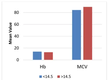
Table 3: Red Cell Distribution Width vs Blood Parameters
RDW <14.5 (n= >14.5 P Value
Hb 14.05±1.82 13.08±1.95 0.014
MCV 84.32±10.09 89.6±11.23 0.016
In studying the relationship between the Red Cell Distribution Width and Blood Parameters, the values like MCV is more when compared to the RDW value <14.5. Hemoglobin value is less when the RDW value is >14.5 with a P value less 0.05. This shows that there is a positive relationship between the Blood parameters and the RDW values with patients with COPD.
Figure 1: Red Cell Distribution Width vs Blood Parameters
Table 4: Echocardiography findings in relationship with RDW
ECHO parameters
RDW <14.5
RDW>14.5 P VALUE
LVEF 57.58
+/- 2.45 55 +/- 4.02 0.001
RVD1
2.66+/-0.41
3.36 +/- 0.57
0.001
RVD2 3.32
+/- 0.36 3.75 +/- 0.38 0.001
RVD3 7.82
+/- 0.43 8.48 +/- 0.53 0.001
S Velocity 9.73
+/- 0.85 7.84 +/- 1.72 0.001
TAPSE 2.04
+/- 0.34 1.73 +/- 0.32 0.001 TRICUSPID E/A 1.15 +/- 0.28 1.03 +/-
0.21 0.02
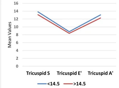
Tricuspid S 13.82
+/- 1.68
13.10 +/-
1.65 0.037
0 20 40 60 80
Hb MCV
M e an Val u e
Tameem Imran et al JMSCR Volume 06 Issue 11 November 2018 Page 216
Tricuspid E' 8.78
+/- 0.84
8.36 +/-
0.91 0.91
Tricuspid A' 13.06
+/- 2.90
12.26 +/-
2.93 0.187
E’/A’ 0.85
+/- 0.36
0.72 +/-
0.19 0.041
Tricuspid E/E'
3.80 +/- 0.35
7.26 +/-
3.04 0.001
RV systolic pressure
26.16 +/- 1.19
28.55 +/-
3.38 0.001
RV wall
thickness
3.99 +/- 1.03
6.25 +/-
1.27 0.001
There is a statistically significant difference were observed with parameters RV basal diameter, RV systolic pressure, RV wall thickness, RV mid cavity diameter, RV longitudinal diameter, Tricuspid E/A, Tricuspid S, Tricuspid e’,’ Tricuspid e’/a’,. And Tricuspid E/e’, S velocity and TAPSE except for Tricuspid E' and Tricuspid A'.
26 members had RV Diastolic Dysfunction with RDW > 14.5 whereas only 4 members with RDW <14.5 and this is statistically significant. (Chi-square value of 37.6).
Figure 2: Echocardiography findings
Figure 3: Echocardiography findings
Discussion
COPD has been accepted as a component of systemic inflammatory syndrome and the mortality mostly depends on cardiovascular diseases rather than respiratory failure.(9) The red cell distribution width (RDW) blood test measures the amount of red blood cell variation in volume and size which reflects impaired erythropoiesis and abnormal red blood cell survival. In the recent past, there is an increased focus of research going on as this is an easily done and less costly parameter and is a strong and independent risk factor for death. There are several potential mechanisms to explain elevated RDW values in heart failure.
Previous studies have addressed inflammation, ineffective erythropoiesis, malnutrition, impaired renal function, and neurohormonal activation in heart failure patients.(10) Dharmesh Kumar Patel et al they found that increased RDW levels were associated with COPD severity and a higher mortality.(11)
In the present study, there are abnormal values in the echocardiography as there is an increase in RV systolic pressure, RV wall thickness, Tricuspid E/A, Tricuspid S, Tricuspid e',' Tricuspid e'/a' and Tricuspid E/e'. RV wall thickness & RV systolic pressure were higher in patients with higher RDW. Peak velocity of early diastolic tricuspid annular motion was significantly lower in patients with increased RDW. S velocity, TAPSE was decreased 0
2 4 6 8 10 12
RVD1 RVD2 RVD3 S VELOCITY
TAPSE
M
ean
Val
u
es
<14.5 >14.5
0 2 4 6 8 10 12 14 16
Tricuspid S Tricuspid E' Tricuspid A'
M
ean
Val
u
es
Tameem Imran et al JMSCR Volume 06 Issue 11 November 2018 Page 217
and RV dilatation was seen. Above parameters indicative of RV diastolic dysfunction were seen in patients having RDW >14.5. A study conducted by Atac Celik et al while trying to know the relationship between red cell distribution width and echocardiographic parameters in patients with diastolic heart failure observed that there is a significant increase in the ratio of early mitral inflow velocity to early diastolic mitral annular velocity and Pulmonary capillary wedge pressure.(12) Sen Liu et al while Assessing Severity of Chronic Heart Failure in relation to RDW concluded that Elevated RDW is an independent risk factor for mortality. (13) Several studies have shown a relationship with a role of RDW in increase all-cause mortality rate. A Meta-analysis conducted on seven community-based studies with a sample of 11,827 observed that For every 1% increment in RDW, all-cause mortality risk increased by 14% (adjusted HR, 1.14; 95% CI, 1.11–1.17). (14) A study conducted by Riedl J et al among 1,990 patients admitted to the emergency department observed that after doing Multivariate logistic analysis after adjustment for multiple confounders showed that the all-cause in-hospital mortality rate increased by 21.8% for each 1% increase in RDW.(15) Amongst 98 COPD patients 30 (30.61%) had RV diastolic dysfunction and RV failure. 86.6% amongst these had RDW of more than 14.5. Sensitivity and specificity of RDW to predict diastolic dysfunction /RV failure is 86.67% and 79.41% respectively. Therefore Levels of RDW before Echocardiography can be used to predict RV failure.
Conclusion
The present study showed there might be a relationship between the increased level of RDW and RV diastolic dysfunction in COPD patients. Future studies are required to confirm the role of RDW in RV diastolic dysfunction in COPD patients as this is simple and non-invasive test might be used as a preliminary biomarker in the evaluation of diseases severity.
References
1. Lozano R, Naghavi M, Foreman K, et al. Global and regional mortality from 235 causes of death for 20 age groups in 1990 and 2010: a systematic analysis for the Global Burden of Disease Study 2010. Lancet 2012; 380(9859): 2095-128.
2. Gold reports 2018 – Global Initiative for Chronic Obstructive Lung Disease: https://goldcopd.org/gold-reports/. Accessed on 11th Oct 2018.
3. Chronic obstructive pulmonary disease: https://en.wikipedia.org/wiki/Chronic_obstru ctive_pulmonary_disease. Accessed on 5th Sep 2018
4. Sin DD, Man SF. Impact of cancers and cardiovascular disease in chronic obstructive pulmonary disease. Curr Opin Pulm Med. 2008;14(2):115-21.
5. Maclay JD, MacNee W. Cardiovascular disease in COPD: mechanisms. Chest. 2013 Mar;143(3):798-807
6. John D.Maclay, William McNee.
Cardiovascular Disease in COPD: Mechanisms. Chest. Volume 143, Issue 3, March 2013, Pages 798-807
7. Jennifer Quint. The Relationship between COPD and Cardiovascular Disease. Tanaffos. 2017; 16(Suppl 1): S16–S17. 8. Morgan AD, Zakeri R, Quint JK. Defining
the relationship between COPD and CVD: what are the implications for clinical practice? .Ther Adv Respir Dis. 2018 Jan-Dec;12.
9. Celli BR, Zuwallack RL. Pulmonary rehabilitation. In: Broaddus VC, Mason RJ, Ernst JD, et al, eds. Murray and Nadel's Textbook of Respiratory Medicine. 6th ed. Philadelphia, PA: Elsevier Saunders; 2016:chap 105.
Tameem Imran et al JMSCR Volume 06 Issue 11 November 2018 Page 218
inflammation, renal function, and nutritional state. Am Heart J 2009; 158:659e66.
11.Dharmesh kumar Patel, Deepali J. Kamdar. Prediction of the severity of COPD based on red cell distribution width value. Indian Journal of Immunology and Respiratory Medicine, October-December 2016;1(4):84-87.
12.Atac Celik, Fatih Koc, Hasan Kadi, Koksal Ceyhan, Unal Erkorkmaz et al. The relationship between red cell distribution width and echocardiographic parameters in patients with diastolic heart failure. Kaohsiung Journal of Medical Sciences (2012) 28, 165-172.
13.Sen Liu, Ping Wang, Ping-Ping Shen, and Jian-Hua Zhou. Predictive Values of Red Blood Cell Distribution Width in Assessing Severity of Chronic Heart Failure. Med Sci Monit, 2016; 22: 2119-2125.
14.Patel KV, Semba RD, Ferrucci L, et al. Red cell distribution width and mortality in older adults: a meta-analysis. J Gerontol A Biol Sci Med Sci 2010; 65:258-65.