Original Research Article
Bacteriological profile and antibiotic sensitivity patterns of blood
cultures
K. Ashwin Reddy, S. Uday Kanth*
INTRODUCTION
The term neonatal septicemia is synonymous with septicemia Neonatorum or neonatal sepsis. Neonatal septicemia refers to a clinical syndrome characterized by systemic signs and symptoms due to generalized bacterial
infection with positive blood culture in the first 4 weeks of life.1,2
It is an important cause of morbidity and mortality among neonates and is one of the four leading causes of neonatal mortality in India.3 Neonates are particularly vulnerable to infection because of weak immune barrier.4
ABSTRACT
Background: Neonatal septicemia is a significant cause of morbidity and mortality worldwide especially so in developing countries. To reduce the mortality caused by neonatal septicemia, it became vital to diagnose it as soon as possible and treat with administration of appropriate antibiotics. The objective of the study was bacteriological spectrum in blood culture of neonates admitted in a hospital, and antibiotic susceptibility pattern of blood culture positive isolates.
Methods: A total of 593 blood culture sample were received from NICU admissions for a period of 15 months were included for this study. Under aseptic precautions, 1 ml of blood was collected from a peripheral vein and inoculated into a bottle of Brain Heart Infusion broth and was incubated for 7 days. Repeated sub-culturing was done as per standard procedures. Inoculation on blood agar and Mac-Conkey’s agar plates were made. Any growth was subjected for identification by appropriate biochemical tests. Antibiotic susceptibility testing was done by disc diffusion method.
Results: Of the 593 cases studied 12.14 % were blood culture positive. Among the blood culture positive neonates 67% were male neonates. Late onset septicemia (87.5%) was more common than early onset septicemia (12.5%). Gram negative organisms 46 (63.88%) were predominant than gram positive organisms 18 (25%). Klebsiella pneumonia 18 (25%), Citrobacter 10 (13.88%), and Pseudomonas auroginosa was found in 7 (9.72%). The other organisms isolated were Escherichia coli 06 (8.33%), Enterobacter 04 (5.55%), Gram positive organisms were obtained in 18 (25.00%) out of 72 cases. MSSA 06 (8.33%), Enterococci 06 (8.33%) was the commonest organisms isolated. Most of the isolates were more susceptible imipenem, meropenem, and ciprofloxacin to amikacin antibiotics.
Conclusions: Blood culture remains the gold standard for the diagnosis of neonatal septicemia. Periodic surveillance of organisms and their antibiotic sensitivity patterns are essential to understand and to prevent emergence of resistant organisms. Effective/Correct selection of antibiotic is essential to decrease mortality and morbidity in the vulnerable group of neonatal population.
Keywords: Blood culture, Klebsiella pneumonia, Neonatal septicaemia
Department of Pediatrics, Malla Reddy Institute of Medical Sciences, Suraram, Hyderabad, Telangana, India
Received: 02 September 2016
Accepted: 06 September 2016
*Correspondence:
Dr. S. Uday Kanth,
E-mail: [email protected]
Copyright: © the author(s), publisher and licensee Medip Academy. This is an open-access article distributed under the terms of the Creative Commons Attribution Non-Commercial License, which permits unrestricted non-commercial use, distribution, and reproduction in any medium, provided the original work is properly cited.
According to the National Neonatal Perinatal Database (NNPP) report 2002- 2003, the incidence of neonatal septicemia in tertiary care institutions has been reported to be 16% of all mortalities among the hospital born neonate neonates.5
The scoring system by Takkar VP and Bhakoo ONwhich consist of six perinatal risk factors is one of such valuable tool most commonly used by clinicians to screen or treat the neonates for septicemia.6
Several authors categorize neonatal septicemia into two groups, primary and secondary.
Gram negative organisms (65-85%) are found to be frequently responsible for septicemia than gram positive organisms (15%) as evidenced by the Indian studies.1 Microbiological pattern of early onset neonatal sepsis is different from late onset sepsis and its association with several perinatal risk factors.7
The most common isolates in the early onset septicemia were Klebsiella species (25.8%), Enterobacter species (22.4%) followed by Escherichia coli (14.5%). Isolates in late onset septicemia Enterobacter spp. (23.3%) was the major pathogen followed by coagulase negative staphylococcus (23.3%).4
Bacterial isolates and their antibiotic susceptibility have been constantly changing which depend on several factors like gestational age, birth weight, maternal risk factors, place of delivery, mode of delivery etc.8
The antibiotic misuse has resulted in further confusion in diagnosis and emergence of drug resistant bacterial strains in the neonatal units with grave sequelae. Thus the successful treatment with a favourable outcome of the neonate depends on an on-going review of the causative organisms and their antibiotic susceptibility pattern.4,8 The present study has been under taken to isolate and identify the aerobic bacteria responsible for neonatal septicemia from blood with their antibiotic susceptibility pattern.
METHODS
Source of data
Data collected retrospectively. Blood samples were collected from 593 cases of neonates admitted in the Neonatal intensive care unit in Lotus children Hospital. Period was October 2014 to December 2015.
Inclusion criteria
The study includes all blood culture positive cases received from neonatal intensive care unit who were admitted in NICU.
Exclusion criteria
All negative blood cultures negative and contaminant growth were excluded from the study.
In the present study the following investigations were adopted to achieve the objective of the study.
Blood culture
Antibiotic susceptibility testing of the isolated pathogen from the blood.
Sample collection
About one ml of venous blood was drown by venous puncture following strict aseptic precautions i.e. suitable site veni-puncture was cleaned with 70% alcohol rubbing vigorously and allowed to dry. Starting in the centre of the circle povidine iodine was applied in widening circles until the entire circle has been saturated with Iodine and allowed to dry on the skin for at least one min. After blood was drown, 70% alcohol was applied to the veni-puncture site as many patients may be sensitive to iodine.
Blood culture
About one ml of blood was drawn aseptically and inoculated into a blood culture bottle infusion broth, thus making dilution of 1 in 10 to nullify the natural bacteriostatic or bactericidal activity of blood. Brain Heart infusion broth was prepared using the commercially available ready to use powder supplied by Hi-Media Laboratories Pvt. Limited, Mumbai, India. The broth was distributed into 10 ml quantity in McCartney bottles and sterilized by autoclaving at 121°C for 15 minutes.
After inoculation, the blood culture bottles were incubated at 37°C under aerobic conditions in the incubator for 7 days. The first subculture was done after 24 hours of incubation the second on the 3rd day and a final on the seventh day. Subcultures were done onto chocolate agar, 5% sheep blood agar and Mac-conkey agar plates. The inoculated plates were incubated aerobically in the incubator at 37°C for 24 hours and the plates were observed for growth. The growth was identified by colonial characteristics, Gram’s stain and standard biochemical tests, described in Mackie and McCartney, Practical Medical Microbiology and Bailey and Scott’s Diagnostic Microbiology.
Culture which did not yield any growth following three subcultures was reported negative at the end of 7 day.
Antibiotic susceptibility testing
using CLSI guidelines for interpretation as resistant, intermediate sensitive and sensitive. Control strains of Escherichia coli ATCC 25922, Pseudomonas auroginosa ATCC 27853 and Staphylococcus aureus ATCC 25923 were used.
RESULTS
Table 1: Distribution of cases according to gender.
Gender Culture positive
Number Percentage
Male 45 62.5
Female 27 37.5
Total 72 100
Among 72 culture positive neonates 45 (62.5%) were male and 27 (37.5%) were females. Male were higher in numbers compared to females with a ratio of 1.67.
Table 2: Distribution of cases according age.
Age (days) Culture positive
Number Percentage
1-7 30 41.6
8-14 22 30.5
15-21 06 8.3
22-28 04 5.5
> 29 10 13.8
Total 72 100
Mean age = 13.56±14.669 days (Mean ±SD)
Out of 72 culture positive cases 30 (41.6%), 22 (30.5%), 06 (8.3%), 04 (5.5%), 10 (13.8%) belonged to 1st, 2nd, 3rd, 4th weeks, and more than 29 days respectively. Majority of culture positive neonates i.e. 30 (41.6%) belonged to their 1st week of life.
Table 3: Distribution of early and late onset sepsis.
Blood culture status
Cases
Total
Early onset septicaemia Late onset septicaemia
Number Percentage Number Percentage Number Percentage
Positive 09 12.5 63 87.5 72 100
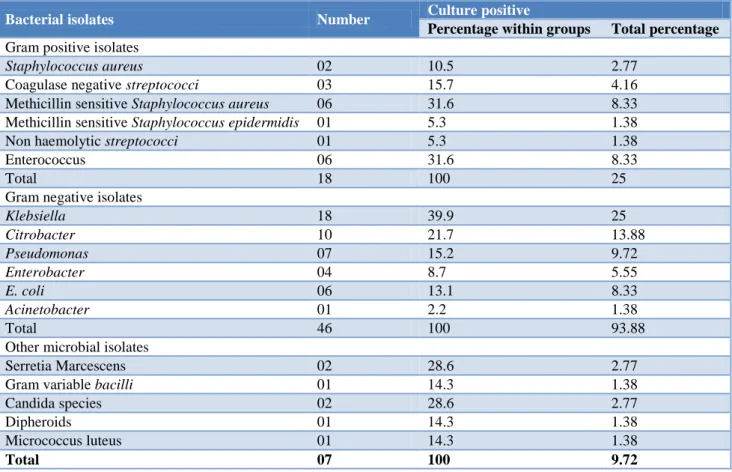
Table 4: Spectrum of bacterial isolates.
Bacterial isolates Number Culture positive
Percentage within groups Total percentage
Gram positive isolates
Staphylococcus aureus 02 10.5 2.77
Coagulase negative streptococci 03 15.7 4.16 Methicillin sensitive Staphylococcus aureus 06 31.6 8.33 Methicillin sensitive Staphylococcus epidermidis 01 5.3 1.38 Non haemolytic streptococci 01 5.3 1.38
Enterococcus 06 31.6 8.33
Total 18 100 25
Gram negative isolates
Klebsiella 18 39.9 25
Citrobacter 10 21.7 13.88
Pseudomonas 07 15.2 9.72
Enterobacter 04 8.7 5.55
E. coli 06 13.1 8.33
Acinetobacter 01 2.2 1.38
Total 46 100 93.88
Other microbial isolates
Serretia Marcescens 02 28.6 2.77
Gram variable bacilli 01 14.3 1.38
Candida species 02 28.6 2.77
Dipheroids 01 14.3 1.38
Micrococcus luteus 01 14.3 1.38
Of the 72 culture positive cases, early onset septicaemia was seen in 9 (12.5%). Cases and 63 (87.5%) cases showed late onset septicaemia.
Majority i.e. 46 (63.88%) of the isolates were Gram negative organisms, Klebsiella being the commonest isolated in 18 (25.00%) of the 72 culture positive cases followed by Citrobacter 10 (13.88%) Pseudomonas auroginosa was found in 7 (9.72%). The other Gram negative organisms isolated were Escherichia coli 06 (8.33%), Enterobacter 04 (5.55%). Gram positive organisms were obtained in 18 (25.00%) out of 72 cases. MSSA 06 (8.33%), Enterococci 06 (8.33%). were commonest isolates followed by Coagulase negative staphylococci 3 (4.16%) and Staphylococcusaureus in 02 (2.77%) cases. Other isolates are Candida species 02 (2.77%), Serretia Marcescens 02 (2.77%), Gram variable bacilli 01 (1.38%), Dipheroids 01 (1.38%), Micrococcus luteus 01 (1.38%) Polymicrobial isolates i.e. Klebsiella Species with Enterococci and Klebsiella with E.coli were obtained in only 2 cases.
MSSA was one of the major Gram positive isolate showing 100% susceptibility to vancomycin Linezolied, Imipenem and pipecillion + Tazobactum, Amikacin and ampicillin + Sulbactum and less susceptibility (50%) to Ampicillin, Ciprofloxacin.
Enterococcus is the another common isolate which was showing 100% susceptibility to vancomicin and 75% susceptibility to Ofloxacin and less than 60% to other antibiotics.
DISCUSSION
The ratio of culture positive neonatal septicemia cases were higher among males 45 (62.5%) than the females 27 (37.5%), showing a ratio of 1.6:1. These results are comparable with the observations made by Tallur et al (63.60%)Uddin Ahmed et al (63.00%)Movahedin AH et al (71.2%).9-11 Our results differed from that of Betty Chacko et al who observed an equal proportion of cases among both males and females, because their study involved only early onset septicemia cases.12 The male preponderance in neonatal septicemia may be linked to the X- linked immuno-regulatory gene factor resulting in the host’s susceptibility to infections in males.13
Maximum culture positive cases were seen in neonates more than 1 week of age 41.6% and Late onset septicemia 87.5% as compared to neonates aged less than 1 week 41.6% and early onset septicemia 12.5% in the present study. Our results correlates from the study by G. Karthikeyan et al who reported an equal proportion of EOS (50%) and LOS (50%) cases in their study, while the study by R S Jaswal et al (74%) and Shrestha P et al (66.9 %) showed a higher proportion of LOS cases.14-16 Our results differ with higher percentage of EOS was seen in the studies done by Tallur et al (83.47%)andRoy et al (71.30%).4,9This could be due to ascending infection
following rupture of membranes or during the passage of baby through the infected birth canal or at the.
In the present study, 72/593 cases studied were culture positive, giving a culture positivity rate of 12.14%. Our study was differs with the studies conducted by Uddin Ahmed et al 35%,Roy et al (47.50%),Betty Chacko et al (43.10%),Tallur et al (64.87%) and G. Karthikeyan et al (51.34%) showed high culture positivity rate.4,9,10,14,15 Most of the NICU admissions blood cultures were sent, at the time of admission. This could be reason for the low culture positive percentage in our study.
In the present study, Klebsiella pneumoniae 18 (25%) was the predominant isolate, followed by Citrobacter 10 (13.88%). Gram negative organisms formed the majority of the isolates as compared to Gram positive organisms (63.88% v/s 25%).
Similarly a higher proportion of Klebsiella pneumoniae were isolated by Tallur et al (53.50%), Roy et al (24.60%).4,9 Studies conducted by G. Karthikeyan et al (61.50%), showed Staphylococcus aureus as the commonest isolate, while Pseudomonas aeruginosa was the commonest pathogen isolated by Betty Chacko et al (60%) and A. H. Movahedian et al (36%), in their study and Coagulase negative Staphylococcus (54.9%) was the commonest pathogen isolated by Edwis Dias et al in their study.11,12,14
In the present study majority of the isolates were Gram negative organisms accounting for 63.88% of the isolates with Klebsiella pneumonia being the commonest isolated in 18 (25%) of the 72 culture positive cases.
Among the Klebsiella pneumonia maximum isolates i.e. (93.33%) were susceptible to Imipenem followed by Meropenem (85.7%), Colistin have 80% while only around 50% susceptibility was noted to Amikacin, Gentamicin, and Ciprofloxacin. Very less sensitivity to Ceftriaxone, Cefotaxime have only 7%. Resistance was seen to Ampicillin was 100%. A varied resistance pattern was seen to Ceftazidime, Aztreonam.
It is evident from the present study that maximum susceptibility was noted to Imipenem, Meropenem and Colistin. Majority of the isolates were less susceptible to third generation cephalosporin.
In a study done by Tallur et al 2000, Klebsiella pneumonia was the commonest pathogen found which were more susceptible to Gentamicin, Amikacin and third generation cephalosporin.9
Gram negative organisms continue to be a menace to the sick, fragile and debilitated newborns. Multidrug resistance of the causative organisms of septicemia is a rapidly emerging, potentially disastrous problem. Our data is no exception to the worldwide antimicrobial emergency. Among these, Klebsiella septicemia continues to be a challenge to the neonatologist and microbiologists.
One of the reasons for the predominance of an organism in causing septicemia in the neonatal units is the selective pressure of antimicrobial agents, so that resistant microorganisms tend to colonize and proliferate in the neonatal units. Cross contamination and nosocomial transmission may play significant role in the etiology of Klebsiella septicemia.9
Also in the present study, 12 (14.5%) isolates were Pseudomonas aeruginosa which was 100% susceptible to Amikicin, Ofloxacin, ciprofloxacin, around 50% to Ceftriaxone, and Cefotaxime and, 100% resistance to Amipicillin, Ampillicin+Sulbactum.
Study of AH Movahedian et al 2006 observed 70% of susceptibility of Pseudomonas aeruginosa to Amikicin followed by Gentamicin 53% and lower susceptibility to Ampicillin, Cephalexin, Ceftriaxone andcefotizoxi.11 In the present study 100% of Escherichia coli were susceptible to Meropenem, Colistin, and 83.3% to Imipenem, around 50% to Amikacin, Ciprofloxacin and Cefotaxime. Citrobacter isolates were susceptible to Tobramycin and Gentamicin 88.8% to Imipenem and Amikicin, and Colistin it is around 80% and Ceftriaxone is 100% resistant.
Overall the Gram negative organisms were susceptible to Imipenem, Meropenem and Amikacin. A variable susceptibility was seen to Colistin, Quinolones and third generation cephalosporins. Almost all isolates showed resistance to Ampicillin, Aztreonam and cefoxitin.
In the present study MSSA, Enterococci was the major Gram positive organism isolated constituting 6 (8.33%) by each; coagulase negative staphylococcus 3 (4.56%) of the isolates and Staphylococcus aureus being 2 (2.77%). All the gram positive isolates were 100 % susceptible to Vancomycin, Linezolid, Imipenem, Meropenem, Amikacin, and Ofloxacin except Enterococci.
Around 50% resistance to Ampicillin was observed to MSSA, CONS, Enterococci.
Enterococci have variable sensitivity pattern compare to other Gram positive isolates, 40-50 resistance is observed to all antibiotics except Vancomycin (100% Sensitive), Linezolid, Ofloxacin.
Tallur et al noted a high incidence of Staphylococcus spec. in their study, pointing towards its nosocomial
origin.9 All these isolates were 100% susceptible to Vancomycin. In the study by G. Karthikeyan et al 2001 major isolate was Staphylococcus aureus and most of these isolates belonged to early onset septicemia group; 24/59 (40.7%).14
The significant proportions of these cases were presumed to occur as a result of vertical transmission thereby implying maternal colonization. Multidrug resistant Staphylococcus aureus has been accepted as an indication for the use of vancomycin, which is the drug of choice in these situations.
Similar observations has been made in the study by Roy et al who found Staphylococcus aureus isolates to be resistant to Penicillin, Erythromycin, Gentamicin and Ciprofloxacin.4 Coagulase negative Staphylococci are emerging as causative organisms of neonatal septicemia and recovery of these pathogens from the blood culture of newborn infants should no longer be consider as a contaminant especially in the preterm and low birth weight neonates with relatively longer hospital stay and excessive exposure to diagnostic and supportive procedures Anand et al.
CONCLUSION
Neonatal septicemia is still leading cause of mortality and mortality and morbidity in developing countries like India. It is presents with non-specific signs and symptoms. It is more common in male, low birth weight and term neonates. A positive blood culture is the one definitive method of confirming a case of septicemia, which is a “Gold standard” test and help in prompt and timely administration of antibiotics which could be lifesaving. Gentamicin, Ampicillin, Amikacin and third generation cephalosporin are the commonly used antibiotics, but these drugs lost their ground in controlling septicemia because of development of resistance in causative organisms. A rational use of antibiotics is necessary for treatment of neonatal septicaemia.
Funding: No funding sources Conflict of interest: None declared
Ethical approval: The study was approved by the Institutional Ethics Committee
REFERENCES
1. Gotoff SP, Behrman RE. Neonatal septicaemia. J Pediatr. 1970;76(1):142-53.
2. Aggarwal R, Sarkar N, Deorari AK. Sepsis in the Newborn. Indian J Pediatr. 2001;68(12):1143-7. 3. Desai KJ, Malek SS, Parikh A. Neonatal
4. Roy I, Jain A, Kumar M. Bacteriology of Neonatal Septicemia in Tertiary care Hospital of Northern India. Indian J Med Microbiol. 2002;20(3):156-9. 5. National Neonatal Perinatal Database. Report for
the year 2002-03. National Neonatology Forum, India.
6. Takkar VP, Bhakoo ON, Narang A. Scoring system for the Prediction of Early Neonatal Infections. Indian Pediatr. 1974;11(9):597-60.
7. Paul VK, Singh M. Diagnosis and Treatment of Neonatal Sepsis. Indian Pediatr. 1986;23:1023-35. 8. Mathur NB. Neonatal Sepsis. Indian Pediatr.
1996;33:663-74.
9. Tallur SS, Kasturi AV, Nadgir SD. Clinico bacteriological Study of Neonatal Septicemia in Hubli. Indian J Pediatr. 2000;67(3):169-74.
10. Nawshad Uddin Ahmed ASM, Chowdhury AMAK, Hoque M. Clinical and bacteriological profile of neonatal septicaemia in a tertiary level pediatric hospital in Bangaldesh. Indian Pediatr. 2002;39:1034-9.
11. Movahedian AH, Moniri R, Mosayebi Z. Bacterial
Culture of Neonatal Sepsis. Iranian J Public Health. 2006;35(4):84-9.
12. Chacko B, Sohi I. Early Onset Neonatal Sepsis. Indian J Pediatr. 2005;72(1):23-6.
13. Khatua SP, Das AK, Chatterjee BD. Neonatal septicemia. Indian J Pediatr. 1986;53(4):509-14. 14. Karthikeyan G, Premkumar K. Neonatal Sepsis:
Staphylococcus aureus as the Predominant Pathogen. Indian J Pediatr. 2001;68(8):715-7. 15. Darmstdt L. Clinical and Bacteriological Profile of
Neonatal Septicemia in a Tertiary level Pediatric Hospital in Bangladesh. Indian Peditr. 2002;39:1034-8.
16. Shrestha P. Clinical and Bacteriological Profile of Blood Culture Positive Sepsis in Newborns. J Nepal Paediatr Society. 2007;27(2):64-7.
17. Jaswal RS, Kaushl RK, Goel A. Role of C - reactive protein in Deciding Duration of Antibiotic Therapy in Neonatal Septicemia. Indian Pediatr. 2003;40:880-3.