i
CHANGES IN INFRASPINATUS CROSS-SECTIONAL AREA AND ECHO INTENSITY IN RELATION TO SCAPULAR DYSKINESIS AND OVERHEAD TRAINING VOLUME IN
COLLEGIATE VOLLEYBALL PLAYERS
Kimberly Chase
A thesis submitted to the faculty at the University of North Carolina at Chapel Hill in partial fulfillment of the requirements for the degree of Masters of Arts in the Department of Exercise & Sport Science (Athletic Training) in the College of Arts & Sciences.
Chapel Hill 2016
Approved by:
D. Padua
T. Blackburn
iii ABSTRACT
Kimberly Chase: Changes in Infraspinatus Cross-Sectional Area and Echo Intensity in Relation to Scapular Dyskinesis and Overhead Training Volume in Collegiate Volleyball Players
(Under the Direction of Darin Padua)
During the pre-season injury risk in volleyball is highest at 6.1 injuries/1000 exposures.
Injury to the posterior shoulder, specifically the infraspinatus, has been attributed to repetitive
eccentric loading. The high volume of overhead activity in the collegiate volleyball player has
been identified is a risk factor for upper extremity injury. Cross sectional area and echo intensity
of the infraspinatus muscle were assessed using diagnostic ultrasound to measure muscle
damage. Visual observation of scapular dyskinesis was completed at baseline and 24 hours
following the pre-season training period to assess changes due to pre-season training volume.
Correlations between cross sectional area, echo intensity, scapular dyskinesis, and swing count
were also examined to assess relationships between these variables. Acceptable reliability was
established for all measurements. This study found no significant outcomes in cross sectional
area or echo intensity, in the severity of scapular dyskinesis, or in relationships between the
iv
TABLE OF CONTENTS
LIST OF TABLES……….v
LIST OF FIGURES………..vi
CHAPTER I: INTRODUCTION………..1
Research Questions and Hypothesis ... 5
CHAPTER II: LITERATURE REVIEW ... 6
Overuse Injury at the Shoulder……….7
Risk Factors for Shoulder Injury ... 9
Scapular Dyskinesis ... 11
Ultrasound and Cross-sectional Area ... 18
CHAPTER III: METHODOLOGY ... 20
Participants ... 20
Instrumentation ... 21
Procedures ... 23
Data Analysis ... 28
CHAPTER IV: MANUSCRIPT……….31
Introduction………..33
Methods………...34
Results ... 40
Discussion ... 44
v
LIST OF TABLES
TABLE 1: Participant demographics……….21
TABLE 2: Scapular Dyskinesis Test Statistics: Inter-rater and Intra-rater reliability…………...22
TABLE 3: ICC for CSA and EI: Intra-rater reliability………22
TABLE 4: Overhead volume by position group……….25
TABLE 5: Operational definitions for scapular motion………..26
TABLE 6: Grading Criteria for Scapular Dyskinesis Test……….39
TABLE 7: Descriptive Statistics for CSA and EI in the pre-season………...40
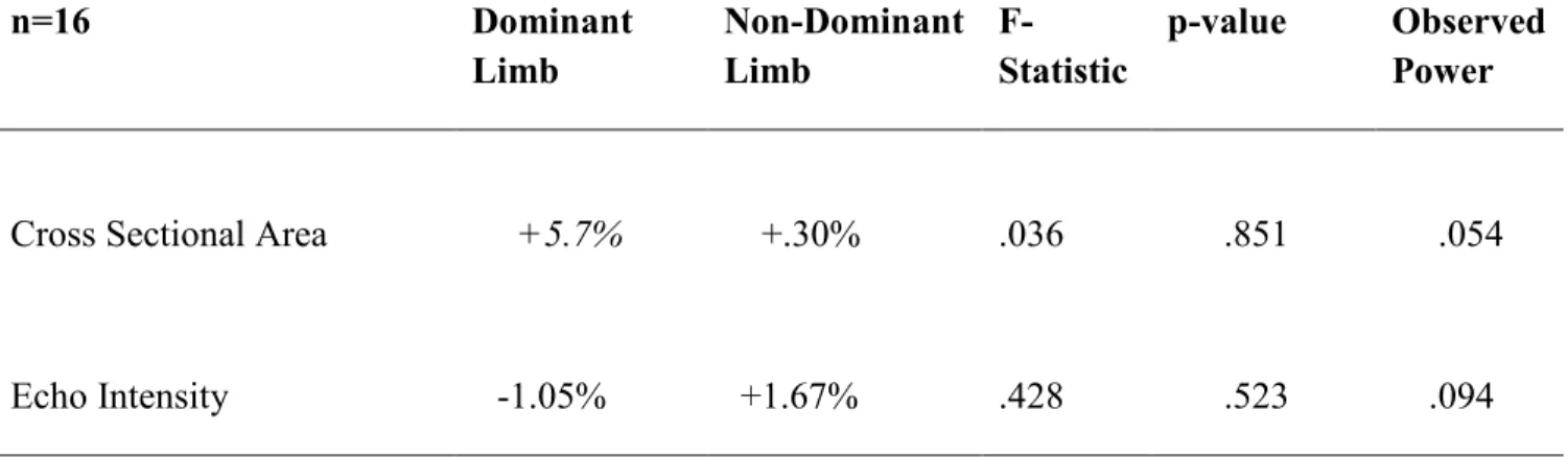
TABLE 8: Changes in CSA and EI during the pre-season………41
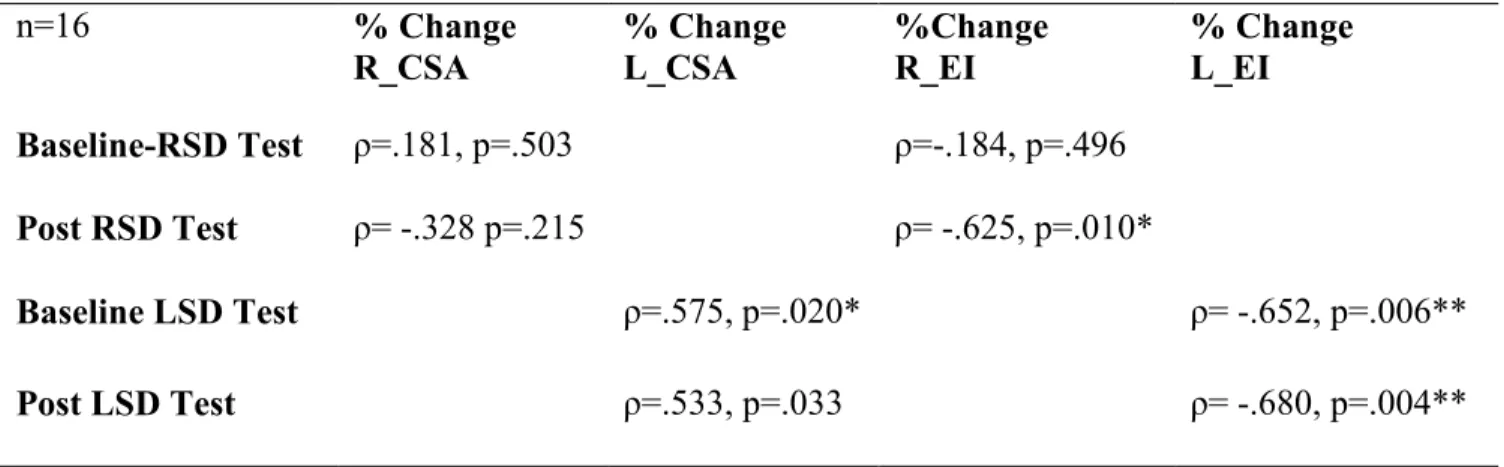
TABLE 9: Correlations between CSA, EI, and Scapular Dyskinesis Test Scores………43
TABLE 10: Overhead Volume by Position………...43
vi
LIST OF FIGURES
FIGURE 1: Scapular Dyskinesis Test positioning………23
FIGURE 2: Landmarks for identification of the infraspinatus………..26
FIGURE 3: US Probe Placement………...27
FIGURE 4: Ultrasound Imaging Analysis……….30
1
CHAPTER I: INTRODUCTION
The shoulder is the third mostly commonly injured area of the body, following only the
ankle and knee, in the sport of volleyball.1-5 Additionally, overuse injury at the shoulder occurs
most frequently among all body regions in the sport.1-5 The incidence of injury in volleyball
athletes at the collegiate level is highest during the preseason with overall injury rate of 6.5 per
1000 athlete exposures.4 The high incidence of injury during the preseason is likely due to the
high volume of training completed by the athletes during a relatively short period of time, as
teams often have multiple training sessions in a day during this phase of the season. The
pre-season for collegiate volleyball occurs in the late summer when athletes are returning to school
from home and conditioning levels are varied. The high volume of overhead training during the
pre-season places significant stress on the glenohumeral joint and surrounding musculature. The
repetitive overhead motions necessary to successfully compete in volleyball, can lead to tissue
damage, pain, and injury.6-8 The literature has shown that volleyball athletes typically experience
their first injury earlier in the season, compared to other sports where injury tends to occur
during the later portions of the season.9
Overuse is the most common mechanism of shoulder injury in volleyball as overhead
swings are made during both the attacking swing and the serve.1,3,5,6,10-14 The volume of swings is
often dependent on the athlete’s playing position as well as average playing time during practice
and competition.6 More than 40,000 swings by a single athlete has been observed during a single
volleyball season.11 Epidemiological reports show that outside hitters suffer the most injuries
2
%) and opposite/diagonal players (7.5%). These numbers correlate well with the amount of
overhead swings taken by players in these positions.1
Eccentric overload and repetitive stresses placed on the rotator cuff musculature and
posterior joint capsule during serving and spiking, are believed to be the main causes of overuse
injuries at the shoulder.1,3,6,10,11,15,16 During the overhead spiking motion, the infraspinatus and
teres minor muscles contract eccentrically to prevent anterior translation of the humerus during
the acceleration phase of the swing. 17As the volume of swings increases, these muscles fatigue
and may experience intra-muscular damage, including muscular edema, micro-tearing, and a
subsequent increase in cross-sectional area of the muscle.16,18-20 Without sufficient recovery time,
muscle damage can develop from overuse of the shoulder musculature. Damage to the
infraspinatus can be measured through diagnostic ultrasound, which is used to determine
cross-sectional area (CSA) and echo intensity (EI) to identify edema and muscle damage.18,20-22
Persistent increased CSA due to damage and inflammation has the potential to create
degeneration and failure of the tendon.20,21
The infraspinatus was chosen as a marker of muscle damage due to its critical role in
providing stabilization at the glenohumeral joint during the overhead swing motions used in
volleyball. It is often the site of muscle damage in athletes who compete in overhead athletic
activities and is easily accessible for evaluation using diagnostic ultrasound. Evidence of
infraspinatus damage has been observed in subjects after a single bout of eccentric external
rotation protocol using an isokinetic dynamometer in a study completed using healthy college
age students in 2011 by Oyama et al.18 With the intensity and volume of swings during
pre-season training, volleyball athletes likely experience similar levels of fatigue with overuse, and
3
The relationship between abnormal scapular mechanics and shoulder dysfunction in
athletes has been observed in a number of overhead sports with a greater incidence of shoulder
dysfunction found in athletes who have poor scapular mechanics and posture.6,9,23-26 With a high
prevalence of overuse injuries in volleyball the interaction of the scapular and rotator cuff
musculature becomes very important during dynamic motion. The musculature of the
glenohumeral joint serves to provide a stabilizing force to the humeral head within the glenoid
fossa. Several muscles act together to create force couples to stabilize the shoulder and prevent
instability. The synergistic motion of the infraspinatus and lower trapezius facilitates normal
motion of the scapula on the thorax during external rotation of the shoulder. This is an important
force couple as the rotator cuff tendon can become overloaded if the scapular stabilizing muscles
do not activate correctly to guide motion of the scapula and maintain the subacromial space.27,28
Changes in subacromial space can lead to tendinitis, impingement, and rotator cuff tendon strain
and rupture. The association between abnormal scapular positioning, glenohumeral motion, and
rotator cuff trauma in overhead athletics supports the need to create training programs which
train the athlete to improve movement efficiency of the upper extremity which may prevent
overuse injury at the shoulder. 15,24,29,30
Scapular mechanics play an important role in determining the efficiency of motion and
transfer of forces at the shoulder girdle during an overhead swing. When the arm is abducted
above 90°, the movement of the scapula along the thorax to bring the arm into an overhead
position is guided by several stabilizing muscles at the posterior shoulder.31 Alterations in
scapular mechanics is termed scapular dyskinesis, and has been implicated as a factor in shoulder
pain and disability in overhead athletics.29,32-34 The leading complaints of shoulder pain in
4
which have been linked to scapular dyskinesis and overuse of the shoulder by the
athlete.5,6,10,11,14,30
Scapular dyskinesis has been studied heavily in baseball, which has a similar overhead
motion in comparison with the volleyball swing with regards to high eccentric loading in
external rotation and quick forward acceleration of the arm. 13,18,27,28,35,36 However, there have
been few studies that examine scapular dyskinesis in volleyball players, and none that focus on
rotator cuff trauma and associated scapular dyskinesis. Therefore, the purpose of this study is to
identify changes in the infraspinatus CSA and EI following participation in pre-season volleyball
training as well as assess the effects of training on scapular mechanics and associated
development of trauma in the infraspinatus muscle.
Inefficient scapular motion influences the glenohumeral and scapulothoracic muscles
which work to bring the arm up overhead to perform the spiking and serving motions utilized in
volleyball. 24-26,30,32,35,37 All rotator cuff muscles have an attachment to the scapula, thus the
scapula must be able to elevate properly so that these muscles may act along the correct lines of
pull. 24 The coupling mechanics of the rotator cuff with movement of the scapula during the first
90 degrees of abduction is essential for efficient glenohumeral motion.15,38
For this reason we hypothesize that scapular dyskinesis in the volleyball athlete may
contribute to infraspinatus muscle trauma. Due to the high volume of overhead swings completed
during the pre-season, it is expected that infraspinatus CSA will increase due to overuse and
resultant muscle damage and edema. We hypothesize that there will be a relationship between
scapular dyskinesis, high training volume and muscular damage to the infraspinatus during the
5 Research Questions and Hypotheses:
1) Is there a significant change in CSA and EI of the infraspinatus following pre-season
volleyball training?
We hypothesize that the training volume of pre-season volleyball will lead to a significant
increase in CSA and EI of the infraspinatus.
2) Does the severity of scapular dyskinesis change following pre-season volleyball training?
We hypothesize that the severity of scapular dyskinesis will increase over the course of the
pre-season.
3) Is there a relationship between the presence of scapular dyskinesis, training load, and changes
in CSA and EI?
It is also believed that the presence of scapular dyskinesis will correlate with high
6
CHAPTER II: LITERATURE REVIEW Incidence of Shoulder Injury in Volleyball
In the sport of volleyball, athletes are subject to repeated loads from jumping and landing
as well as from overhead swings and making defensive plays. The variety of movements
required to be successful in the sport requires high levels of neuromuscular control, power, and
endurance. Injury is an inherent risk of all sports with volleyball ranking as the eighth highest in
rates of injury/exposures in youth sports.39 An athletic exposure is defined as one athlete
competing in a single game or practice session.1,9 Injury in sport is defined as an incident that
results in lost time participating in practice or competition by the athlete. Injuries can be
classified into two categories: acute and overuse. Acute injuries have a sudden onset with a
known cause, while overuse injuries develop insidiously over time or as a result of repeated
traumas. Overall, the shoulder is the third most frequently reported area of injury in volleyball,
following the ankle and the knee, respectively.1,3 However; the shoulder has the highest rate of
overuse injury reported. 1
Overuse conditions in volleyball are reported at 0.6 injuries for 1000 athletic exposures
with the shoulder being most frequently reported area of complaint.5 Along with being the most
frequently reported area for overuse injury it also accounts for the most time loss, reported at an
average of 6.2 weeks per injury.5 Training overload is cited as a leading cause of overuse injury,
and with the sudden increase in practice time during the pre-season, athletes often experience
7
The incidence of shoulder injury in volleyball athletes during the preseason has been
recorded at 6.5 injuries per 1000 athlete exposures as compared to 2.82 injuries per 1000 athletic
exposures during the regular season during a 16 year epidemiological study conducted from
1988-2004 by the NCAA.1 This study encompassed all divisions of the NCAA and surveyed a
large population within the volleyball community that is representative of the sport as a whole.
Several authors have reported overall injury rates during over one competitive season at similar
rates to that found by the NCAA. Bahr et al.2 found rates of 3 injuries per 1000 exposures in
beach volleyball. Verhagen et al.5 found rates of injury throughout the season of 2.6 injuries per
1000 exposures. From this data it is apparent that the pre-season is a period of increased risk for
injury, due to a sudden increase in activity and heavy training loads. It is estimated that all rates
reported do not fully represent injuries sustained as many athletes have injuries that are not yet
symptomatic or are not severe enough for the athlete to seek medical attention.1,2,6,10,11
Overuse Injuries at the Shoulder
In volleyball, overuse mechanisms rather than acute incidents account for approximately
20-32% of all shoulder injuries sustained in practices and games.1,5 Overuse injuries lead to
chronic pain and inability to perform at the desired level of participation throughout the entirety
of a season.1,2 Early intervention with training programs and awareness of risk factors for injury
may help to prevent overuse injuries and increase the longevity of the athlete throughout the
season.
Overuse conditions are defined as an inability to tolerate repeated stresses over a period of time,
resulting in symptomatic overuse injury.7,8,40 In sports medicine, overuse injury is categorized
into four types; (1) pain in the affected area after physical activity; (2) pain during the activity,
8
chronic, unremitting pain even at rest.7 Often an athlete will not complain of their pain until it
inhibits performance in their chosen activity or affects activities of daily living.1,6,41
Injury generally occurs as a result of multiple risk factors that are both intrinsic and
extrinsic. Intrinsic factors are non-modifiable and include anatomy, age, sex, and previous injury.
Extrinsic factors are modifiable and include training type, volume of training, technique,
equipment, and environment.8 Strains of the shoulder musculature are one of the most commonly
seen injury in overhead sports and accounted for 5.2%-10% of volleyball injuries that received
medical attention during two prospective epidemiological studies of elite level volleyball
athletes.1,2 Other studies have reported rates of shoulder overuse injury as high as 30% during a
season.10 This high value of injury rate may actually be more realistic as overuse injuries often
come on insidiously and do not result in lost time from competition until the problem has
become chronic and is causing disability.13
The literature cites several possible factors which contribute to overuse injury at the
shoulder. The first risk factor is the spiking and serving method used by the athletes.2,3,12,13 The
overhead motion of spiking and serving has several key components that include the arm
cocking, arm acceleration, and follow through phases. Between the cocking and acceleration
phases there is a critical instance of maximum external rotation of the glenohumeral joint before
the arm is brought forward. To attain this position, the infraspinatus is the primary agonist
muscle and the teres minor acts as a secondary mover. As the athlete accelerates the arm forward
to strike the ball the glenohumeral joint must internally rotate and flex while the elbow extends.13
The posterior shoulder musculature must then act to prevent anterior displacement of the
humeral head by contracting eccentrically to control the forward motion of the arm.37 This
9
muscle damage. During the cocking phase of the spike and serve, the athlete experiences 54-71%
of maximum voluntary contraction at the rotator cuff musculature which serves to counteract the
distractive forces on the humerus, that have been reported as upwards of 80-120% of the
athlete’s body weight.17,37 The stresses imposed through repetitive overhead swings can place
significant strain on the musculature, which can lead to tissue damage which displays through
edema, muscle soreness, and increased CSA of the muscle.
Risk Factors for Shoulder Injury
In the sport of volleyball, there are several types of overhead swings that attackers use to
spike the ball: straight ahead, cross court, and the roll shot, which is an off-speed ball hit over the
net. Each of these swings requires a significant force to be generated through the trunk to the upper
extremity to contact the ball with a high velocity. Of these methods the roll shot requires the least
amount of force.13 Reeser et al.13 did not find a statistical difference between the straight ahead
and cross court spikes with respect to joint position during kinematic analysis throughout the
swing. However; a study by Mitchinson et al.12 found that the trunk and upper arm positions did
have an altered position at impact, with the arm forward of the trunk due to rotation of the trunk
during the swing. The authors believe that increased axial rotation of the trunk during a cross court
shot places increased stress on the shoulder during follow through which increases risk of injury
compared to a straight ahead or roll shot spike. The forward position of the shoulder increases the
eccentric load on the posterior shoulder muscles. The positions which hit more cross court shots
are the outside and right side attackers. Outside attackers have the highest rates of injury (37%) of
all positions.4
Serving style has also been attributed to a difference in shoulder injury prevalence. Athletes
10
performs a standing serve.6 This difference is likely due to the increased force generated by the
trunk and legs during the jumping approach as compared to a stationary service method.
Mechanically the jump serve is similar in motion to an overhead spiking motion, while the standing
serve generally requires less external rotation of the shoulder when the arm is brought overhead.
The forces are transferred along the kinetic chain to the glenohumeral joint. The forces cause a
distractive mechanism which must be resisted by contractions of the rotator cuff muscles.37
Muscle Recruitment Patterns During Overhead Motion
Balance between the internal rotator muscles which work to accelerate the arm forward as
the athlete goes to strike the ball and the external rotators which serve to decelerate the arm during
follow through is important to dynamic stabilization of the shoulder.42 The highest peak muscle
activity for the internal rotator muscles (subscapularis, teres major, pectoralis major, latissimus
dorsi) is seen during the acceleration phase.37 The infraspinatus and supraspinatus muscles are
most active during the deceleration phase of the spiking and serving motion after the athlete has
made contact with the ball and is controlling the forward momentum of the arm (37% and 45%
MVIC respectively)17 as well as during the critical instant of external rotation. With the extreme
ranges of external rotation available to volleyball attackers must have strong posterior shoulder
musculature with sufficient endurance to avoid injury associated with repeated overhead activity
During EMG analysis the serve generated the highest peak EMG readings for the infraspinatus
and supraspinatus during the deceleration phase.37 The authors noted a strong similarity between
the forces applied to the shoulder in volleyball and that seen in baseball pitching which
demonstrates a high prevalence of shoulder overuse injuries as well.37
The rate of shoulder pain in attacking positions (middle blockers, outside hitters, and
11
difference is expected as the attacking positions perform a much higher number of overhead
swings compared to defensive specialists.6 Practicing 16-20 hours a week can result in
approximately 40,000 swings during a season for an attacker.11 When paired with dysfunctional
mechanics, swing volume may contribute to the development of an overuse injury.14
Scapular Dyskinesis
A second factor that contributes to shoulder pain in overhead athletes is scapular
dyskinesis. Scapular dyskinesis is defined as an alteration in scapular movement patterns during
arm motion. It has been shown that the presence of scapular dyskinesis can lead to shoulder
problems including sub-acromial impingement, instability, and damage to the rotator cuff
tendons.6,14,15,24-26,30,32,35,41,43,44 It can be assessed through visual observation of the scapula during
upper extremity motion either digitally or by a trained clinician. Both methods have been
validated and are comparable in identification of dyskinesis in the overhead athlete.1,33,34,45,46 In
the sport of volleyball, scapular dyskinesis has been identified as risk factor for developing
shoulder pain and disability in performing overhead motion in the sport.
Scapular dyskinesis contributes to poor kinematics at the glenohumeral and scapulothoracic
joints, leading to pain and disability during the overhead athletic movements.6,14,25,29,32,34,37,38
When the arm is abducted beyond 90 degrees the scapular stabilizing muscles must coordinate
the gliding motion of the scapula on the thoracic wall to bring the arm overhead in a smooth arc
pattern.25,26,31,32 This pattern allows elevation of the acromion, and prevents impingement of the
supraspinatus tendon and long head of the biceps tendon which run in the subacromial space.
Normal scapulohumeral rhythm is defined as having the coordination and timing of these two
12
abduction and flexion, and finally, downward rotation during humeral adduction and extension.
No evidence of winging is present.33
Scapular dyskinesis has been described to include scapular winging, side to side asymmetry,
and prominence of the medial scapula border.33,34 Several types of grading systems have been
developed to describe dyskinesis based on bony landmark prominences or movement plane
asymmetry.25,29 The current study will utilize the methods employed by McClure and Tate33,34
which has been validated to identify scapular dyskinesis.
Scapular Dyskinesis in the Overhead Athlete
Overhead athletes show increased presence of scapular dyskinesis in comparison to
non-overhead athletes.35 The literature has also shown that this change in scapular kinematics tends to
occur over time with repeated overhead activity.47 It is unclear whether or not the dyskinesis
leads to shoulder pain or if the shoulder pain causes an adaptation in muscle activation patterns
which predisposes overhead athletes to developing dyskinesis. These athletes may or may not
present with symptomatic shoulder pain during initial evaluation; however, an increase in
prevalence of shoulder pain is correlated with patients with scapular dyskinesis. 6,10,14,24-26,30,32,33,35,43,44,48,49
There are several possible factors which lead to scapular dyskinesis, including changes in the
activation of the scapular stabilizing muscles, damage to the neurological structures which
innervate these muscles, and reduced length of the pectoralis minor muscle.33 Innervation for the
scapulothoracic muscles comes from the long thoracic, dorsal scapular, and spinal accessory
nerves.33 Proper neuromuscular function of the scapula assists in maintaining muscle length
tension relationships between the scapulothoracic and glenohumeral musculature that create
13
motion between these joints assists in maintaining the glenohumeral and subacromial joint
spaces during overhead activity.46 When this space is decreased due to swelling or mechanical
obstruction the athlete often experiences subacromial impingement syndrome that can lead to
muscular damage to the rotator cuff and degeneration of the tissue over time.25,26,45 Other
complications from scapular dyskinesis include muscle imbalance, single sided dominance, and
body alignment problems, which can in turn make the athlete more prone to injury or worsening
of the condition.29,32,33
Despite many of injuries associated with scapular dyskinesis, athletes who present with
scapular dyskinesis are not always symptomatic; many individuals maintain the ability to
function normally in their daily lives and in athletic competition. When completing an evaluation
for pain in the upper extremity the clinician should observe for dyskinesis and determine if it
may be a factor in the injury or condition assess scapular movement patterns and determine if a
dyskinetic pattern is present.29,32 In the sport of volleyball the presence of dyskinesis should be
taken seriously, as the athlete may develop symptoms as the volume of training increases from
what the athlete has experienced previously in his or her career. Changes in the athlete’s playing
position may also effect the volume of overhead swings taken by the athlete during a season.
Measuring Scapular Dyskinesis
Measuring scapular motion can be completed through video analysis, 3-D motion
assessment or clinical real-time observation. 3-D motion assessment is completed by attaching
reflective markers directly on the skin above the bony landmarks of the scapula and analyzing
the motion through electromagnetic imaging. This method is time consuming, and would not be
indicated for the population being studied in this project, or for clinical practice.46 For this reason
14
identifying scapular dyskinesis. Clinical observation of scapular motion has been shown to be
valid and reliable through comparison to 3-D motion assessment. A moderate inter-rater
reliability was found when trained clinicians viewed video of an athlete performing the test
motions, Κw=.57.33 This method of assessment is a tool that can be used in sports medicine
settings to evaluate altered scapular movement patterns without the time consuming and
expensive method of 3-D motion analysis.29,33,34
Visual observation of the shoulder/scapula by a trained clinician has been validated as an
appropriate method for determining the extent of dyskinesis, and can be used to determine the
most appropriate intervention to correct dyskinetic motion patterns.33,46 In the validation study,
motion was assessed through bilateral active shoulder flexion and abduction in a weighted
position while the patient is viewed from a posterior angle.33 The participants were then assessed
through a 3-D motion analysis which reflected the findings of the clinicians; those participants
with observed dyskinesis possess different kinematics which include scapular upward rotation,
posterior tilting and external rotation, and clavicular retraction and elevation when the arm is
raised overhead.34 The rating system for video observation describes scapular motion patterns as
normal, subtle dyskinesis, and obvious dyskinesis.33
Infraspinatus activation and scapular motion patterns
The infraspinatus and teres minor are highly active during external rotation, which occurs
as part of the arm cocking phase during the spike and serve.37 As the athlete fatigues, these muscles
lose the ability to externally rotate the shoulder at the same magnitude to maintain the force that
can be applied to the ball when spiking or serving.36 Poor neuromuscular control, as a result of
fatigue at the shoulder girdle may lead to or perpetuate many common shoulder overuse injuries
15
activity in the infraspinatus increases the scapular stabilizing muscles (lower trapezius, serratus
anterior) fatigue which leads to an upward rotation of the scapula and altered shoulder
kinematics.27,28 An important force couple relationship exists between the lower trapezius and
infraspinatus; in this interaction the lower trapezius functions to maintain scapular contact with the
thorax while the infraspinatus performs external rotation of the shoulder.27,28 Maintaining muscular
balance between the scapulothoracic and glenohumeral muscles is important in prevention of poor
mechanics which may lead to overuse injury pathologies.35,41
Overuse pathologies
During the volleyball pre-season the athletes often participate in two practices a day,
along with strength and conditioning workouts. During this time the athlete is subject to much
higher intensity loads than their body is used to experiencing. The result of this increased
physical activity is muscle soreness and fatigue. Muscle tissue has the ability to adapt to sudden
changes in activity levels by breaking down and rebuilding over a relatively short period of time,
allowing strength gains. The literature has shown that a single bout of high intensity eccentric
exercise which initiates delayed onset muscle soreness (DOMS) symptoms provides a protective
effect against future high intensity exercise bouts.16,19,51-53 The protective effect has been thought
to be due to a repair of micro-tearing of the weakest muscle tissues which occurs as a result of
eccentric exercise. The muscle is then re-built stronger so that it may tolerate a similar intensity
of exercise in subsequent repetitions of the same motion.16 During the pre-season, the athlete
experiences these effects during the first practices; however, without sufficient time for recovery
and synthesis of new muscle fibers the athlete can experience muscle soreness and fatigue for the
duration of pre-season training without the protective benefits and building of new muscle fibers.
Oyama et al.18 demonstrated that the adaptive changes in the CSA of the infraspinatus
16
significant increase in infraspinatus CSA, following eccentric external rotation protocols has
been observed in work with healthy college students.18 Due to similar overhead motion required
for a volleyball spike we hypothesize that a comparable increase in CSA would be found in
volleyball players. In volleyball, there is no limit on the number of swings an athlete may
perform in any given practice or competition. Unlike baseball pitchers, volleyball players do not
have a designated time for recovery between heavy bouts of overhead swings. Therefore, we
could expect that over the course of a pre-season training camp, volleyball players would sustain
trauma to the infraspinatus similar to that seen in other overhead sports.
Effects of Fatigue on Muscle Tissue
Through panoramic ultrasound measurement of the shoulder the quality of the muscle
tissue can be assessed through echo-intensity (EI).54-58 EI is measured by assessing the grey scale
quality of each pixel within a specified area of the ultrasound image. Decreased values on the
grey scale indicate improved quality of the muscle tissue.54 The scale depicts a number between
0 and 255.This method of determining muscle quality has been validated through comparison of
panoramic ultrasound to single transverse measurements.55 It has been demonstrated in previous
works that presence of intramuscular fat content and fibrous tissue decreases the muscle quality
and negatively influences the power generation of the muscle during activity.59 In the short term,
measurement of EI would not show an increase in fatty infiltration, but rather would show
changes in echo intensity due to infiltration of swelling and metabolites in the muscle.Young
individuals have a significantly lower EI than elderly counterparts due to muscle loss and
increased adipose tissue gain which occurs with aging.60,61 Thus for comparison of data between
individuals in the current study it is important that subjects are within a similar age range.
17
from micro-trauma and overuse. During a pre-season training period, the effects of delayed onset
muscle soreness (DOMS) and repetitive motions causing fatigue are likely to elicit these changes
and influence the EI observed in diagnostic ultrasound.16,51-53
The speed of muscular contraction required for volleyball is quick as the athlete must be
able to perform explosive motions (jumping, spiking, serving etc.) to be successful on the court.
A linear relationship exists between speed of contraction and muscle damage with repetitive
contractions at a high velocity. Chapman et al.19 found that there was a 450% increase in peak
creatine kinase (CK) levels following a bout of fast velocity contractions as compared to slow
velocity contractions. CK levels indicate damage to the muscle tissue as it is released when
muscle tissue breaks down. Subjectively, the participants experienced an increase in DOMS
symptoms after the fast velocity exercise bout compared to slow velocity training.19 This data is
supported by Shepstone et al.62 who found that repeated bouts of high velocity isokinetic
exercise lead to an increase in muscular CSA and increased muscle protein remodeling due to
micro-trauma sustained in the tested muscle group. Over the course of the pre-season levels of
CK are likely to increase, as muscle does not have sufficient time to recover from the repetitive
loading that occurs during training. These factors and infiltration of muscle proteins due to
damage can be observed through changes in the EI of the muscle. In addition, CSA of the muscle
assists in assessment of muscle condition by tracking changes of muscle volume due to atrophy,
hypertrophy, disease, and injury.63-66
Ultrasound measurement of Cross-sectional Area
The use of ultrasound technique to identify abnormalities in the musculature of the
shoulder has been validated and is a useful tool in diagnosis of musculoskeletal muscle damage
18
been validated against MRI arthrogram, which is considered the gold standard in
musculoskeletal diagnoses.64,65,67,69,70 The reliability and validity of panoramic ultrasound gives
clinicians another tool to use which may assist in diagnosis of injury and identification of
physiologic changes in the muscle tissue due to overuse training, or injury.63-65 Benefits of
ultrasound measurements over MRI include the ease of use in clinic, low cost, no exposure to
radiation, and availability. Ultrasound is a relatively inexpensive and efficient method of
determining changes to the muscle and has been verified with 92.3% sensitivity and 94.4%
specificity for full-thickness and 66.7% sensitivity and 93.5% specificity for partial-thickness
tears.20-22
The infraspinatus is one of the four rotator cuff muscles which functions to stabilize and
control motion at the glenohumeral joint. The infraspinatus has a larger CSA than the
supraspinatus or teres minor which perform similar function as the infraspinatus.18 The average
width of the infraspinatus tendon is about 22mm, compared to 20mm for the supraspinatus, and
inserts into the entire middle facet of the greater tuberosity.21 In previous studies the
infraspinatus has been used as a marker of overall damage to the shoulder due its location and
ease of measurement via diagnostic ultrasound. Prior studies have shown that US measurement is
a reliable method of determining infraspinatus CSA and EI, CSA reliability in a previously
conducted study showed US can be used to measure muscle area with good reliability, (ICC2, 1
=.984).18 Primarily US imaging to evaluate EI is completed to measure fatty infiltration
indicating a decrease in muscle quality however in the case of short term muscle damage it can
be used to assess the infiltration of swelling, metabolites, and other biologic material within the
19
The infraspinatus has been recorded to have a mean thickness of 4.4mm in healthy
female subjects through diagnostic ultrasound imaging. This measurement is similar to the
thickness of the other rotator cuff muscles, supraspinatus (4.9mm), and subscapularis (3.8mm).71
These researchers did not find any significant difference between the muscular width and
thickness of the infraspinatus of the dominant and non-dominant arm in their subjects.71 For the
current study the measurements of infraspinatus CSA and EI were taken bilaterally prior to the
20
CHAPTER III: METHODS
Participants
Seventeen NCAA division I varsity female volleyball players participated in the current study which took place during the pre-season training period for the 2015 NCAA Division I
volleyball season. All participants were between the ages of 18-21 years old and were registered
as student-athletes at the University of North Carolina at Chapel Hill for the 2015 fall semester.
This group consisted of two setters, three defensive specialists, and twelve athletes who played
attacking positions. There were three right side players, four middle hitters, five outside hitters.
Complete participant demographics appear in Table 1. Inclusion criteria for the current study
included membership on the varsity volleyball team at the University of North Carolina at
Chapel Hill for the 2015 season, and ability to complete all testing sessions. Exclusion criteria
for the current study included inability to participate in pre-season training, or testing sessions
due to current injury or illness. All participants completed a health history questionnaire,
underwent pre-participation screening for the upcoming season, and signed an informed consent
document approved by the University of North Carolina at Chapel Hill Institutional Review
21
Height (cm) Mass (kg) Age (years) Limb Dominance
Position Volleyball Experience
(Years)
170.14±9.87 77.09±8.68 19.71± 1.16 n=17 Right n=0 Left
2 setters 3- DS/Libero 12-Hitter/Blocker
7.82± 2.24
Table 1. Participant Demographics
Instrumentation and Set Up
A single video camera (Casio Computer Co., Ltd., Tokyo, Japan) was used to record the
scapular dyskinesis test, and play back the recorded motions. Video was analyzed by the
principal investigator (K.C) to observe for dyskinetic patterns of motion bilaterally. Each
shoulder was rated separately and given a score of 0, 1 or 2 to quantify the amount of dyskinesis
present. Reliability for the principal investigator was determined for both intra-rater and
inter-rater reliability with observation of video of 17 participants. The videos were watched two weeks
apart and were graded without viewing previously determined scores during that time period.
Time between video grading sessions was determined based on the length of the average
pre-season. For the right limb, intra-rater reliability was assessed through ICC. Excellent intra-rater
reliability was determined. The principal investigator also was assessed for inter-rater reliability
against a trained grader. Kappa coefficients were determined to be substantial with Kavg =.853
22 Pre Right Scapular Dyskinesis Tests Pre Left Scapular Dyskinesis Test Post Right Scapular Dyskinesis Test Post Left Scapular Dyskinesis Test Inter-rater reliability (K)
.809 .815 .884 .906
Intra-rater reliability (ICC)
.822 .969 .895 .969
Table 2. Scapular Dyskinesis Test Statistics: Inter-rater and Intra-rater reliability
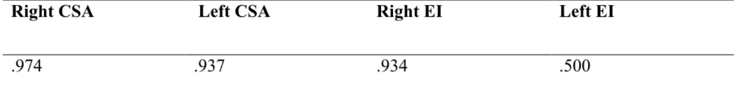
Similar to scapular dyskinesis, three images of the infraspinatus were collected bilaterally
and saved for analysis of CSA and EI after all testing was completed. A pilot testing session with
the principal investigator (K.C.) was completed with 11 participants to establish repeatability and
precision of the investigator of measuring the infraspinatus. Excellent intersession reliability and
precision was established for measurement of the CSA for the right (ICC=.974) and left
(ICC=.937) infraspinatus respectively. Excellent intersession reliability and precision was also
established for EI on the right shoulder (ICC= .934), while a poor reliability and precision was
established for the left shoulder (ICC=.500). The principal investigator collected all images
during data collection to ensure reliability and precision across all measurements and all subjects.
Right CSA Left CSA Right EI Left EI
.974 .937 .934 .500
23 Procedures
Prior to data collection all participants signed an informed consent approved by the IRB
at the institution and were educated on the objectives of the project. Data collection sessions for
assessment of scapular kinematics as well as panoramic ultrasound measurements occurred prior
to the start of pre-season practice as well as after the conclusion of the pre-season. Measurements
were taken with the participant at a resting state 24 hours prior to the start of pre-season training,
and 24 hours after the conclusion of pre-season training. During testing, each participant
performed the scapular dyskinesis test and was recorded using a portable video camera mounted
on a stationary tripod. Participants were asked to stand facing a blank wall away from the
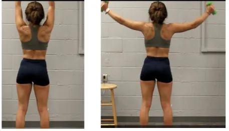
camera, and were videotaped from a posterior view (Figure 1). Adjustments were made to the
camera lens between participants to ensure that the view captured the participant from the head
to the waist throughout the entire motion and the camera was level at the time of filming.
Instructions for the motions performed are described in the section below entitled Scapular
Dyskinesis Test.
*forward flexion * abduction
24
Panoramic ultrasound measurements were taken by a trained investigator (K.C) for all
participants during both measurement sessions to ensure reliability and accuracy of the image
obtained. Panoramic ultrasound of the infraspinatus of both upper limbs were taken 24 hours
prior to the start of the first pre-season training session and 24 hours after the conclusion of the
last pre-season training session. Ultrasound measurements were completed prior to testing of
scapular dyskinesis to ensure a resting state of the muscle. Description of testing position is
described below in section below entitled Ultrasound Measurements.
Volume of swings was assessed daily during the pre-season training period. Each practice
session was filmed and viewed by trained investigators at a later date to assess overhead activity
volume experienced by the participants. There are four types of overhead motions which occur
frequently in volleyball. The first of these motions is blocking, which is defined as two arms up
overhead when the participant is attempting to stop the ball from crossing the net after an
opponent contact. A second overhead motion which occurs as part of the offensive strategy is
tipping which is defined by the participant contacting the ball with one hand and flicking the
wrist to change the trajectory of the ball upon contact. A third overhead motion is setting in
which the participant contacts the ball with both hands equally overhead. The final and most
frequently occurring motion is the overhead swing, which consists of spiking and serving. These
motions are classified as a single handed contact with the ball in which the shoulder comes into a
position of full external rotation before accelerating forward to make contact with the ball. A
strong relationship exists between overhead swings and shoulder pain in volleyball athletes, due
to the extreme forces applied to the shoulder joint during this motion.2,3,6,10 All overhead contacts
were counted for the study however, only overhead swings were statistically assessed in this
25
investigators collected swing volume data using the definitions described above. Swing volume
was used as a co-variate to changes in muscle CSA and EI during statistical analysis.
Position Swing Volume
N=17
Attack 844.33±97.015 12
Defensive Specialist 580.67±60.962 3
Setter 536.50±47.376 2
Table 4. Overhead Volume by Position Group
Scapular Dyskinesis Test
Participants were instructed to wear a sports bra for screenings to allow observation of
the posterior thorax. Screenings were performed using the procedures described by McClure and
Tate et al.33,34 Each participant was videotaped with a single camera from the posterior view and
performed five repetitions of both weighted bilateral active shoulder flexion and shoulder
abduction (ten total repetitions). Prior to performing the test, participants were instructed on the
movement by the examiner and were allowed to practice each movement once if they were
unsure of how to complete it. Participants were instructed to keep the arms at the side of the
body to start the test, then elevate both simultaneously into flexion completely overhead in the
neutral rotation position (thumbs up) over a three second count, then lower back to the starting
position over a three second count. Adduction was completed next with the arms resting at the
side of the body, the both arms raised with the thumbs pointed up in a neutral rotation into
complete abduction overhead. The motion occurred over a three second count up, and a three
26
body weight: participants < 150 lbs. (68.18kg) used 3lb dumbbells, while those weighing > 150
lbs. (68.18 kg) used 5lb dumbbells.34
Ultrasound Measurements: CSA & EI
Participants were asked to wear sports bra which allows for access to the posterior
shoulder that allows the ultrasound probe head to directly contact the skin overlying the
infraspinatus muscle. Participants were asked to lie prone on the examination table with the arms
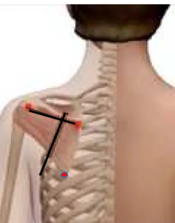
positioned to the participant’s sides with the palms of the hands facing up. The borders of the
infraspinatus were marked with a permanent pen after palpation of landmarks. The landmarks
included the acromial angle, trigonum spinae, and the inferior angle of the scapula. A straight
line was marked from the acromial angle to the trigonum spinae using a straight edge ruler. The
second line was drawn perpendicularly, using a standard goniometer, and was marked to the
length of the inferior angle of the scapula. This line was drawn at 1/3 the distance between the
acromial angle and the trigonum spinae on the medial side (Figure 2).
27
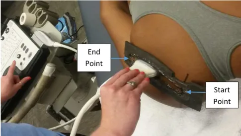
A custom template was created to fit the ultrasound (US) head and ensure a straight line
was followed along the muscle (Figure 3). This template was secured to the patient along the
second line drawn using clear tape. The inside of the template was filled with US gel to ensure
adequate conduction of the US head. The ultrasound device was consistently set with gain of 60,
depth of 5cm, and frequency 12 MHz. Depth of 5 cm was determined based upon pilot testing
which determined this to be the depth which would capture the entire muscle in most
participants. Three serial images of the infraspinatus were taken bilaterally for each participant
during each testing session, by moving the US probe head from a superior to inferior position
along the perpendicular line within the template. Images were analyzed using Image J software
(National Institutes of Health, Bethesda, MD, USA). The trial means for CSA and EI were
calculated and used for analysis of muscle trauma markers including edema, and decreased
muscle quality.
Figure 3. US probe placement End Point
28 Data Reduction and Analysis
Visual rating of the scapular dyskinesis test was determined at a later viewing time and
was rated by the principal investigator on a scale of 0-2. The investigator is a certified athletic
trainer who rated the videos for normal motion, subtle dyskinesis, and obvious dyskinesis. Each
shoulder was rated separately and given an independent score. Criterion for each category
(normal motion, subtle dyskinesis, obvious dyskinesis) are listed in Table 4 and has been shown
to be reliable for determining the extent of dyskinesis.33,34 Each shoulder was rated individually
in both flexion and abduction, then the participant was given an overall motion score of normal,
subtle or obvious dyskinesis for each shoulder. Operational definitions for normal, subtle
dyskinesis, and obvious dyskinesis are outlined in Table 5. For the overall scoring participants
was assessed for dysrhythmia and winging in both flexion and abduction separately however the
scoring was a combination of these evaluations. A rating of normal on both flexion and
abduction gave a normal score. A rating of normal for one motion and subtle for another gave a
normal score. Rating of subtle dyskinesis for both motions gave a subtle score, and if either score
29
Normal Motion Subtle Dyskinesis Obvious Dyskinesis
Operational Definition
Both test motions are rated as
normal or one of the motions is
rated as normal and the other as
having subtle abnormality.
Both flexion and abduction are
rated as having subtle
abnormalities which are not
consistently present.
Either flexion or
abduction has striking,
clearly apparent
abnormality, evident on at
least 3/5 trials
(dysrhythmias or winging
of 1 in [2.54 cm] or
greater displacement of
scapula from thorax)
Table 5. Operational Definitions for Scapular Motion33
Analysis of the ultrasound images was completed using Image J software (National
Institutes of Health, Bethesda, MD, USA) by tracing the inside border of the epimysium of the
infraspinatus using Image J polygon function to assess the CSA. EI measurements were
calculated by Image J based upon the color quality (black to white) 0-255, of the pixels within
the traced area. One participant was excluded from the data set for this analysis due to severe
infraspinatus atrophy on her dominant shoulder which made the image unable to be analyzed.
The procedure was repeated for all three images taken for each participant (n=16). Means of the
CSA and EI were calculated in Excel to represent the value of CSA and EI for each participant
30 Figure 4: Ultrasound Imaging Analysis
Statistical Analysis
The CSA and EI were assessed using a two-way within-subjects ANCOVA analyses with
the swing volume as the covariate variable. Means were compared between the dominant and
non-dominant arms and within each limb prior to the pre-season and following the conclusion of
the pre-season training period for both CSA and EI of the infraspinatus.
A Wilcoxon Signed Ranks Test was completed to assess changes in scapular dyskinesis
severity from baseline testing prior to the start of pre-season, to the end of the pre-season training
period.
Spearman’s correlation statistic were utilized to examine the relationships between
scapular dyskinesis and muscular damage (% change in CSA and EI). Another correlation
between the presence of dyskinesis and swing count was run to determine if swing volume may
affect the development of dyskinesis over the course of the pre-season. Dyskinesis was set as the
categorical variable, while the change in CSA, EI, and swing count were set as the continuous
31
CHAPTER IV: MANUSCRIPT
Background: During the pre-season training period, volleyball athletes experience a high training volume and have little time for recovery. It has been shown that athletes are more likely
to develop overuse injuries during the pre-season training period. Forces placed on the shoulder
through repetitive overhead activity in volleyball is a risk factor for injury due to the
development of muscle damage and changes in scapular kinematics which have been shown to
occur with repetitive overhead training.
Hypothesis: We hypothesized that the training volume of pre-season volleyball would lead to a significant increase in CSA and EI of the infraspinatus. We also hypothesized that the
severity of scapular dyskinesis would increase over the course of the pre-season training period.
Thirdly we hypothesized that the severity of scapular dyskinesis would be associated with high
training load, determined via swing count, as well as greater changes in CSA and EI.
Study Design: A descriptive observational study
Methods: 17 female Division One collegiate female volleyball athletes between the ages of 18 and 21 years old (age: 19.71 ±1.16 years old, height 170.14: ± 9.87 cm, mass 77.09
±8.68kg), who participated in the 2015 volleyball season were recruited for this study. All
participants were right hand dominant with 12 athletes playing attacking positions, three
32
Results: No significant findings were noted for changes in infraspinatus cross sectional area (F=.036, p=.851) or echo intensity in the dominant limb (F=.428 p=.523) across the
pre-season period. There were also no significant changes in the severity of scapular dyskinesis of
the dominant limb across the pre-season period (z= -.289 p=.779). No significant correlations
(ρ> .05) between ultrasound measurements (CSA or EI), scapular dyskinesis score, and swing
count were found in the dominant limb. Significance was set a priori at (p=.05).
Conclusions: Pre-season volleyball training volume did not have a significant effect on the development of muscle damage to the infraspinatus muscle. Additionally, there were no
significant changes in scapular dyskinesis over the course of this training period. Finally, there
was no significant relationship between swing count, indicators of muscular damage and the
severity of scapular dyskinesis.
Clinical Relevance: Information from this study provides athletes, coaches, strength and conditioning staff, and athletic trainers with potential risk factors, such as training load and
scapular kinematics, for the development of shoulder injury during the pre-season. While no
significant findings were made in regards to changes in muscle size and quality, or in the severity
of scapular dyskinesis, it is concerning to see such a high prevalence of scapular dyskinesis in
this sample of athletes. The prevalence in this sample may be representative of the overall
population and would indicates the need for preventative programming to prevent the
33 INTRODUCTION
Shoulder injury in volleyball is the most common overuse injury in the sport and third
most frequently injured area in the whole body.1-6,10,14,40,42,44 The pre-season training period
places significant stress on the shoulder due to high training load and extensive overhead volume
experienced by the athletes. During a single volleyball season, greater than 40,000 swings may
be taken by a single player.41 During each swing, the shoulder joint experiences high levels of
eccentric demand on the external rotator muscle group as the arm swings forward to strike the
ball.13,16,19,37,72,73 These forces place significant strain at the musculature of the posterior shoulder
due to the eccentric forces required to keep the shoulder from distracting during the deceleration
phase of the swing. These muscles must also contract to create the necessary external rotation to
provide velocity to the ball during the loading phase. In the current study the infraspinatus
muscle was selected for study due to the eccentric stress it faces as well as its use as an external
rotator. It has been used successfully in prior research also evaluating the effects of repeated
loading on muscle damage.18 Over time, chronic muscle pain and acute muscle damage,
including muscle strains, can occur if volume remains high and recovery time is not permitted.
The scapula plays a critical role in coordinated motion of the glenohumeral joint. The
rotator cuff musculature of the shoulder functions to provide dynamic support to the relatively
unstable glenohumeral joint. Many of the dynamic glenohumeral stabilizers also influence the
motion of the scapula due to force couple relationships that facilitate efficient motion at the
scapulothoracic and glenohumeral articulations.25-28,32,37,43,48 A greater incidence of shoulder
injury has been found in athletes who have scapular dyskinesis and poor resting posture. The
purpose of a scapular dyskinesis assessment is to identify motion patterns which may lead to
34
scapular dyskinesis has been quantified through kinematic analysis as well as visual observation,
which is used frequently in clinical practice and has been shown to be equally
reliable.27,29,33,34,46,74
The combination of scapular dyskinesis and high training load has been suggested as a
risk factor for the development of injury to the shoulder joint. Evidence of muscle damage can be
identified through ultrasound imaging, specifically by measuring muscular cross sectional area
(CSA) and muscle quality through echo intensity (EI). These markers of muscle damage have
been shown by Oyama et al (2011) to be reliable outcome measures that indicate damage to the
infraspinatus muscle18. This ultrasound imagining technique is a cost-effective and time efficient
screening solution for the trained clinician to use when monitoring muscle damage in the
overhead athlete.22,57,63-65,67,70,75
The purpose of our study was to identify changes in the infraspinatus CSA and EI
following participation in season volleyball training, as well as to assess the effects of
pre-season training on scapular mechanics. We also sought to determine if there was a relationship
between, swing count, damage in the infraspinatus muscle, and when scapular dyskinesis.
Identification of factors such as training load and scapular dyskinesis is important for the
clinician, coach, and athlete to prevent the development of overuse injury during the pre-season
which may last into the season and have a negative effect on the performance of the athlete.
METHODS
Participants
Seventeen collegiate female volleyball players (age: 19.71 ±1.16 years old, height
35
study. Dominant limb was defined as the arm that the athlete strikes the ball with during the
overhead swinging motion. Only females were recruited due to the presence of only a female
team on campus. Average volleyball experience in this group was 7.82 ±2.24 years of
competitive volleyball participation. The cohort consisted of two setters, three defensive
specialists and twelve athletes who play an attacking position. The attacking players can further
be categorized into four middle hitters, three opposite side hitters, and five outside hitters.
Exclusion criterion for the study was an inability to complete the two testing sessions before and
after the pre-season training period, as well as missing greater than three practice sessions during
the data collection period.
Instrumentation
A single video camera (Casio Computer Co., Ltd., Tokyo, Japan) was used to record the
scapular dyskinesis test, and play back the recorded motions. It was placed and leveled on a
tripod at a distance from the participant that allowed visualization from the waist up of the
participant. The camera was placed so that the upper extremity could be viewed throughout the
range of motion. The ultrasound measurements were taken using LogicE software ultrasound
device (General Electric, Waukesha, WI, USA) with a 4cm linear array transducer. Settings were
standardized for all participants in all trials with a frequency of 12 Hz, Depth 5.0cm and Gain 60.
Landmarks for identification of the infraspinatus were determined and a template was secured
over the skin. Panoramic images were collected drawing the transducer superiorly to inferiorly
along the template placed on the posterior shoulder over the infraspinatus. Images were analyzed
using Image J software (Bethesda, MD, USA) for CSA and EI. Reliability and precision of the
36 Procedures
Data collection sessions for assessment of scapular kinematics and panoramic ultrasound
measurements occurred in a biomechanics laboratory at two time points: 24-hours prior to the
start of pre-season training and 24-hours following the conclusion of pre-season training. Prior to
testing, each participant was given an informed consent as required by the institutional review
board for the university, demographic variables were collected, and participants completed a
health history questionnaire.
For the panoramic ultrasound measurements, participants were positioned in a prone
lying position with their arms to the side of the body and palms facing upwards. The area for
assessment of the infraspinatus was identified through palpation of the trigonum spinae,
acromion process, and inferior angle of the scapula. The first line was drawn horizontally from
the trigonum spinae to the acromion. A second line, drawn vertically at a 90 degree angle to the
first line, was measured 1/3 the distance from the trigonum spinae to the acromion and was
drawn to the length of the inferior angle of the scapula. A template which fit the transducer head
was secured to the participant along the second line using athletic tape. Three images of both the
dominant and non-dominant arm were taken by the same investigator for all trials. The dominant
arm was measured first in all cases.
For the scapular dyskinesis test, each participant performed five repetitions of weighted
flexion and abduction with the thumbs positioned in a neutral plane (thumbs up). Weights used
were based upon body weight, participants < 150 lbs. (68.18 kg) used 3# weights, and those >
150 lbs. (68.18kg) used 5# weights. The testing order for each participant was flexion followed
37
range of motion at a tempo of three seconds to raise and three seconds to lower. The participants
were cued by the clinician to maintain this pace of motion. Five repetitions for each motion were
performed. Sufficient inter-rater reliability was determined for the principal investigator through
comparison to a trained grader in accordance with the procedures described by McClure and Tate
(k=.853).33,34
The principal investigator assessed swing count by viewing the practice film captured
throughout the pre-season. An overhead swing was defined as a motion in which the shoulder
comes into full external rotation and accelerates forward to contact the ball. These motions
consisted of the attacking spike and serve. Practice film was viewed at a later date and swings for
each participant were tallied by the investigator. All overhead motions including sets and blocks
were counted; however, only the total attacking spikes and serves were summed to get a final
overhead swing volume (Table 10).
Data Reduction
Analysis of the ultrasound images was completed using Image J software Bethesda, MD,
USA). Borders of the target muscle for analysis of CSA was assessed by tracing the inside
border of the epimysium of the infraspinatus using Image J polygon function. EI measurements
were calculated by Image J based upon the color quality (black to white) 0-255, of the pixels
within the traced area. Whiteness in the image, indicated by higher EI values, indicates poor
muscle quality due to edema, and fatty infiltration. Each image was scaled in Image J with the
reference of one cm prior to evaluation of area to ensure accuracy and precision of measurement.
A mean value for the three trials of the CSA and EI was calculated to represent the value of CSA
38
The principal investigator rated scapular dyskinesis on a scale of 0, 1, and 2 indicating
normal scapular motion, subtle dyskinesis, or obvious dyskinesis, respectively. Each shoulder
was rated separately and given an independent score. Criteria for each category (normal, subtle,
and obvious dyskinesis) is listed in Table 5 and was rated separately in flexion and abduction.
The overall score given to each participant is a combination of the flexion and abduction scores,
and follows the criteria established by McClure & Tate 2009.33,34 Rating of normal on both
flexion and abduction represented a normal score. A rating of normal for either motion and
subtle dyskinesis for the other represented a normal score. Rating of subtle dyskinesis for both
motions represented a subtle score, and if either motion showed obvious dyskinesis the
participant was given a rating of obvious dyskinesis. Measurements were taken at a baseline 24
39
Normal Motion Subtle Dyskinesis Obvious Dyskinesis
Operational Definition
Both test motions are rated
as normal or one of the
motions is rated as normal
and the other as having
subtle abnormality.
Both flexion and abduction
are rated as having subtle
abnormalities which are not
consistently present.
Either flexion or
abduction has striking,
clearly apparent
abnormality, evident on
at least 3/5 trials
(dysrhythmias or
winging of 1 in [2.54
cm] or greater
displacement of scapula
from thorax)
Table 6. Grading Criteria for Scapular Dyskinesis Test
Data Analysis
Comparison between non-dominant and dominant limbs as well as within each limb was
conducted between pre-test and post-test measures. Separate between and within subjects
repeated measures designs was completed for both CSA and EI using an ANCOVA with the
co-variate being swing count. The Wilcoxon signed ranks test was conducted to evaluate changes
in scapular dyskinesis ratings from baseline testing to post pre-season training period. Finally
Spearman’s rho correlations were conducted to evaluate relationships between changes in CSA
and EI, scapular dyskinesis scores pre and post pre-season training period, and total swing