POLYUNSATURATED FATTY ACIDS, GENETIC SUSCEPTIBILITY, AND BREAST CANCER INCIDENCE AND SURVIVAL
Nikhil K. Khankari
A dissertation submitted to the faculty of the University of North Carolina at Chapel Hill in partial fulfillment of the requirements for the degree of Doctor of Philosophy in the
Department of Epidemiology in the Gillings School of Global Public Health.
Chapel Hill 2014
ABSTRACT
Nikhil K. Khankari: Polyunsaturated Fatty Acids, Genetic Susceptibility, and Breast Cancer Incidence and Survival
(Under the direction of Marilie D. Gammon)
Laboratory studies have demonstrated that ω-3 polyunsaturated fatty acids (PUFAs) inhibit inflammatory eicosanoids generated by ω-6 PUFAs metabolism. Additionally, ω-3 PUFAs have been shown to induce a cytotoxic environment thereby increasing apoptosis and reducing cell growth in breast cancer cells. Despite this biologic plausibility, epidemiologic investigations of dietary PUFA intake and breast cancer are inconclusive among Western populations. This ancillary study examined the impact of dietary PUFA and fish (a primary source of beneficial long-chain (LC) ω-3 PUFAs) intake, and genetic susceptibility in
biologically relevant pathways (i.e., inflammation, oxidative stress, and estrogen metabolism) on: the risk of breast cancer (Aim 1); and survival following a first, primary breast cancer diagnosis (Aim 2). To address these aims, resources from the Long Island Breast Cancer Study Project (LIBCSP), a case-control study of 1463 breast cancer cases and 1500 controls were utilized. Additionally, vital status for the population-based cases was determined through 2011, yielding a median follow-up time of 14.7 years and 485 deaths. Adjusted odds ratios (ORs) and hazard ratios (HRs), and corresponding 95% confidence intervals (CIs), were estimated using unconditional logistic regression and Cox-proportional hazards
breast cancer risk, though the CIs for the joint exposure of low ω-3 and high ω-6 compared to high ω-3 and low ω-6 intake were imprecise (OR=1.21; 95% CI=0.86, 1.70). No interactions were observed with polymorphisms considered, but odds were elevated for low ω-3/ω-6 ratio across genotypes. All-cause mortality was reduced by 25-29% among women with breast cancer reporting the highest quartile of intake (compared to never) for: tuna (HR=0.71, 95% CI=0.55, 0.92); other baked/broiled fish (HR=0.75, 95% CI=0.58, 0.97); and dietary long-chain ω-3 PUFAs docosahexanoic (DHA, HR=0.71, 95% CI=0.55, 0.92) and
ACKNOWLEDGEMENTS
This dissertation would not have been possible without the support and guidance of several people. First, I would like to acknowledge my magnificent advisor, Dr. Marilie Gammon, for whom I have had the privilege of working with over the past several years. Her brilliance, creativity, and enthusiasm for cancer epidemiology are remarkable and
inspirational.
I would like to additionally thank Dr. Patrick Bradshaw, who has been a wonderful mentor over the years. His ingenuity in the field of epidemiologic methods, related to
nutritional exposures and cancer outcomes, has helped shape my thinking regarding complex epidemiologic questions.
I would like to additionally acknowledge the other members of my exceptional dissertation committee, including Drs. Andrew Olshan, Regina Santella, and Susan Steck. Their advice and thoughtful comments on this dissertation have been invaluable and greatly appreciated.
TABLE OF CONTENTS
LIST OF TABLES ... xi
LIST OF FIGURES ... xviii
ABBREVIATIONS ...xx
CHAPTER 1: BACKGROUND AND INTRODUCTION ...1
1.1 Epidemiology of Breast Cancer Incidence and Mortality ...1
1.2 Breast Cancer Risk Factors ...3
1.2.1 Reproductive risk factors ...3
1.2.2 Family History ...4
1.2.3 Obesity and Physical Activity ...5
1.2.4 Non-Steroidal Anti-inflammatory Drugs...7
1.2.5 Alcohol ...9
1.2.6 Dietary Fat ...10
1.2.7 Summary ...11
1.3 Prognostic Factors for Breast Cancer ...12
1.3.1 Age, Socioeconomic Status, and Race ...12
1.3.2 Tumor Size and Stage at Diagnosis ...13
1.3.3 Breast Cancer Subtypes ...13
1.3.4 Treatment ...14
1.3.6 Dietary Fat and NSAIDs...16
1.3.7 Summary ...18
1.4 PUFAs ...19
1.4.1 Structure...19
1.4.2 PUFA Sources and Biosynthesis ...20
1.5 Metabolism of PUFAs ...22
1.5.1 Cyclooxygenase pathway ...22
1.5.2 Lipoxygenase pathway ...23
1.5.3 Cytochrome p450 pathway ...25
1.5.4 Inflammation ...26
1.5.5 Beneficial effects of ω-3 fatty acids ...27
1.5.6 Cytotoxic environment induced by ω-3 fatty acids ...29
1.5.7 Summary ...31
1.6 PUFA Assessment in Epidemiologic Studies of Breast Cancer ...31
1.6.1 Issues to Consider in the Evaluation of the PUFA-Breast Cancer Studies ...31
1.6.2 Geographic Variation in PUFA Intake ...32
1.6.3 Self-reported Dietary Assessment of PUFAs ...33
1.6.4 Biochemical Markers of PUFAs...35
1.6.5 Study Design and Timing of Exposure Assessment ...36
1.6.6 Summary ...38
1.7 Epidemiology of PUFAs and Breast Cancer ...39
1.7.1.1 Case-Control Studies ...39
1.7.1.2 Cohort Studies ...43
1.7.2 PUFAs Assessed using Dietary Intake Measures and Survival ...48
1.7.3 Summary ...49
1.8 PUFA Biomarkers ...50
1.8.1 Epidemiology of PUFA Biomarkers and Breast Cancer ...50
1.8.2 Summary ...52
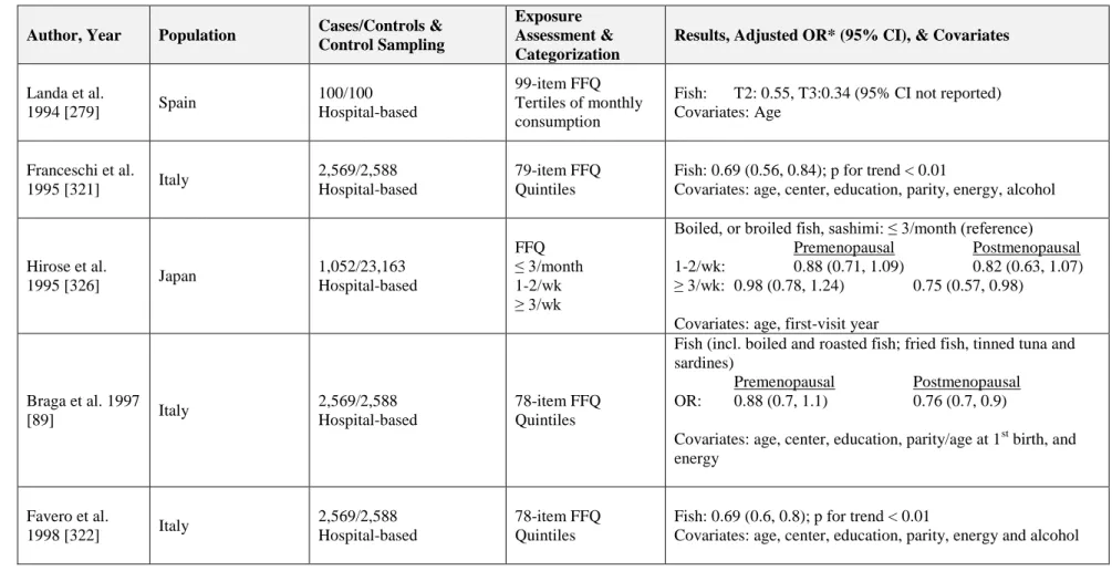
1.9 Epidemiology of Fish Intake and Breast Cancer ...52
1.9.1 Fish Intake and Incidence ...52
1.9.1.1Case-Control Studies ...53
1.9.1.2Cohort Studies ...57
1.9.2 Fish Intake and Survival & Mortality ...58
1.9.3 Summary ...60
1.10Epidemiology of PUFA-Gene Interactions and Breast Cancer ...61
1.11Background and Introduction Summary ...62
CHAPTER 2: METHODS ...89
2.1 Study Overview ...89
2.1.1 Specific Aims ...89
2.1.2 Importance of Knowledge to be gained ...91
2.2. Long Island Breast Cancer Study Project (LIBCSP) ...92
2.2.1 Case-Control Study...92
2.2.2 Follow-up Study ...93
2.3 Outcome and Exposure Assessments...95
2.3.1 Outcome Assessment ...95
2.3.2 Dietary Assessment ...96
2.3.3 PUFA Exposure Assessment ...97
2.3.4 Genotyping ...98
2.4 Results from Previous Analyses ...100
2.4.1 Inflammation genes and breast cancer risk in LIBCSP ...100
2.4.2 Oxidative stress genes and breast cancer risk in LIBCSP ...101
2.4.3 Aromatase genes and breast cancer risk in LIBCSP ...102
2.5 Data Analysis ...103
2.5.1 Statistical Methods ...103
2.5.2 Multiple Comparisons ...105
2.5.3 Confounding and Effect Measure Modification ...108
2.5.4 Energy Adjustment ...109
2.6 Study Power ...110
2.7 Data Interpretation Issues ...111
2.8 Study Purpose ...113
CHAPTER 3: INTERACTION BETWEEN PUFAs, GENETIC POLYMORPHISMS, AND BREAST CANCER RISK: A POPULATION-BASED, CASE-CONTROL STUDY ON LONG ISLAND, NEW YORK ... 119
3.1 Introduction ...119
3.2 Materials and Methods ...121
3.4 Discussion ...129
CHAPTER 4: DIETARY INTAKE OF FISH, PUFAs, AND SURVIVAL AFTER BREAST CANCER: A POPULATION-BASED, FOLLOW-UP STUDY ON LONG ISLAND, NEW YORK ... 143
4.1 Introduction ...143
4.2 Methods...145
4.3 Results ...150
4.4 Discussion ...153
CHAPTER 5: CONCLUSIONS ... 167
5.1 Summary of Study Aims and Results ...167
5.2 Summary of Public Health Impact ...168
5.3 Study Strengths ...169
5.4 Study Limitations ...171
5.5 Future Directions ...174
5.6 Conclusion ...175
APPENDIX: TABLES & FIGURES ... 176
LIST OF TABLES
Table 1.1 Polyunsaturated Fatty Acids and Sources ...64
Table 1.2 Summary of Case-Control Studies of Self-reported
PUFA Intake and Breast Cancer Incidence ...65
Table 1.3 Summary of Cohort Studies of Self-reported PUFA
Intake and Breast Cancer Incidence ...66
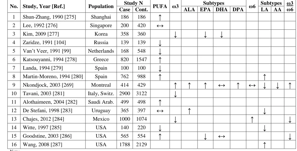
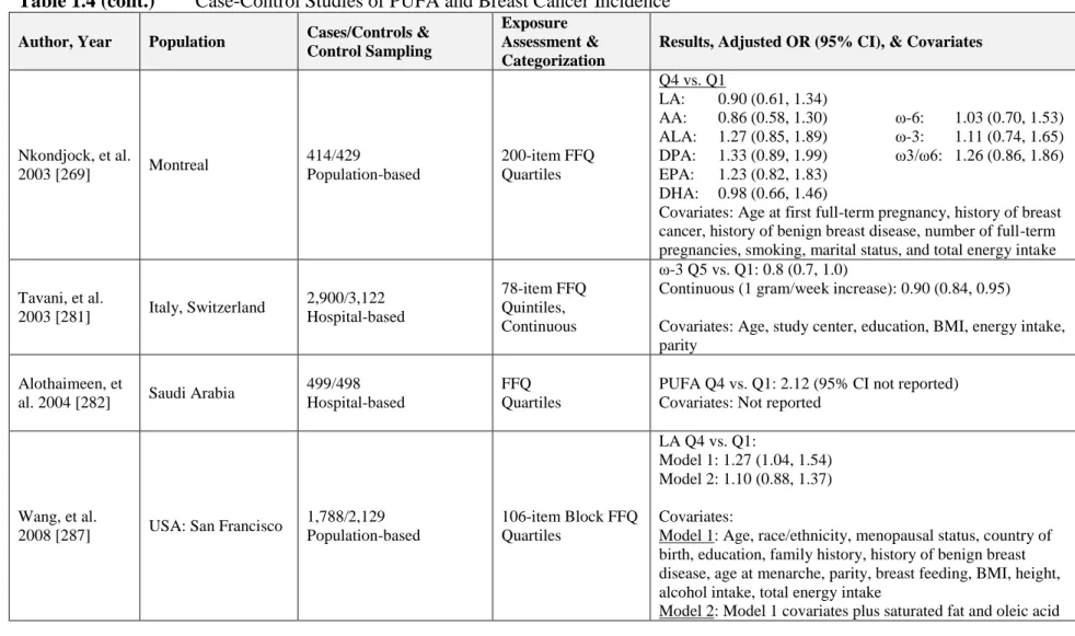
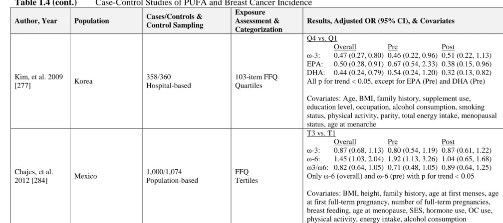
Table 1.4 Case-Control Studies of PUFA and Breast Cancer Incidence ...67
Table 1.5 Cohort Studies of PUFA and Breast Cancer Incidence ...71
Table 1.6 Case-Control Studies of Dietary Fish Intake and
Breast Cancer ...77
Table 1.7 Cohort Studies of Dietary Fish Intake and Breast Cancer ...82
Table 1.8 Studies of Fish Intake and Breast Cancer Survival
and Mortality ...85
Table 2.1 Example calculation of PUFA intake using USDA ...114
Table 2.2 Summary of putatively functional SNPs and LIBCSP results ...115
Table 3.1 Characteristics of polyunsaturated fatty acid intake (PUFA) and fish intake among the population-based sample of
control women (N=1500) in the LIBCSP, 1996-1997 ...135
Table 3.2 Age- and multivariate-adjusted ORs and 95% CI for the association between dietary PUFA intake and the
risk of breast cancer in the LIBCSP, 1996-1997 ...136
Table 3.3 Age- and multivariate-adjusted ORs and 95% CI for the association between fish intake and the risk of breast
cancer in the LIBCSP, 1996-1997 ...137
Table 3.4 Age- and multivariate-adjusted ORs and 95% CI for the additive interaction between dietary ω-3 and ω-6 (high and low intake) and the risk of breast cancer
in the LIBCSP, 1996-1997 ...138
cancer for the hypothesized highest risk additive
interaction between ω-3/ω-6 ratio and putatively functional
genetic polymorphisms in the LIBCSP, 1996-1997 ...139
Table 3.6 Assumed variant allele function based on previous literature ...140
Table 3.7 Age- and multivariate-adjusted ORs and 95% CI for the multiplicative interaction between dietary ω-3 and ω-6 (high and low intake) and the risk of breast cancer
in the LIBCSP, 1996-1997 ...141
Table 3.8 Multivariate-adjusted odds ratios (ORs) and 95% confidence intervals (95% CIs) for the risk of breast cancer for the hypothesized highest risk multiplicative interaction between ω-3/ω-6 ratio and putatively functional genetic polymorphisms
in the LIBCSP, 1996-1997 ...142
Table 4.1 Distributions of intakes of polyunsaturated fatty acid (PUFA) and fish at baseline among a population-based sample of women newly diagnosed with breast cancer (N=1463),
LIBCSP, 1996-1997 ...158
Table 4.2 Age- and multivariate-adjusted HRs and 95% CI for the association between dietary PUFA intake and all-cause mortality among a population-based sample of women with breast cancer, LIBCSP, 1996/1997 through 2011
(an average of 14.7 years of follow-up) ...159
Table 4.3 Age- and multivariate-adjusted HRs and 95% CI for the association between fish intake and all-cause mortality among a population-based sample of women with breast cancer, LIBCSP, 1996/1997 through 2011
(an average of 14.7 years of follow-up) ...160
Table 4.4 Age- and multivariate-adjusted HRs and 95% CI for the association between dietary PUFA intake and
breast cancer-specific mortality among a population-based sample of women with breast cancer, LIBCSP, 1996/1997
through 2011(an average of 14.7 years of follow-up) ...162
Table 4.5 Age- and energy-adjusted HRs and 95% CI for
competing risks analysis for all-cause, breast cancer-specific, other cause mortality, among a population-based sample of women with breast cancer, LIBCSP, 1996/1997
Table 4.6 Age- and multivariate-adjusted hazard ratios (HRs) and 95% confidence intervals (CI) for all-cause mortality among a population-based sample of women with breast cancer for the interaction between dietary ω-3 and ω-6 intake, LIBCSP, 1996/1997 through 2011
(an average of 14.7 years of follow-up) ...164
Table 4.7 Hazard ratios (HRs) and 95% confidence intervals (95% CIs) for all-cause mortality for the association with putatively functional genetic polymorphisms using dominant model (N=1463) among a population-based sample of women with breast cancer, LIBCSP, 1996/1997 through 2011
(an average of 14.7 years of follow-up) ...165
Table 4.8 Age- and energy-adjusted hazard ratios (HRs) and 95% confidence intervals (95% CIs) for
all-cause mortality for the hypothesized highest risk interaction between ω-3/ω-6 ratio and putatively
functional genetic polymorphisms among a population-based sample of women with breast cancer, LIBCSP, 1996/1997
through 2011(an average of 14.7 years of follow-up) ...166
Table A.1 Pearson correlation between PUFA subtypes
among 1,500 LIBCSP controls ...176
Table A.2 Pearson correlation between PUFA subtypes
among 1,463 LIBCSP cases ...177
Table A.3 Age- and energy-adjusted ORs and 95% CI for
the interaction between total PUFA intake (low or high)
and genetic variants on breast cancer incidence ...178
Table A.4 Age- and energy-adjusted ORs and 95% CI for the interaction between total ω-3 intake (low or high)
and genetic variants on breast cancer incidence ...179
Table A.5 Age- and energy-adjusted ORs and 95% CI for the interaction between ALA intake (low or high)
and genetic variants on breast cancer incidence ...180
Table A.6 Age- and energy-adjusted ORs and 95% CI for the interaction between DPA intake (low or high)
Table A.7 Age- and energy-adjusted ORs and 95% CI for the interaction between DHA intake (low or high)
and genetic variants on breast cancer incidence ...182
Table A.8 Age- and energy-adjusted ORs and 95% CI for the interaction between EPA intake (low or high)
and genetic variants on breast cancer incidence ...183
Table A.9 Age- and energy-adjusted ORs and 95% CI for the interaction between total ω-6 intake (low or high)
and genetic variants on breast cancer incidence ...184
Table A.10 Age- and energy-adjusted ORs and 95% CI for the interaction between LA intake (low or high)
and genetic variants on breast cancer incidence...185
Table A.11 Age- and energy-adjusted ORs and 95% CI for the interaction between AA intake (low or high)
and genetic variants on breast cancer incidence...186
Table A.12 Age- and multivariate-adjusted hazard ratios (HRs) and 95% confidence intervals (CI) for all-cause mortality among a population-based sample of women with breast cancer for the multiplicative interaction between dietary ω-3 and ω-6 intake, LIBCSP, 1996/1997
through 2011(an average of 14.7 years of follow-up) ...187
Table A.13 Age- and energy-adjusted hazard ratios (HRs) and
95% confidence intervals (95% CIs) for all-cause mortality for the hypothesized highest risk multiplicative interaction between ω-3/ω-6 ratio and putatively functional genetic polymorphisms among a population-based sample of women with breast cancer, LIBCSP, 1996/1997
through 2011(an average of 14.7 years of follow-up) ...188
Table A.14 Age- and energy-adjusted HRs and 95% CI for the interaction between PUFA intake (low or high)
and genetic variants on all-cause mortality ...189
Table A.15 Age- and energy-adjusted HRs and 95% CI for the interaction between ω-3 intake (low or high)
and genetic variants on all-cause mortality ...190
Table A.16 Age- and energy-adjusted HRs and 95% CI for the interaction between ALA intake (low or high)
Table A.17 Age- and energy-adjusted HRs and 95% CI for the interaction between DPA intake (low or high)
and genetic variants on all-cause mortality ...192
Table A.18 Age- and energy-adjusted HRs and 95% CI for the interaction between DHA intake (low or high)
and genetic variants on all-cause mortality ...193
Table A.19 Age- and energy-adjusted HRs and 95% CI for the interaction between EPA intake (low or high)
and genetic variants on all-cause mortality ...194
Table A.20 Age- and energy-adjusted HRs and 95% CI for the interaction between ω-6 intake (low or high)
and genetic variants on all-cause mortality ...195
Table A.21 Age- and energy-adjusted HRs and 95% CI for the interaction between LA intake (low or high)
and genetic variants on all-cause mortality ...196
Table A.22 Age- and energy-adjusted HRs and 95% CI for the interaction between AA intake (low or high)
and genetic variants on all-cause mortality ...197
Table A.23 Multiple comparisons adjustment via FDR for
statistically significant (p<0.05) multivariate-adjusted
associations (Aim 1a) ...198
Table A.24 Multiple comparisons adjustment via FDR for
statistically significant (p<0.05) multivariate-adjusted
associations for individual PUFA-gene interactions (Aim 1b) ...199
Table A.25 Multiple comparisons adjustment via FDR for statistically significant (p<0.05) additive interactions
(RERI) for PUFA-gene interactions (Aim 1b) ...200
Table A.26 Multiple comparisons adjustment via FDR for
statistically significant (p<0.05) multivariate-adjusted
associations (Aim 2a) ...201
Table A.27 Multiple comparisons adjustment via FDR for statistically significant (p<0.05) gene associations
Table A.28 Multiple comparisons adjustment via FDR for
statistically significant (p<0.05) multivariate-adjusted
associations for individual PUFA-gene interactions (Aim 2c) ...203
Table A.29 Multiple comparisons adjustment via FDR for statistically significant (p<0.05) additive interactions
(RERI) for PUFA-gene interactions (Aim 2c) ...205
Table A.30 Age-adjusted ORs and 95% CIs for the potential association between PUFA and breast cancer incidence
stratified by supplement use ...206
Table A.31 Age-adjusted ORs and 95% CIs for the potential association between PUFA and breast cancer incidence
stratified by menopausal status ...207
Table A.32 Age-adjusted ORs and 95% CI for potential
association between PUFA intake and breast cancer
according to breast cancer tumor subtype ...208
Table A.33 Age-adjusted HRs and 95% CIs for the potential association between PUFA and all-cause mortality
stratified by supplement use ...209
Table A.34 Age-adjusted HRs and 95% CIs for the potential association between PUFA and all-cause mortality
stratified by menopausal status ...210
Table A.35 Age-adjusted HRs and 95% CIs for the potential association between PUFA and all-cause mortality
stratified by hormone therapy treatment ...211
Table A.36 Age-adjusted HRs and 95% CIs for the potential association between PUFA and all-cause mortality
stratified by chemotherapy treatment ...212
Table A.37 Age-adjusted HRs and 95% CIs for the potential association between PUFA and all-cause mortality
stratified by radiation treatment ...213
Table A.38 Models for adjusting for total energy intake ...214
Table A.39 Comparison of different energy adjustment methods
Table A.40 Comparison of different energy adjustment methods
(alcohol adjustment) for breast cancer incidence ...216
Table A.41 Comparison of different energy adjustment methods (no alcohol adjustment) for breast cancer survival ...217
Table A.42 Comparison of different energy adjustment methods (alcohol adjustment) for breast cancer survival ...218
Table A.43 Sensitivity analyses examining PUFA adjustment for breast cancer incidence ...219
Table A.44 Sensitivity analyses examining PUFA adjustment for breast cancer survival ...220
Table A.45 Power for PUFA/Fish intake & Incidence (Aim 1a) ...221
Table A.46 Power for PUFA-gene interactions & incidence (Aim 1b) ...222
Table A.47 Power for PUFA/Fish intake & Survival (Aim 2a) ...223
Table A.48 Power for Genotypes & Survival (Aim 2b) ...224
Table A.49 Age- and multivariate-adjusted ORs and 95% CI for the association between dietary fish intake (by various cooking methods) and breast cancer incidence ...225
LIST OF FIGURES
Figure 1.1 Age-specificbreast cancer incidence rates (per 100,000)
for Western and Asian populations in 2002 ...86
Figure 1.2 PUFA Biosynthesis ...87
Figure 1.3 Metabolism of Arachidonic Acid...88
Figure 2.1 Directed Acyclic Graph (DAG) of potential confounders
of the PUFA and breast cancer incidence association ...117
Figure 2.2 Directed Acyclic Graph (DAG) of potential confounders
of the PUFA and breast cancer mortality association ...118
Figure 4.1 Kaplan-Meier survival curves for dietary intake (quartiles) of long-chain ω-3 fatty acids DPA, DHA, and EPA,
among a population-based sample of women with breast cancer, LIBCSP, 1996/1997 through 2011
(an average of 14.7 years of follow-up) ...161
Figure A.1 Dose-response between total PUFA intake (g/day) and
the age-adjusted log-odds of breast cancer in the LIBCSP...227
Figure A.2 Dose-response between total ω-3 intake (g/day) and
the age-adjusted log-odds of breast cancer in the LIBCSP...228
Figure A.3 Dose-response between total ω-6 intake (g/day) and
the age-adjusted log-odds of breast cancer in the LIBCSP...229
Figure A.4 Kaplan-Meier Survival curves for total PUFA,
total ω-3, total ω-6, and ω-3/ω-6 intake (quartiles) and
all-cause mortality ...230
Figure A.5 Kaplan-Meier Survival curves for ALA, DPA, DHA, and
EPA intake (quartiles) and all-cause mortality ...231
Figure A.6 Kaplan-Meier Survival curves for LA and AA intake
(quartiles) and all-cause mortality ...232
Figure A.7 Dose-response between total PUFA intake (g/day) and the age-adjusted difference in log-hazards of all-cause
Figure A.8 Dose-response between total ω-3 intake (g/day) and the age-adjusted difference in log-hazards of all-cause
mortality among women with breast cancer in the LIBCSP ...234
Figure A.9 Dose-response between total ω-6 intake (g/day) and the age-adjusted difference in log-hazards of all-cause
ABBREVIATIONS
AA arachidonic acid
ALA α-linolenic acid
BMI body mass index (kg/m2)
BRCA Breast Cancer Gene
CI confidence interval
COX cyclooxygenase
CYP cytochrome p450
DAG directed acyclic graph DCIS ductal carcinoma in-situ
DHA docosahexanoic acid
DNA deoxyribonucleic acid
DPA docosapentaenoic acid
EPA eicosapentaenoic acid
ER estrogen receptor
FFQ food frequency questionnaire
g gram
GST glutathione S-transferase
HR hazard ratio
HCFA Health Care Finance Administration HER human epidermal growth factor receptor HETE hydroxyeicosatetraenoic acid
IL interleukin
IRR incidence rate ratio
kg kilogram
LA linoleic acid
LC ω-3 long-chain ω-3
LCIS lobular carcinoma in-situ
LOX lipoxygenase
LIBCSP Long Island Breast Cancer Study Project LRT likelihood ratio test
m meter
miRNA micro ribonucleic acid
NCI National Cancer Institute
NDI National Death Index
NHANES National Health and Nutrition Examination Survey NSAID non-steroidal anti-inflammatory drug
nsSNP non-synonymous single nucleotide polymorphism
OR odds ratio
PGE2 prostaglandin E2
PLA2 phospholipase A2
PPAR peroxisome proliferator-activated receptor
PR progesterone receptor
RERI relative excess risk due to interaction
RR risk ratio
RNA ribonucleic acid
SHBG sex hormone binding globulin SNP single nucleotide polymorphism TFBS transcription factor binding site TNF tumor necrosis factor
U.S. United States
USDA United States Department of Agriculture
VITAL VITamins and Lifestyle
CHAPTER 1:
BACKGROUND AND INTRODUCTION
This dissertation examines the association between polyunsaturated fatty acids (PUFAs) and the risk of breast cancer incidence and mortality, and explores whether these associations vary by PUFA class (ω-3, ω-6, the relative balance of ω-3 and ω-6, and ω-3 and ω-6 subtypes), or by fish (the major source of long-chain ω-3), or by genetic polymorphisms in biologically related pathways (inflammation, oxidative stress, and estrogen metabolism). This first chapter provides the rationale for the dissertation aims, including a review of: the descriptive and analytic epidemiology of breast cancer incidence and mortality; the biologic and epidemiologic characteristics of the primary study exposure, PUFAs, as well as the primary group of effect modifiers of interest, polymorphisms in biologically related pathways; and critically evaluates the previously reported epidemiologic studies that have examined the putative PUFA-breast cancer association to underscore the novel aspects of the dissertation aims.
1.1 Epidemiology of Breast Cancer Incidence and Mortality
highest among Caucasian white women, followed by African American, Asian/Pacific Islanders, American Indian/Alaska Native, and lowest among Hispanic women [1].
Majority of breast cancers are invasive, where the tumor spreads past the lobules and ducts into the surrounding tissue. In situ breast cancer occurs when the tumor is contained within either the duct (ductal carcinoma in situ, DCIS) or the lobule (lobular carcinoma in situ, LCIS). It is estimated that about 58,000 new cases of in situ breast cancer were diagnosed in 2013, and approximately 83% of these were DCIS [2].
Breast cancer rates are much lower among Asian populations than among Western populations (see Figure 1.1 below). Among Western populations, breast cancer incidence increases with age, with rate of increase slowing around age fifty, but continuing to increase after age fifty [2]. However, among Asian women, the incidence rate of breast cancer increases with age until fifty, and then stabilizes (Figure 1.1). This distinct pattern seen among Western and Asian populations points to the direct role of reproductive factors in breast cancer etiology [3]. Reproductive risk factors for breast cancer include: early age at menarche; late age at menopause; nulliparity; late age at first full-term pregnancy; no lactation; and use of hormone replacement therapy. Also, migration studies of Asian immigrants have shown that the breast cancer incidence patterns begin to reach those of Western countries after only a few generations after migration [4-7], suggesting a role for other environmental risk factors for breast cancer. As discussed in more detail below, other risk factors for breast cancer include: post-menopausal obesity; alcohol use; physical inactivity; and family history of breast cancer [8].
older (90%) [2]. Five-year relative survival rates are highest for women whose tumors are localized (99%) compared to regional (84%) and distant tumors (23%). Mortality rates are highest and 5-year survival rates are lowest among African American women [1]. As
discussed below, prognostic factors for breast cancer include: tumor size; tumor stage; breast cancer treatment; breast cancer subtypes; and obesity at diagnosis [9].
1.2 Breast Cancer Risk Factors
1.2.1 Reproductive risk factors
There are several established breast cancer risk factors that are associated with reproduction, including early age at menarche, late age at menopause, late age at first full-term pregnancy, nulliparity, lack of or short duration of breast-feeding, and hormone replacement therapy. Many of these reproductive risk factors are thought to affect breast cancer via regulation of long-term exposure to endogenous hormones, including estrogen and progesterone, which are thought to play an important role in breast cancer etiology [8]. The tumor inducing and promoting potential of estrogens has been demonstrated [10, 11], and removal of the ovaries or administration of an anti-estrogenic drug can prevent this effect [12]. Early age at menarche and late age at menopause maximizes a women’s lifetime exposure to estrogen, and this prolonged exposure to estrogen has been shown to increase breast cancer risk among many different populations [13-18].
and these terminally differentiated cells have been shown to have lower proliferation due to longer cell cycles and increased time spent in the resting phase (G1) of the cell cycle [19]. A similar mechanism has been proposed for the risk reductions observed among women who have increased duration of lactation, which has been reported in a number of studies [15, 16, 18, 20, 21]. This risk reduction conferred by breastfeeding may be due to the increase in terminal differentiation of the breast epithelium, as well as the delay in the restoration of the ovulatory cycle post-pregnancy [14].
Many women are prescribed hormone replacement therapy in order to help ameliorate the symptoms associated with menopause. However, the use of hormone replacement
therapy has been demonstrated to affect breast cancer risk, and is dependent upon the type of hormones that are prescribed. In 2001, among participants in the Women’s Health Initiative trial, there was a reported 26% (95% CI = 1.00, 1.59) increased hazard for breast cancer among postmenopausal women on estrogen and progestin replacement therapy compared to those taking placebo [22]. Increased risks also were observed among the Million Women Study in the United Kingdom, where breast cancer risk was doubled for estrogen-progestin replacement therapy [23].
In sum, examination of the reproductive risk factors for breast cancer has helped to elucidate an important role of endogenous and exogenous estrogen and progesterone in breast cancer carcinogenesis.
1.2.2 Family History
cancer risk. Women, whose mothers or sisters have breast cancer, are 1.5 to 3 times as likely to have breast cancer compared to other first-degree relatives without breast cancer [24]. A meta-analysis reported the risk for breast cancer was nearly double for women with any relative, first-degree relative, mother, or sister with breast cancer [25]. The highest risk was observed for women who had a mother and sister with breast cancer (RR = 3.6; 95% CI = 2.5, 5.0).
In the mid-1990s, breast cancer gene 1 (BRCA1) on chromosome 17q21 and breast cancer gene 2 (BRCA2) on chromosome 13q12-13 were identified [26]. These two genes are suggested to act as tumor suppressors, and mutations in these genes are highly penetrant and account for 2-5% of breast cancer risk [24]. In the general population, estimated prevalence of BRCA1 and BRCA2 mutation carriers range from 0.1- 0.2% [26, 27], and these mutations may be responsible for early-onset breast cancer among high risk families [27].
However, family history cannot solely explain geographic variation in breast cancer rates. Studies conducted among Asian immigrants have shown that the breast cancer incidence patterns for these populations mimic those of Western countries only a few
generations after migration [4-7]. Therefore, other environmental risk factors, for which the prevalence varies by geographic residence, may help to explain this observed geographic variation in breast cancer rates.
1.2.3 Obesity and Physical Activity
metabolically active estrogen that is not produced by the ovaries [28, 29]. These laboratory findings are supported by epidemiologic results. For example, a pooled analysis based on data collected primarily among women of European descent examined the effect of body mass index (BMI) on breast cancer risk and reported that increasing BMI among
postmenopausal women (BMI exceeding 28 kg/m2) was associated with an increase in breast cancer risk of 26% (95% CI = 1.09, 1.46) [30]. Changes in weight (greater than 15 kg since age 20) compared to maintaining weight, has been reported to increase risk for
postmenopausal breast cancer (OR = 1.6; 95% CI = 1.11, 2.26) [31], and this increased risk has been consistently reported among several other studies [32-34]. Asian studies also reported a similar increased risk for postmenopausal breast cancer with increases in body weight [35] or BMI [36, 37].
seen among premenopausal women, and include obesity-triggered anovulation [41], which can result in lower levels of exposure to both progesterone and estradiol [42].
Physical activity also is thought to reduce breast cancer risk via a variety of mechanisms, including lower levels of estrogens and increased levels of sex hormone binding globulin (SHBG) resulting from reduced adipose tissue and visceral fat [43, 44], improved immune response [45], and lower inflammatory markers [46, 47]. A systematic review reported risk reductions ranging from 20-80% were observed for postmenopausal breast cancer with increasing levels of physical activity [48]. A smaller risk reduction (15-20%) was observed for premenopausal women [48-50]. Similar risk reductions were observed for recreational physical activity among Chinese women [51]. A recent study examined the joint effects of physical activity, weight gain, and body size on breast cancer risk, and reported risk reductions for breast cancer and recreational physical activity during both reproductive years and postmenopausal [52]. However, it also was reported that excessive weight gain during postmenopausal years may negate any of the beneficial effects of any physical activity.
In summary, physical activity, obesity, and weight maintenance provide a potential opportunity for breast cancer risk reduction. However, increasing physical activity, reducing obesity or maintaining weight may not be an easily implemented option for all women. Therefore, other opportunities for breast cancer risk reduction need to be explored, including nutritional factors.
1.2.4 Non-Steroidal Anti-inflammatory Drugs
anti-inflammatory drugs (NSAIDs), such as aspirin and ibuprofen. NSAIDs are drugs that inhibit cyclooxygenase (COX) activity and thereby result in reduced levels of prostaglandins, which have been implicated in breast carcinogenesis [53]. There is evidence from animal studies that NSAIDs may also have an inhibitory effect that is independent of prostaglandin synthesis. Animal and laboratory studies suggest that NSAIDs may inhibit the effects of estrogen in the pituitary gland [54] and may inhibit binding of estradiol to the estrogen receptor [55]. Observational studies have reported a modest risk reduction (10-20%) for NSAID use and breast cancer risk [56], though this inverse association is not consistently reported across all studies [56, 57]. Risk reductions were slightly stronger among case-control studies compared to cohort studies, which may reflect differential recall bias present in case-control studies resulting in exaggerated effect estimates. Recent studies have been inconsistent regarding NSAID use and breast cancer, with one reporting risk reduction [58], another reporting null effects [59], and yet another reporting increased risk [60]. NSAID use was reported to decrease breast cancer risk by 20% among postmenopausal women [58], but had no effect among premenopausal women [61]. A recent study reported stronger risk reductions for NSAIDs that were selective COX-2 inhibitors (e.g., celecoxib, rofecoxib, and valdecoxib) in comparison to non-specific NSAIDs [62]. Inconsistencies in the literature may arise from differences in control selection (e.g., population- versus hospital-based), differences in exposure assessments (e.g., questionnaire versus health care prescription data), or differential recall in case-control studies versus cohort studies.
reductions for aspirin use among those with the variant allele rs4648261 (C-to-T base pair change), a single nucleotide polymorphism (SNP) in COX-2. In the Long Island Breast Cancer Study Population (LIBCSP), risk reductions were reported for regular use of aspirin and breast cancer [64], but largely no interactions were seen for COX-2 polymorphisms and NSAID use [65].
In sum, although many studies have reported risk reductions for NSAID use and breast cancer, the results are not conclusive. NSAIDs inhibit cyclooxygenase activity thereby reducing levels of inflammatory prostaglandins. It has also been noted that long-term use of NSAIDs could have potentially adverse outcomes on health [66, 67]. Similar to NSAIDs, ω-3 PUFAs are also known to competitively inhibit binding of ω-6 PUFAs to cyclooxygenase enzyme (see below), thereby reducing levels of inflammatory prostaglandins [68]. Thus, dietary intake of ω-3 PUFAs may provide a safer alternative for breast cancer risk reduction compared to NSAIDs.
1.2.5 Alcohol
Alcohol is thought to affect breast carcinogenesis via multiple mechanisms, including increasing levels of endogenous estrogen, production of acetaldehyde and reactive oxygen species resulting from alcohol metabolism, and interference with absorption of essential nutrients [69]. An early meta-analysis [70] reported a 24% increased risk (95% CI = 1.15, 1.34) for women consuming two drinks per day compared to non-drinkers. Women
per day, respectively [71]. More recent studies examining alcohol intake and breast cancer have reported similar effect estimates. The Million Women Study reported a 12% increase in breast cancer risk for a 10-gram increase [72]. The Nurses’ Health Study reported a 15% increase in breast cancer risk for 5-10 gram increase in alcohol consumption [73], and the American Cancer Society Nutrition Cohort reported a 26% increase for 15 gram per day increase in alcohol consumption [74]. However, among the European Prospective
Investigation into Cancer and Nutrition (EPIC) cohort, no effects were observed for 10-gram per day increase in alcohol intake and breast cancer risk [75].
In sum, alcohol is an established breast cancer risk factor which acts via multiple mechanisms to affect breast cancer risk, including increased estrogen production. Thus, epidemiologic and supporting laboratory evidence has demonstrated that an ingested
compound can influence estrogen levels among adult women and increase breast cancer risk.
1.2.6 Dietary Fat
Dietary fat and breast cancer risk has been extensively examined, however, there is limited evidence suggesting an association between total fat intake and breast cancer. Dietary fat is known to increase endogenous estrogen production [76]. Also, dietary fat can increase levels of serum-free fatty acids thereby displacing estradiol from serum albumin, and thus resulting in increased levels of free estradiol [77]. A null association has been reported for total fat intake among all ages or premenopausal breast cancer [78]. Numerous studies have examined the effect of total fat on postmenopausal breast cancer, where
postmenopausal breast cancer [79-82, 82-85], whereas two other studies reported risk reductions [86, 87]. The majority of case-control studies report an increased risk for postmenopausal breast cancer with total fat intake [88-100], with a relatively fewer number of studies reporting a decrease in risk [89, 101-104]. A summary estimate was derived from seven case-control studies reporting a modest 11% increase in risk for total fat intake [78]. This difference in effect estimates derived from the case-control and prospective studies may reflect differential recall bias of total fat intake, which would exaggerate effects in case-control studies. Therefore, there is limited evidence regarding total fat intake and breast cancer, with no associations reported for premenopausal women, and a suggested increase in risk for postmenopausal breast cancer. However, conclusions regarding total fat and breast cancer risk are still unclear.
The inconsistent reported effects of the association between breast cancer incidence and fat intake may be due to type of dietary fat consumed. A pooling project reported differential effects for different types of fat, where saturated and monounsaturated fats increased risks by 9% and 5%, respectively [105]. Modest risk reductions (2-13%) were reported when substituting either saturated or monounsaturated fats with PUFAs [105]. In order to better understand the effects of fat on breast cancer risk, it may be important to examine the different types of fat in relation to breast cancer, such as PUFAs.
1.2.7 Summary
and select nutritional factors such as obesity, physical activity, and alcohol. With the exception of these select nutritional factors, few risk factors are easily modifiable. Identification of other potentially modifiable nutritional factors, such as dietary intake of PUFAs, may be warranted in order to provide additional avenues for breast cancer prevention.
1.3 Prognostic Factors for Breast Cancer
1.3.1 Age, Socioeconomic Status, and Race
A few established prognostic factors for breast cancer survival include age at
diagnosis, socioeconomic status, and race. Women with younger age at diagnosis have been reported to have reduced survival and increased relapse compared to women who are
status have poor prognosis, irrespective of race [111].
1.3.2 Tumor Size and Stage at Diagnosis
The size of the tumor is known to predict survival. Women with larger tumors have worse prognosis than those with smaller tumors [107, 112]. Women with tumors greater than two centimeters are more likely to die from breast cancer five years post diagnosis [107], and are more likely to have distant metastatic disease [113]. Metastases to regional lymph nodes are a strong prognostic indicator, among whom women with greater number of lymph nodes with worse survival than those with negative lymph nodes [112, 114]. Tumor stage is also a strong indicator of disease survival, where women with higher stage have poor prognosis compared to those with lower stage [114, 115]. Thus, tumor size and stage are established breast cancer prognostic factors.
1.3.3 Breast Cancer Subtypes
relative survival rates than women with ER- or PR- tumors [120].
Gene expression analysis has documented multiple intrinsic subtypes, in addition to the ER/PR subtypes. The ER+/ER- subtypes can be further classified into separate groups, which include: (1) luminal A: ER+ and/or PR+, human epidermal growth factor receptor-2 negative (HER2-); (2) luminal B: ER+ and/or PR+, HER2+; (3) HER2+/ER-: ER-, PR-, (HER2+); and (4) basal-like: ER-, PR-, HER2-, cytokeratin 5/6+, and/or HER1+ [109]. Basal-like tumors are more prevalent among African American women (premenopausal), and both basal-like and HER2+/ER- tumors were more likely to be of higher grade, greater mitotic index, higher prevalence of p53 mutations, and more likely to be poorly differentiated [109]. These factors may account for the poor prognosis observed among women with basal-like and HER2+/ER- tumors [109]. Understanding the prognostic influence of breast cancer subtypes is not only important for identifying successful treatments for women diagnosed with breast cancer, but also could help to identify women who would benefit from additional interventions to help improve survival.
1.3.4 Treatment
compared to chemotherapy alone [125]. Supplementary treatment strategies, that are both cost-effective and easily-implemented, need to be considered to further improve survival for women diagnosed with breast cancer. Nutritional factors that enhance treatment efficacy, such as PUFAs, should be considered as they may provide an additional means for improving survival among women diagnosed with breast cancer.
1.3.5 Obesity and Physical Activity
There is a strong indication that obesity and physical activity play a role in breast cancer prognosis, though they are not considered established breast cancer prognostic factors. A recently published pooling project reported increased hazards for overall mortality and breast cancer mortality for underweight (BMI < 18.5 kg/m2) and morbidly obese women (BMI ≥ 40 kg/m2) [126]. More recently, Cleveland et al. also reported increased mortality among women who were obese at diagnosis among both pre- and post-menopausal women [127]. Also, among premenopausal women, those who gained greater than 16 kg between age 20 and one year prior to diagnosis had more than double the hazard for mortality when compared to those who maintained a stable weight (± 3 kg) [127]. Post-diagnosis
maintenance of weight is also important for improving survival [128]. Bradshaw et al. reported more than two-fold increase in mortality for those who gained more than 10% after diagnosis, and this effect was more pronounced in the first two years after diagnosis [128]. Thus, maintenance of weight, whether it is prior to diagnosis or after, is suggested to improve breast cancer survival for both premenopausal and postmenopausal breast cancer patients.
women who engaged in nine or more metabolic equivalent task hour (MET-hour) per week of lifetime recreational physical activity from menarche to diagnosis compared to women who did not exercise [129]. Similar results were reported among the California Teachers Study [130]. Additionally, exercise within three years of diagnosis was inversely associated with both overall and relapse/disease-specific mortality [131, 132]. Similarly, Bradshaw et al. reported improved survival among women who were highly active (>9 weekly MET-hours) diagnosis [133]. Studies suggest that both pre-diagnostic and timely post-diagnostic engagement in exercise may help to improve survival among women diagnosed with breast cancer.
1.3.6 Dietary Fat and NSAIDs
The relation between dietary fat intake and breast cancer survival has been
increasingly reported by a number of investigators. The majority of studies regarding dietary fat and survival showed an increased risk of dying with higher fat intake [134-137], with the exception of one study showing a risk reduction [138]. An early study examined the relation between dietary fat and breast cancer survival among both Caucasian and Japanese women with breast cancer diagnoses in Hawaii [139]. The study reported more than triple the risk of death for high versus low total fat intake for Caucasian women. In comparison, nearly 40% mortality risk reduction was reported among women of Japanese ancestry for high versus low total fat intake. Though both estimates were imprecise, this difference in the direction of the reported associations among Caucasian and Japanese women may highlight the importance of examining different types of fat on breast cancer, such as ω-3 and ω-6 PUFA.
fat intake. Two large randomized trials were conducted in the mid-1990s in order to examine the effect of reductions in dietary fat intake on prognosis among breast cancer survivors in the U.S. The Women’s Intervention Nutrition Study (WINS) was focused primarily on the efficacy of reduction in dietary fat intake [140]. The WINS study included approximately 2,400 postmenopausal women who had completed primary treatment and had been diagnosed with stage one breast cancer in the previous year. Study participants were randomized to receive an intervention targeted to reduce fat intake. The study reported slight reductions in the hazard for overall survival (HR = 0.89; 95% CI = 0.65, 1.21) [140]. In contrast, the Women’s Healthy Eating and Living (WHEL) trial focused on the efficacy of changes in dietary pattern, which included reduction in dietary fat intake [141]. The WHEL study included approximately 3,000 women, among whom nearly 80% were postmenopausal, and had been diagnosed with stage one breast cancer within the previous four years. The
intervention focused on implementing a dietary pattern with high fruit, vegetable, and fiber intake and low total fat intake. The WHEL study reported null associations for the dietary pattern intervention on overall survival (HR = 0.97; 95% CI = 0.78, 1.22) [142]. A modest reduction in the hazard for recurrence was reported in the WINS study (HR = 0.76; 95% CI = 0.60, 0.98), but not for the WHEL study (HR = 0.99; 95% CI = 0.83, 1.17).
study would focus on more short-term prognosis, or women who could have a breast cancer event within five years of diagnosis. In contrast, the WHEL study under sampled women who had a breast cancer event within four years of the diagnosis, and therefore are unable to examine the effect of the intervention on early recurrence and death. This may explain the differences in the reported HRs for breast cancer recurrence in the two studies.
These previous studies on breast cancer progression and mortality focused on
reductions in total fat intake without any consideration given to the type of fat. It is possible that ω-3 and ω-6 PUFAs will have differential effects on survival, and that reduction in dietary intake of the unfavorable ω-6 fatty acids and increases in the more favorable ω-3 fatty acid could improve breast cancer survival. Therefore, examination of these different types of PUFAs and their relative balance could further elucidate the relation between dietary fat and breast cancer survival.
Non-steroidal anti-inflammatory drugs could also improve survival by reducing inflammatory metabolites resulting from metabolism of arachidonic acid via cyclooxygenase enzymes. However, only two studies on NSAID use in relation to survival from breast cancer have been reported to date, and results were conflicting [143, 144]. Thus, other avenues for improving breast cancer survival should be considered.
1.3.7 Summary
growing evidence that lifestyle factors such as weight maintenance, increased physical activity, and reduced fat intake before and after diagnosis improve survival. The promising findings for nutritional factors underscore the hypothesis that some factors may be modified in an effort to improve survival, and support the examination of other possible nutritional factors that could influence breast cancer survival. One possibility is dietary PUFA intake, which may help to provide an opportunity for improving survival among women diagnosed with breast cancer.
1.4 PUFAs
1.4.1 Structure
1.4.2 PUFA Sources and Biosynthesis
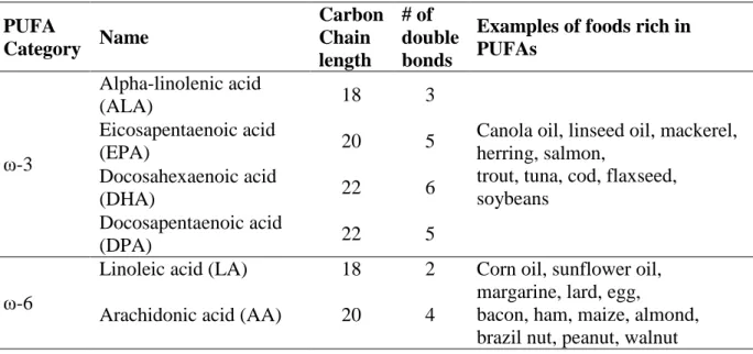
Mammalian cells are unable to endogenously produce ω-3 and ω-6 PUFAs and, thus the primary source of PUFAs in humans is diet. As shown in Table 1.1 (see below), fatty fish (e.g., halibut, mackerel, herring, and salmon) are the primary source of long-chain ω-3 (LC ω-3) PUFAs. Vegetable oils are the major source of ALA [146]. Other sources
contribute only minor quantities of ω-3 to the diet and include nuts and seeds, vegetables and some fruit, egg yolk, poultry and meat [146]. Different cooking methods (e.g., deep frying) have been shown to substantially reduce LC ω-3 PUFA content [147]. Vegetable oils are major sources of ω-6 fatty acids. Corn oil, peanut oil, sunflower oil, safflower oil,
margarine, lard, bacon, ham, nuts are sources of LA [145, 148].
subtypes in vivo. For example, low intake of ω-3 PUFAs that are precursors (e.g., ALA) in fatty acid biosynthesis may affect the bioavailability of downstream LC ω-3 PUFAs (e.g., EPA, DPA, DHA) [150]. Therefore, in addition to considering dietary intake of specific subtypes of ω-3 and ω-6 PUFAs separately, it may be equally important to consider total dietary intake of ω-3, ω-6, and the relative balance of ω-3 and ω-6 PUFAs. Figure 1.2
shown below provides an overview PUFA biosynthesis.
It is suggested that human beings evolved on an ω-3:ω-6 ratio approximately
equivalent to one, and since then our diets have evolved to include more sources of ω-6 fatty acids [151]. This increased consumption of ω-6 fatty acids was due in large part to the development of technology during the early 1900s marking the beginning of the vegetable oil industry, and modern agriculture with the emphasis on feeding domestic livestock with grains rich in ω-6 fatty acids [151]. During the 20th
century, the consumption of soybean oil increased dramatically [152]. Thus, the availability of LA (as a percentage of total energy) has increased dramatically in Western populations; whereas, the availability of LC ω-3 PUFA has remained stable [152]. The relative intake of ω-3 and ω-6 PUFA varies
geographically, with western diets having an intake ratio of ω-3:ω-6 of 1:15-20, compared to the ratio of 1:5-6 in India and 1:4 in Japan [153]. Also, it has been reported that serum levels of LCω3-PUFA are lower among U.S. Whites when compared to other Asian populations [154]. Since ω-3 and ω-6 fatty acids compete for the same enzymes, it is important to consider the relative balance of the two when considering their effects on chronic disease.
In summary, examination of the relative balance of ω-3 and ω-6 fatty acid intake may be vital to understanding the effects of PUFA on breast cancer, and for identifying an
1.5 Metabolism of PUFAs
PUFAs are incorporated into the cellular membrane. After incorporation within this lipid bilayer, PUFAs are then available for metabolism which occurs via the cyclooxygenase, lipoxygenase, and cytochrome p450 pathways. Both ω-6 and ω-3 fatty acids are metabolized via these pathways and compete with one another for the same enzymes, and they have demonstrated effects on the hallmarks of cancer [155]. The differential effect of ω-6 and ω-3 fatty acids on breast cancer etiology has been well demonstrated in animal and laboratory studies. Figure 1.3 shown below provides an overview of the biologic pathways involved in arachidonic acid metabolism.
1.5.1 Cyclooxygenase pathway
Metabolism of AA via the cyclooxygenase pathway results in the formation of
inflammatory intermediates, known as eicosanoids. A key metabolic enzyme in this pathway is prostaglandin synthase 2, or COX-2. Aberrant upregulation of COX-2 expression has been reported in breast tumors [156-158]. COX-2 is known to be overexpressed in most human epithelial cancers, and is overexpressed in 40-50% of human invasive breast cancers [159-166]. Additionally, local COX-2 expression in the mammary gland of mice has been suggested to be sufficient for in situ tumor initiation and progression [167]. COX-2
overexpression has been shown to enhance lymphatic invasion of breast cells [168], increase metastasis [165, 166, 169], inhibit apoptosis and differentiation [170], and has been
172-176].
A key prostaglandin resulting from the cyclooxygenase pathway includes
prostaglandin E2 (PGE2), which has been implicated in breast cancer etiology in animal and laboratory studies. Prostaglandin E2 is known to be a primary eicosanoid of COX-2 derived AA metabolism [177, 178], and is known to have tumorigenic properties [177]. Overall, PGE2 has been shown to influence Hanahan and Weinberg’s hallmarks of cancer [179], which include the following: evasion of apoptosis; autonomy in growth signals; promotion of angiogenesis; and increased cell migration eventually leading to tissue invasion and
metastasis.
Specifically, in breast tissue and breast cancer cell lines, PGE2 has been shown to increase angiogenesis, metastasis, and invasiveness. Chang et al. [180] demonstrated the harmful effects of COX-2 derived PGE2 on mammary gland tumor progression by inducing mammary gland angiogenesis in mouse models. Similar metastatic effects of PGE2 in breast cancer have been demonstrated in human breast cancer cell lines [181]. A proposed
mechanism for PGE2’s contribution to enhanced metastatic activity in breast cells is via the suppression of natural killer cell function. Increasing concentrations of PGE2 resulted in inhibition of natural killer cell function in mouse models [182]. In breast cancer cells, PGE2 was also shown to increase Id-1 gene expression leading to increased invasiveness [183]. Thus, inhibition of COX-2 enzyme, and resulting PGE2 production, may help to inhibit the tumorigenic effects of this metabolic pathway.
1.5.2 Lipoxygenase pathway
enzymes involved in the lipoxygenase pathway include 15-lipoxygenase-1 and -2 (15-LOX-1 and 15-LOX-2), 5-lipoxygenase (5-LOX), and 12-lipoxygenase (12-LOX). 12-LOX has been shown to be overexpressed in breast tumors [163, 184, 185] and both animal and laboratory studies have observed the tumorigenic and metastatic potential of ω-6 fatty acids such as AA. These enzymes metabolize either LA or AA, both ω-6 fatty acids, and result in different sets of inflammatory eicosanoids. 15-LOX-1 metabolizes LA into the mitogenic metabolite, 13S-hydroxyoctadeca-9Z, 11E-dienoic acid (13-S-HODE). The potential mitogenic activity of 13-S-HODE is considered to be epidermal growth factor (EGF) dependent. Increasing formation of 13-S-HODE has been observed to augment the EGF receptor signaling pathway, and thus increase cellular proliferation in breast cells [186]. Therefore, reduction in LA may help to reduce the tumorigenic effects of 13-S-HODE in breast cells. In addition to increased cellular proliferation, 13-S-HODE has been shown to influence metastasis by decreasing E-cadherin expression in breast cancer cells [187].
The lipoxygenase pathway also has enzymes that metabolize AA, including, 15-LOX-2, 5-LOX, and 12-LOX. The eicosanoids resulting from these enzymes include the
mammary tumorigenesis [190].
1.5.3 Cytochrome p450 pathway
In addition to the production of prostaglandins and leukotrienes via the
cyclooxygenase and lipoxygenase pathways, respectively, AA can also be metabolized via the cytochrome p450 pathway. The eicosanoids produced via the cytochrome p450 pathway include HETEs and epoxyeicosatrienoic acids (EETs) [191]. The principal pro-inflammatory product derived from arachidonic acid via the cytochrome p450 pathway is 20-HETE [192]. This metabolite has been implicated in cardiovascular disease [192-194] and renal cell carcinoma proliferation and growth [195, 196]. However, the effects of 20-HETE has not been elucidated in animal and laboratory studies with respect to breast cancer.
1.5.4 Inflammation
AA metabolism is influenced by different inflammatory enzymes, in addition to the enzymes involved in the cyclooxygenase, lipoxygenase, and cytochrome p450 pathways. The primary cytokines and receptors that influence AA metabolism include: tumor necrosis factor alpha (TNF-α) and peroxisome proliferator-activated receptors (PPAR-α, PPAR-γ). Phospholipase A2 (PLA2) is responsible for releasing the membrane bound-form of AA into the cytosol, and therefore making it available for metabolism via the different pathways [53]. TNF-α has been shown to indirectly influence AA release by inducing PLA2 activity in human tumor cells [205, 206]. In human breast adipose cells, TNF-α was also shown to increase expression of COX-2 and production of PGE2 [207]. FAS and FAS-L, a ligand-receptor system part of the TNF family, is known to increase apoptosis; however, increased production of FAS-L is seen in many cancer types, including breast cancer [208-211]. PGE2, the cyclooxygenase-derived metabolite resulting from AA metabolism, has
demonstrated effects on increasing FAS-L production [212], which may lead to the aberrant regulation of apoptosis in cancer cells. FAS/FAS-L expression may provide an advantage for tumor cells (both late and early in the carcinogenic process) by facilitating tumor immune escape [209, 211, 212]. Thus, in addition to its apoptotic properties, TNF-α and family members, could potentially influence carcinogenesis by increasing cytosolic levels of AA, or increasing expression of FAS/FAS-L thus giving tumor cells the ability to evade immune response.
PPAR-γ can inhibit breast cancer growth [215], promote apoptosis [216], and invasion of human breast cancer cells [213]. Ligands required for PPAR-γ activation include
eicosanoids from AA metabolism, namely 15-deoxy-Δ12,14-prostaglandin J2 (PGJ2) derived from the cyclooxygenase pathway, and leukotriene B4 derived from the lipoxygenase pathway [217]. Also, long-chain fatty acids are known ligands for PPAR-activation, which inhibits vascular inflammation and induces apoptosis via NFkb and AP1 signaling [218, 219].
1.5.5 Beneficial effects of ω-3 fatty acids
Animal models have demonstrated the beneficial effects of cyclooxygenase inhibition on mammary tumorigenesis by reducing cell migration, invasiveness, cell proliferation, and angiogenesis [220]. Similarly, reducing LA intake, an ω-6 fatty acid and precursor to AA, induced tumor apoptosis in mouse models [221]. Also, in the same study conducted by Connolly et al. increasing intake of DHA in combination with reduction of LA induced greater levels of apoptosis then reducing LA intake alone. Other animal studies regarding ω-3 supplementation (EPA, DHA, alone or combination of the two) have echoed these results regarding reduced tumor growth [222, 223], prevention of human breast cancer cell
metastasis [224], and suppression of human breast cancer cell proliferation [225]. The beneficial effects of ω-3 intake within the cyclooxygenase pathway occur via the decreased production of the harmful COX-2 derived metabolite, PGE2 [68]. Thus, ω-3 fatty acids can competitively inhibit metabolism of AA via COX-2, and potentially reduce the tumorigenic effects of harmful COX-2 derived metabolites.
nordihydroguaiaretic acid (NDGA) can reduce S-HODE production, and thus, reduce 13-S-HODE induced cellular proliferation in breast cells [226]. Specifically, ω-3 fatty acids have been shown to reduce 13-S-HODE production in liver cancer cells [227] and breast cancer cell lines [225]. Similarly, inhibition of the 5-LOX and 12-LOX enzymes has been shown to inhibit apoptosis and thereby reduce human mammary cancer growth [228-230]. DHA has been shown to inhibit linoleic acid-derived 12-S-HETE and 15-S-HETE production in mice [68]. Also, ω-3 fatty acids have been demonstrated to suppress human breast cancer cell line growth by reducing leukotriene B production in vitro [222]. Thus, ω-3 fatty acids have been shown to reduce production of harmful eicosanoids resulting from the
lipoxygenase pathway, namely 13-S-HODE, 12-S-HETE, and 15-S-HETE.
Animal and laboratory studies have also examined the beneficial effects of fish oil, a major source of ω-3 PUFAs, on breast cancer tumorigenesis. Studies conducted in animals have shown that fish oil can have beneficial effects on breast cancer via multiple
mechanisms, including: inhibition of breast cancer growth [231-233]; increased apoptosis [234]; down-regulation of anti-apoptotic gene activity [235]; increased expression of tumor suppressor molecule (syndecan-1) [216]; decreased cell proliferation [234]; and prevention of metastasis [232, 236, 237]. Other dietary supplements high in ω-3 fatty acid content
(including walnuts, flaxseed oil, seal oil) have been shown to inhibit breast cancer growth, induce apoptosis, and prevent metastasis as well [238-241].
The beneficial effects of ω-3 fatty acids are not limited to the prostaglandin and leukotriene pathways. Omega-3 fatty acids have been shown to differentially activate PPARs in human breast cancer cells compared to ω-6 fatty acids. Omega-3 PUFAs were shown to inhibit transactivation of PPAR-γ, whereas ω-6 PUFAs were shown to stimulate activity in breast cancer cells [251]. Also, ω-3 fatty acids have been shown to inhibit progestin-driven invasiveness in human breast cancer cells [252]. Additionally, ω-3
supplementation was also shown to suppress the synthesis of interleukin-1 (IL-1) and TNF, inflammatory proteins that promote cell growth and differentiation [253].
1.5.6 Cytotoxic environment induced by ω-3 fatty acids
The beneficial effects of ω-3 fatty acids occur via a number of mechanisms mentioned previously, and they include the following: inhibition of cell proliferation;
increased apoptosis; reduced angiogenesis; and prevention of metastasis. In addition to these mechanisms, lipid peroxidation has also been suggested to contribute to the beneficial effects of 3 fatty acids. Apoptosis in mammary cancer cells involves lipid peroxidation of both ω-3 and ω-6 fatty acids [254]. However, the level of lipid peroxidation and resulting cellular oxidative stress depends upon the number of double bonds within the fatty acids chain [255]. The number of double bonds found in ω-3 fatty acids, particularly in EPA and DHA, are greater than those found in ω-6 fatty acids, such as AA. This cytotoxic environment induced by lipid peroxidation of PUFAs can inhibit breast cancer growth [254, 256-258], and
greater reactive oxygen species (ROS) scavenging capabilities may lead to reduced levels of oxidative stress within the cell, and thus remove the potential benefit of this cytotoxic cellular environment on breast cancer [254, 256, 260].
The beneficial effect of ω-3 fatty acids on increasing apoptosis via lipid peroxidation may be limited to women with early stages of breast cancer. It has been previously reported that the ω-3 induced cytotoxicity was significantly less in normal cells compared to tumor cells [261]. It is possible that dietary supplementation of ω-3 fatty acids could increase lipid peroxidation resulting apoptosis in transformed or malignant mammary cells [262]. Thus, ω-3 fatty acid intake could have an impact on late stage promotion of breast cancer among women at high risk for breast cancer.
suggested greater chemotherapy efficacy among those with high levels of DHA [267, 268]. Thus, lipid peroxidation via ω-3 fatty acids could potentially enhance the cytotoxic
environment induced by breast cancer treatment, regardless of type of treatment (e.g., chemotherapy, radiation, and hormone therapy).
1.5.7 Summary
Metabolism of AA can occur via three different pathways, including cyclooxygenase, lipoxygenase, and cytochrome p450, as illustrated in Figure 1.2 (see below). The resulting eicosanoids produced via AA metabolism have been demonstrated to influence breast carcinogenesis in experimental studies, and include: 13-S-HODE, 12-S-HETE, 15-S-HETE, PGE2, and 20-HETE. These AA-derived eicosanoids have been demonstrated to increase cell proliferation, metastatic potential, aromatase activity, angiogenesis, and cell
proliferation. At the same time, these harmful eicosanoids can also reduce apoptosis and cell differentiation. Omega-3 fatty acids also bind to the same enzymes utilized in AA
metabolism, however, the production of the harmful eicosanoids are reduced. In addition to the reduction of inflammatory eicosanoids, ω-3 fatty acids are also known to induce a cytotoxic environment within the cell by increasing levels of lipid peroxidation within the cell, and inducing apoptosis. Thus, intake of ω-3 fatty acids may provide a means for reducing breast carcinogenesis which could affect both incidence and survival.
1.6 PUFA Assessment in Epidemiologic Studies of Breast Cancer
1.6.1 Issues to Consider in the Evaluation of the PUFA-Breast Cancer Studies
studies undertaken to address the potential link between PUFAs and breast cancer. Key issues include: (1) the distribution of PUFA classes varies by geography, and biologic effects are likely to vary by PUFA subtype – thus epidemiologic study results are likely to vary by geographic location; (2) methods used to assess PUFA subtype exposures (self-reports of dietary intake or biomarker levels) provide estimates of individual level exposures that reflect different time periods, and thus findings from epidemiologic studies are likely to vary by the PUFA assessment method; and (3) the epidemiologic study design employed affects the timing of the exposure assessment relative to the diagnosis of the disease, which in turn influences the underlying assumptions regarding the timing of the exposure –and thus epidemiologic findings are likely to vary by study type. These issues will be discussed in more detail below.
1.6.2 Geographic Variation in PUFA Intake
As previously discussed, the distribution of dietary intake of the classes of PUFA varies widely by country. Thus, the relative intake of ω-3 and ω-6 PUFA varies
geographically, with western diets having an intake ratio of ω-3:ω-6 of 1:15-20, compared to the ratio of 1:5-6 in India and 1:4 in Japan [153]. Also, previous investigators have reported that serum levels of LC ω3 PUFA are lower among U.S. Whites when compared to other Asian populations [154]. Thus, if the association between PUFA subtypes and breast cancer varies by exposure dose, then studies conducted in different geographic populations with varying exposure doses could yield varying results. Thus, consideration of the geographic location of the population under study is critical.
effects on breast cancer. Thus, it is important to examine the different subtypes of PUFA intake since the laboratory evidence suggests that the association between PUFA and breast cancer is complex. A comprehensive examination of PUFA intake, including ω-3 subtypes, ω-6 subtypes, and the relative balance of ω-3 and ω-6 classes, is warranted and may help to capture this complexity and help elucidate the PUFA-breast cancer association. However, given this biologic complexity, few studies [269, 270] have comprehensively examined PUFA intake.
Therefore, consideration of the geographic location of the study population and whether assessment of PUFA intake was comprehensive (including ω-3 subtypes, ω-6 subtypes, and the relative balance of ω-3 and ω-6 fatty acids) are both important
considerations when evaluating epidemiologic studies that address the PUFA-breast cancer association.
1.6.3 Self-reported Dietary Assessment of PUFAs
The majority of studies to date have relied upon self-reported measures of dietary PUFA intake. Among those studies using self-reported measures of dietary PUFA intake, the majority used food frequency questionnaires (FFQ) to measure usual dietary intake. Other alternative methods of dietary assessment include short-term recall (e.g., 24-hour recall) and diet records. Short-term 24-hour recall, which reflects foods eaten the day prior to the assessment only, has its limitation as it does not account for day-to-day variations, seasonality of dietary intake, or long-term patterns of intake. Multiple 24-hour recalls