HARNESSING WHAT LIES WITHIN:
PROGRAMMING IMMUNITY WITH BIOCOMPATIBLE DEVICES TO TREAT HUMAN DISEASE
REID AUSTIN ROBERTS
A dissertation submitted to the faculty of the University of North Carolina at Chapel Hill in partial fulfillment of the requirements for the degree of Doctor of Philosophy in the Department of Microbiology and Immunology.
Chapel Hill 2013
Approved By: Jenny Ting, PhD
Joseph DeSimone, PhD Stephen Clarke, PhD
Joseph Duncan, MD, PhD Lishan Su, PhD
ii © 2013
iii ABSTRACT
REID AUSTIN ROBERTS: Harnessing What Lies Within –
Programming Immunity with Biocompatible Devices to Treat Human Disease (Under the direction of Jenny Ting, PhD and Joseph DeSimone, PhD)
Advances in our mechanistic insight of cellular function and how this relates to host physiology have revealed a world which is intimately connected at the macro and micro level. Our increasing understanding of biology exemplifies this, where
cells respond to environmental cues through interconnected networks of proteins which function as receptors and adaptors to elicit gene expression changes that
drive appropriate cellular programs for a given stimulus. Consequently, our deeper molecular appreciation of host homeostasis implicates aberrations of these
pathways in nearly all major human disease categories, including those of infectious,
metabolic, neurologic, oncogenic, and autoimmune etiology.
We have come to recognize the mammalian immune system as a common network hub among all these varied pathologies. As such, the major goal of this
dissertation is to identify a platform to program immune responses in mammals so that we may enhance our ability to treat disease and improve health in the 21st
century. Using advances in materials science, in particular a recently developed particle fabrication technology termed Particle Replication in Non-wetting Templates (PRINT), our studies systematically assess the murine and human immune
iv
biodegradable and biocompatible materials. We then build on these findings and present particle design parameters to program a number of clinically attractive
immune responses by targeting endogenous cellular signaling pathways. These include control of particle uptake through surface modification, design parameters that modulate the magnitude and kinetics of biological signaling dynamics that can
be used to exacerbate or dampen inflammatory responses, as well as particle designs which may be of use in treating allergies and autoimmune disorders. In
total, this dissertation provides evidence that rational design of biocompatible nano- and microparticles is a viable means to instruct therapeutic immune responses that
v
DEDICATION
vi
ACKNOWLEDGEMENTS
This dissertation and my graduate training would not have been possible
without the ceaseless patience and continual support of my advisor, Dr. Jenny Ting. She gave me the freedom to discover my own passion for science with the
resources and intellectual guidance to keep me on track when I so often steered off
course. I am truly indebted to her. I am also grateful to my co-advisor, Dr. Joseph DeSimone, for his belief in my potential as a scientist, his mentorship, and his willingness to always push the limits of what is feasible in order to improve society.
Science is a collaborative effort and I have been fortunate to work, learn and be inspired by numerous talented scientists across multiple disciplines through my
training in the Ting and DeSimone labs. To lab members that contributed directly and indirectly to this dissertation and my development as a scientist, I thank you. My committee members, Drs. Jon Serody, Stephen Clarke, Lishan Su and Joseph
Duncan offered crucial insights and advice to advance my research projects. In particular, Dr. Jon Serody and Dr. Joseph Duncan helped me think far deeper about
my dissertation work through their critical review of my qualifying exam.
I also am appreciative of the funding which supported my training and made this work possible: NIH Director’s Pioneer Award 8-VP1-CA174425-04 and Carolina
vii
Clinical Sciences Institute Grant #100K1202 and the University Cancer Research Fund for a Nanovaccine Project (to Joseph DeSimone and Jenny Ting). I also thank
Dr. Eugene Orringer and the UNC Medical Scientist Training Program (T32-GM008719) for the opportunity to train at UNC.
Lastly, I thank my family and friends for their support of my educational
pursuits and keeping me grounded as to why I am pursuing them in the first place. To my sweetheart, Anna Child: thank you for the joy and inspiration you foster in me
and making life outside the lab so darn fun.
viii
TABLE OF CONTENTS
LIST OF TABLES ...xi
LIST OF FIGURES ... xii
LIST OF ABBREVIATIONS ... xiv
CHAPTER 1. INTRODUCTION ... 1
1.1 Cellular Function from a Network Perspective ... 2
1.2 The Immune System: Providing Context, Branding Responses ... 4
1.3 Innate Immunity: Providing Context to Experience ... 5
1.4 The lessons of TLRS and NLRS ... 7
1.5 Adaptive Immunity: Brands of Response ... 12
1.6 A Call for Immunotherapy ... 15
Non-Communicable Diseases – Our Modern Epidemics ... 15
1.7 Nanomaterial Interactions with Biological Systems... 19
Composition Considerations of NMP – The Inflammasome Example ... 21
Physical Parameter Considerations of Particle Uptake ... 23
ix
1.8 Immunoengineering: Where We Are... 30
Next-Generation Vaccines... 32
Design Control of Nanomaterials ... 34
Cancer Immunotherapy ... 38
Hitchhiking Particles and Inflammation ... 39
Autoimmune Disorders – No Need to Fight the Self ... 41
1.9 Dissertation Overview ... 42
CHAPTER 2. ANALYSIS OF THE MURINE IMMUNE RESPONSE TO PULMONARY DELIVERY OF PRECISELY FABRICATED NANO- AND MICROSCALE PARTICLES ... 44
2.1 Introduction ... 45
2.2 Materials and Methods ... 49
2.3 Results ... 56
2.4 Discussion ... 76
2.5 Contributions ... 79
CHAPTER 3. ANALYSIS OF THE HUMAN IMMUNE RESPONSE TO PRECISELY FABRICATED NANO- AND MICROSCALE PARTICLES WITH CROSS VALIDATION IN A HUMANIZED MOUSE MODEL ... 80
3.1 Introduction ... 81
3.2 Materials and Methods ... 83
x
3.4 Discussion ... 118
3.5 Contributions ... 121
CHAPTER 4. LESSONS IN IMMUNE PROGRAMMING WITH PRECISELY FABRICATED NANO- AND MICROSCALE PARTICLES ... 123
4.1 Introduction ... 124
4.2 Materials and Methods ... 125
4.3 Results ... 135
4.4 Discussion ... 162
4.5 Contributions ... 164
CHAPTER 5. GENERAL CONCLUSIONS ... 165
xi
LIST OF TABLES
xii
LIST OF FIGURES
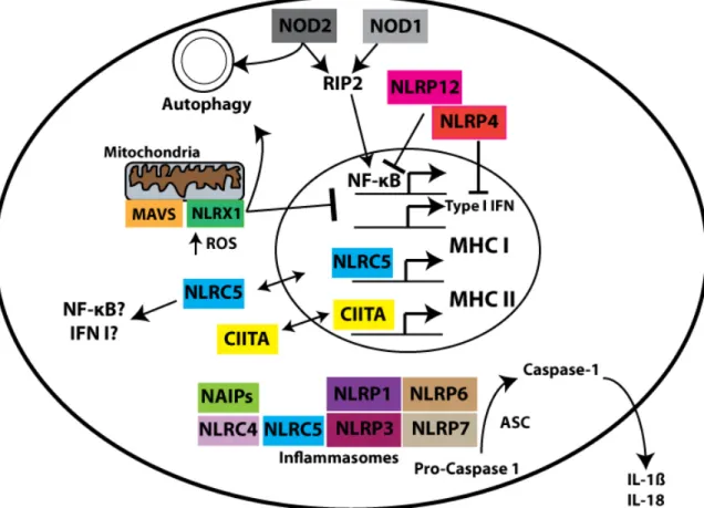
Figure 1-1. NLRs are sensors of the Intracellular Environment. ... 10
Figure 1-2. Generation of Adaptive Immune Classes. ... 14
Figure 1-3. Formation of a Phagocytic Synapse in response to Particulate Ligand. ... 26
Figure 1-4. Schematic of the PRINT fabrication process. ... 29
Figure 2-1. PRINT Particle Characterization. ... 59
Figure 2-2. PRINT particles do not cause inflammation in bone marrow- derived macrophages from BALB/c or C57BL/6 mice. ... 64
Figure 2-3. 80x320nm PLGA particles do not cause lung inflammation in mice. ... 68
Figure 2-4. Hydrogel particles do not cause lung inflammation in mice. ... 72
Figure 2-5. Sustained deposition of hydrogel particles in murine lungs. ... 75
Figure 3-1. PRINT particle characterization. ... 94
Figure 3-2. PRINT particles are non-inflammatory in THP-1 human monocytes. .... 99
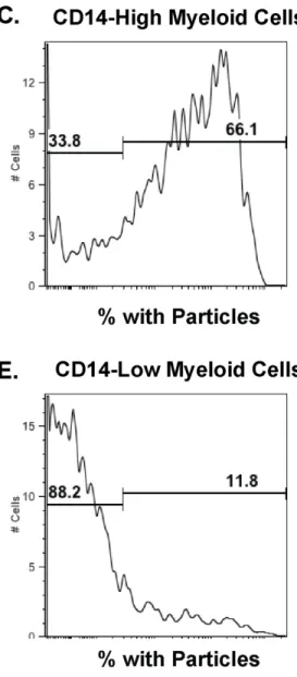
Figure 3-3. 80x320nm PRINT hydrogel particles target myeloid and not lymphocytic human cells. ... 103
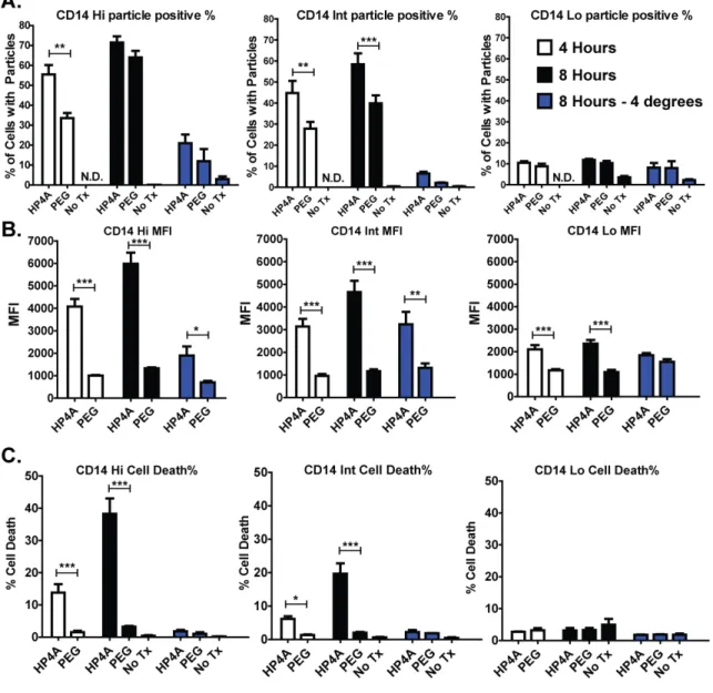
Figure 3-4. Human PBMC differentially process PRINT particles depending on CD14 expression and PEGylation. ... 107
Figure 3-5. Hydrogel nanoparticles do not induce a broad spectrum of cytokines in human PBMC. ... 109
Figure 3-6. Heterogeneity in the Human Immune Response to Nanoparticles. ... 111
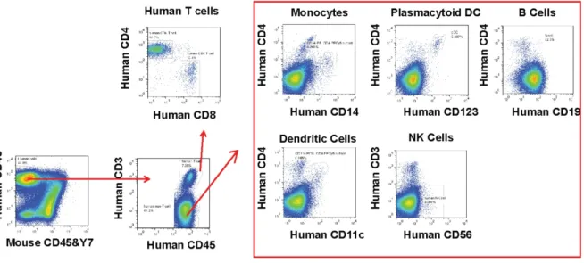
Figure 3-7. Humanized mice reconstitute major types of human immune cells. .... 113
Figure 3-8. Humanized mice recapitulate human PBMC nanoparticle studies. ... 117
Figure 4-1. Design control of PLGA particle uptake in THP-1 cells. ... 137
xiii
Figure 4-3. Design control over the kinetics and magnitude of inflammasome
activation. ... 145 Figure 4-4. Characterization Details for Anti-Inflammatory PRINT particles. ... 149
Figure 4-5. Biodegradable PRINT Particles Complexed with
Natural Ligands are Anti-inflammatory Programming Devices. ... 150
Figure 4-7. OVA Asthma Study Details. ... 155 Figure 4-8. Particulate delivery of OVA reduces allergic responses
to an allergen ... 156 Figure 4-9. Programming Regulatory T cells with
xiv
LIST OF ABBREVIATIONS
ATP – Adenosine triphosphate
BALF – Bronchoalveolar lavage fluid
BMDM – Bone marrow-derived macrophage
CLR – C-type lectin receptor
CNS – Central nervous system
CRD – Carbohydrate recognition domain
DC – Dendritic cell
DHA – Docosahexaenoic acid
DLS – Dynamic light scattering
EAE – Experimental autoimmune encephalomyelitis
EPA – Eicosapentaenoic acid
HP4A – Tetraethylene glycol monoacrylate
HPLC – High performance liquid chromatography
i.p. – Intraperitoneal
i.t. – Intratracheal
xv IL-1β – Interleukin 1β
iPS – Induced pluripotent stem cell
LDH – Lactate dehydrogenase
LPS – Lipopolysaccharide
M0 – Macrophage
MHC – Major histocompatibility complex
MP – Microparticle
MPL – Monophosphoryl Lipid A
MPS – Mononuclear phagocyte system
MSU – Monosodium urate
NCD – Non-communicable disease
NLR – Nod-like Receptor
NMP – Nanoparticles and Microparticles
NP – Nanoparticles
PBMC – Peripheral blood mononuclear cells
PEG – Polyethylene gycol
xvi PFA – Paraformaldehyde
PLGA – Poly(lactic-co-glycolic acid)
PLP – Proteolipid protein
PPS – Preparticle solution
PRINT – Particle Replication in Non-wetting Templates
PRR – Pattern Recognition Receptor
PS – Phosphatidylserine
PVOH – Polyvinyl alcohol
QD – Quantum Dot
RLR – RIG-like Receptor
SCM – Succinimidyl carboxy methyl ester
SEM – Scanning electron microscope
TEA – Triethanolamine
TFF – Tangential filtration flow
TGA – Thermogravimetric analysis
TLR – Toll-like Receptor
CHAPTER 1 INTRODUCTION
This dissertation seeks to define a platform for programming immune responses in order to fundamentally improve how we treat disease in the 21st
century. It is our tenet that therapeutically relevant instruction of the immune system
will require extremely modular and biocompatible systems capable of providing a range of biological signals on the physical scale at which cellular networks
communicate. These nano- and micro-scale devices will thus be capable of
programming host biology in defined manners.
The research presented herein spans the fields of cell signaling biology,
immunology, materials science and bioengineering. To this end, I first provide
background of relevant information for each of these fields to frame this dissertation. Subsequent chapters will expound in more detail work performed and findings
uncovered. While not exhaustive, this introductory overview of immune system function from a cellular network perspective provides the framework from which I
pursued this dissertation. Immune cells precisely sense their environment in order to elicit appropriate responses. These responses are required to defend against infectious diseases but also drive pathology in the setting of non-communicable
2
function in a targeted manner should become a key intervention in the clinician’s arsenal.
1.1 CELLULAR FUNCTION FROM A NETWORK PERSPECTIVE
In order to fully appreciate what is required for a human to maintain health over the course of an average modern lifespan of 67.2 years [3], it is worthwhile to first understand what constitutes a human body at the cellular level. There are trillions of
human cells specialized for their functional purpose, be it detoxifying substances in the liver, perceiving light in the retina of the eye, forming memories in the brain, providing structural support as bone, or patrolling the body for signs of infection to
name but a few examples [4]. Yet all these trillions of cells emerged from a single union between a sperm and an egg, and thus divided and specialized throughout
development to fill all the niches required for a functioning human being. While the details of development and niche specialization are complex, unique and beyond the scope of this dissertation, suffice to say that some simple
repeating themes emerge that we can use to guide our understanding of human biology. Most critical of these themes is the overarching principle that a cell
responds to environmental cues in order to ensure the survival of the organism it constitutes, be it unicellular or a multicellular organism. This response is dictated by signal transduction pathways that first emerged in unicellular organisms responding
to intracellular signals picked up from the environment and then was expanded to extracellular detection in order to support the complexity of higher order multicellular
3
At its most basic, signal transduction requires a cell to sense information encoded in its environment and then relay that information to immediate response
networks such as G-protein coupled receptors as well as the nucleus of the cell, where expression of genes can be modulated in order to better adapt the cell to its current state of experience. A common example is the metabolic response of
humans to glucose, a nutrient taken in from the environment in the form of dietary sugar consumption. Specialized cells in the pancreas - beta cells - bind glucose with
glucose receptors and in response, upregulate expression of insulin and trigger its release into the bloodstream. Systemic insulin can then bind cell-surface bound insulin receptors throughout the body which coordinate uptake of glucose from the
bloodstream for immediate or long-term energy use depending on the needs of the organism [7]. Using this simplified example, we can see all the hallmarks of signal
transduction and network coordination of an appropriate response: 1) sensing of the environment; 2) relaying that information to the nucleus; 3) turning on appropriate gene programs; and 4) production of proteins that enable downstream functions
required for the homeostasis of the organism at large, including secretion of host-derived environmental cues out of a cell.
We must also acknowledge that the human organism is not only integrating signals from its ~10 trillion human cells, but also must maintain order with the
estimated 100 trillion bacterial cells which cohabit each of our bodies [8]. This latter
4
isolated entities in the world at large, but more of an ecosystem of multiple species that shifts depending on the geographic and dietary environment [8-14].
With this basic theme of cellular response to environmental cues in mind, let us now discuss how the human immune system maintains homeostasis of the organism as this information can enable more selective and integrated therapeutic
interventions for the panoply of health ailments that afflict modern man. While we will delve into more explicit details from hereon, it is of utmost importance to recall
the underlying theme that an organism’s survival depends explicitly on its ability to respond appropriately to its environment. As we will see, an appropriate response often requires involvement of numerous signaling pathways and biological networks
within and between cells.
1.2 THE IMMUNE SYSTEM: PROVIDING CONTEXT, BRANDING RESPONSES
While there are thousands of signal transduction pathways in human cells, we will restrict ourselves to discussing some pathways which comprise major network
hubs for function of the immune system. Abundant evidence suggests the immune system has profound effects on mammalian biology and is intimately involved in our
maintenance of health through interactions with all major organ and tissue types [2,15-18]. Thus, clarifying our understanding of signal transduction principles in the immune system will frame our research directions in subsequent chapters.
5
differentiation. This principle task is a hallmark of our evolutionary history which required cells to fend off attack by microbes such as fungi, bacteria and viruses in
order to survive using the innate immune system. As multicellular organisms became entrenched on Earth and expanded in size and complexity, higher order immune systems became necessary, as is evidenced by the development of an
adaptive immune system in jawed vertebrates over 500 million years ago [19]. However, as we have recently come to appreciate, the simple story of the immune
system as defense against infection is only one aspect of the complex role this interconnected network of cells and soluble mediators plays in host homeostasis. To begin to appreciate this complexity, we will briefly outline major players of the
mammalian immune system.
1.3 INNATE IMMUNITY: PROVIDING CONTEXT TO EXPERIENCE
In order to respond appropriately to an environmental encounter, innate immune cells much first identify whether the stimulus is self or non-self. This occurs
through numerous classes of receptors located on the surface and within the cell. While beyond the scope of this dissertation, we briefly mention the critical role of T
cell receptors (TCR) and B cell receptors (BCR) in immune system function as these receptor signaling networks constitute some exciting clinical advances for treatment of cancers and autoimmune disorders as will be highlighted later in this chapter.
6
wall components and viral nucleotides. This occurs through direct binding of these structures or through sensing intracellular responses to microbes, like the production
of metabolites specific to infection with a virus [20,21]. Upon activation of PRRs within a cell, unique gene programs are turned on that result in expression and secretion of soluble mediators that alert other cells to both the presence and quality
of the engaged stimulus [22,23]. These mediators, such as cytokines and
chemokines, drive not only movement of cells into and out of tissues spaces, but
also program stimuli-appropriate responses based on the combination of mediators present.
While many non-immune cells throughout the body have various PRRs,
innate immune cells such as macrophages (M0) and dendritic cells (DC) are two of the primary effectors of innate defense. These innate cells have the capacity to eat
microbes in a process termed phagocytosis that serves two purposes: 1) remove the offending stimuli from the body where it can do more harm, and 2) process the
stimuli so that an adaptive immune response can be elicited against it [24-26]. A key
point is that depending on the context of the stimuli, innate immune cells instruct other cells as to the quality of the stimuli encountered. Thus the innate immune
response to a viral infection is qualitatively different than that to a bacterial infection or encounters with fungi [20,27]. This contextualization of environmental stimuli drives stimuli-appropriate tissue and adaptive immune responses as will be
discussed in more detail shortly. There are a number of other cell types which also play critical roles in innate immunity but we will restrict ourselves to discussing M0
7
nanoparticles and microparticles and thus serve as the frontline for programming immunity in mammals.
Our understanding of innate immunity has come a long way since PRRs were first proposed to exist by Charles Janeway, Jr. in 1989 [28]. Since his prescient prediction of PRRs, we have identified not only multiple classes of PRRs but have
increasingly refined our molecular understanding of these receptor families. There are currently four major classes of PRRs - Toll-like Receptors (TLRs), Nod-like
Receptors (NLRs), C-type Lectin Receptors (CLRs) and RIG-like Helicases (RLHs) – though other individual PRR have recently been identified [29,30].
1.4 THE LESSONS OF TLRS AND NLRS
TLRs are the best studied PRR class and serve as a model for which we
understand the function of other PRRs. Of the 10 TLRs identified in humans, 9 have known functions, with five being cell surface bound and four localized to intracellular compartments termed endosomes [31]. This distribution in various cellular
compartments correlates with function; cell surface TLRs bind microbial motifs present on the surface of microbes whereas endosomal TLRs bind microbial nucleic
acid motifs that are unmasked after successful degradation [32]. Upon binding cognate ligand, TLRs undergo post-translational modifications such as
phosphorylation and relocalization in the cell, thus enabling downstream adaptors to
8
triggering of TLR3 in the endosome by viral double stranded RNA results in activation of the TRIF adaptor and subsequent translocation of two transcription
factors to the nucleus, NF-κB and IRF3, where they drive gene expression of pro -inflammatory cytokines and anti-viral interferons [27]. In summary, the location of the TLR corresponds to where its microbial ligand is perceived and the subsequent
gene programs elicited are appropriate for the microbe encountered.
Whereas TLR signaling pathways have been well characterized, the NLR
family of PRR are far less well known, in part due to their initial identification only in the early 2000s [35]. There are 22 human NLRs which are grouped as a family based on their structural homology, with C-terminal Leucine rich repeat (LRR)
regions, a central nucleotide binding domain (NBD) and a N-terminal effector domain that is thought to confer unique properties among the NLR members [36,37]. As a
beautiful example of convergent evolution, plants defend themselves with the NB-LRR receptor family using very similar protein domain architecture as human NLRs
[38].
Most NLRs are believed to reside in the cellular cytosol, though some
members have been found at the inner leaflet of the plasma membrane (NOD1 and
NOD2) or localized to the outer membrane of the mitochondria (NLRX1) [39-41]. Since their initial discovery, NLRs have been shown to play a broad role in host immunity (Figure 1-1). NLR functions include transcriptional control of MHC class I
and II expression, modulation of autophagy, inhibition and activation of pro-inflammatory pathways including NF-κB-mediated transcription, as well as a
9
activation of the cysteine protease caspase-1 and subsequent maturation of the pro-inflammatory cytokines interleukin-1β (IL-1β) and interleukin-18 (IL-18) [42-45].
NLRs have been implicated in the innate immune responses to fungi, bacteria and viruses, with concomitant effects on ensuing adaptive immune responses [46-56]. Two NLRs, NOD1 and NOD2, have clear roles in the autophagic response to
bacteria and subsequent loading of antigen in MHC class II complexes [40,57,58]. A separate means by which NLRs control adaptive immune responses is through
upregulating expression of MHC I and MHC II genes via NLRC5 and CIITA,
respectively [59-67]. As antigen-specific adaptive immune responses are critically dependent on MHC peptide presentation, these NLR members are thus placed at a
central hub in initiating adaptive immunity.
As discussed shortly in more detail, the quality of an adaptive immune
response is dependent not on just the antigen but also on the context in which that antigen is presented. Context is encoded in part through soluble messengers termed cytokines which greatly augment the quality of adaptive immune responses.
One major type of adaptive immune response involved in defense against fungi and autoimmune pathology, termed Th17, is thought to be at least partially dependent on
activation of inflammasome forming NLR proteins, such as NLRP3. Release of the potent pro-inflammatory cytokine IL-1β from innate immune cells with activated
inflammasomes can initiate Th17 adaptive immune responses. Evidence supporting
this comes from studies in mice deficient in inflammasome components which are more susceptible to fungal infection or protected from Th17-mediated pathology in
10
effects on Th17 immune responses elicited from NLRP3 inflammasome activation, designing vaccines which can trigger this PRR is an avid area of research in the
nanomaterials field and one we will study in more detail in subsequent chapters [71-75].
Figure 1-1. NLRs are sensors of the Intracellular Environment.
While not exhaustive, this schematic overview presents cell locations and functions for NLR family members based on published literature. Of note, NLRs reside
throughout the cell, from the inner leaflet of the plasma membrane (NOD1 and NOD2) to the mitochondria (NLRX1), the cytosol (inflammasome forming NLRs) and
11
Based on work we and others have published, NLRs mediate these wide-ranging immune effects in part through interactions with receptors involved in other
innate immunity signaling pathways, such as the TNF receptor associated factor family (TRAF) [76-79]. Through the network nature of NLR effects on innate
immunity, they have also been implicated in modifying the host microbiota [80-83].
Disease susceptibility to metabolic syndrome and cancer is modified by a number of NLR receptors based on studies using mice deficient in NLR genes [63,80,84-92].
Genome-wide association studies have also uncovered a powerful role for mutations in NLRs in a range of human disease, including bare lymphocyte syndrome, Crohn’s disease, auto-inflammatory disorders such as Muckle-wells and Blau syndrome, and
autoimmune disorders such as vitiligo [37,45,93-104].
We have highlighted the NLR family in detail to serve as an example of how
immune sensors can potently augment human physiology, with either beneficial or detrimental effects depending on the intact nature of these signaling networks. While this dissertation focuses on nanomaterial interactions with mammalian
immunity, it is worthwhile to note that simultaneous research into the basic molecular functions of immune signaling networks will be crucial to broadening and enhancing
the therapeutic potential of immunoengineering. To this end, we note our own research efforts to further molecular understanding of the NLR family via
biochemical studies and bioinformatics approaches that are beyond the scope of this
12
1.5 ADAPTIVE IMMUNITY: BRANDS OF RESPONSE
Perhaps one of the more astounding features of human biology is how unique
immune responses are orchestrated for specific classes of microbes. We have already discussed the role of innate immune cells and PRRs such as TLRs and NLRs in contextualizing environmental encounters but how this context drives
appropriate adaptive immune responses is equally fascinating. At root, an appropriate cellular response to its environment is elicited through networked
orchestration of signal transduction pathways and the immune system is perhaps the clearest example of this concept. Here, we will attempt to synthesize a tremendous amount of immunology research into a few simple concepts reflecting this notion
[2,15,16,18,20,23,24,32,33,56,107-115]:
1) innate immune cells, in particular dendritic cells (DCs), encounter microbes
and process them according to the PRR that are triggered
2) the sum total of PRR signaling pathways triggered within the DC and surrounding tissues will elicit a unique milieu of cytokines and cell surface
receptors that provide context to other cells and regional tissues in order to mount appropriate responses
3) within regional lymph nodes, DCs instruct T cells to mount adaptive
immune responses to specific antigens presented by DC on MHC class I and II receptors to drive either CD8+ or CD4+ T cell effector responses
13
4) the quality and quantity of T cell responses to an antigen depends on the context in which it is presented by a DC, which is encoded in the cytokine
milieu initially triggered by PRRs within the DC and affected tissues
5) these cytokines drive specific T cell gene programs that qualitatively shift the class of effector T cell response to tailor it for the task at hand, in part
through inhibiting production of cytokines associated with inappropriate classes of T cell effector responses
6) in a similar fashion, B cell antibody responses are appropriately tailored through B cell-intrinsic PRR signaling, sampling of antigens, and sensing of the cytokine milieu elicited by innate immune cells and effector T cells
7) in the absence of PRR signaling, anergic or tolerogenic T cell responses are generated, such as would be the case to self-antigens
Over the course of mammalian evolution, the end result of this immune symphony is the generation of general classes of microbe-specific adaptive immune responses as
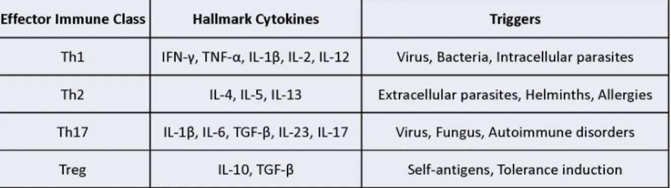
listed in Table 1.1 and visualized in Figure 1-2.
15 1.6 A CALL FOR IMMUNOTHERAPY
With this molecular understanding of innate immunity and the generation of
adaptive immune responses, we have gained unheralded insight into the
pathogenesis of both infectious diseases and those of non-microbial origin (termed non-communicable diseases, NCD). For infectious diseases, this can enable the
production of more efficacious and microbe-specific vaccines. By designing
vaccines that activate innate immune pathways normally elicited during the course of
infection with a pathogen , we can generate adaptive immune responses appropriate to the pathogen we are trying to vaccinate against [18]. For example, we now know that a Th17 response is required to clear infection with the fungus Candida albicans
so efforts are underway to design vaccines which elicit anti-fungal Th17 responses [116-120]. Similarly, using recent gains from immunology research a number of
pathogens with high morbidity and mortality around the world for which there are currently no vaccines are being targeted. These include malaria, Dengue virus, HIV and cholera [121]. As we will discuss, many of these vaccines will rely on
nanomaterials designed specifically to augment appropriate innate immune signaling networks for a given pathogen [74,122-124].
Non-Communicable Diseases – Our Modern Epidemics
A potentially transformative research direction made available through our understanding of immunological networks is the ability to entirely change how we
treat non-communicable diseases (NCD). These illnesses – including heart disease, cancer, chronic obstructive pulmonary disorders and diabetes – have the highest
16
societies [125-127]. It is estimated that 63% of deaths worldwide are due to NCD and the global economic output lost due to NCD over the next 20 years is predicted
to reach over $30 trillion [127]. While NCD have behavioral risk factors that should be first line intervention strategies, specific aberrations in immune function correlated with these diseases are potential therapeutic targets that have yet to be fully
addressed. Immunotherapies for NCD may show better efficacies at lower cost than currently used non-specific treatment approaches.
Cancer is one example of a NCD with immune therapeutic options. Cancer, at its core, is a cell which no longer recognizes its rightful place in the host organism and proliferates without concern for the body by co-opting its microenvironment
[128,129]. It more recently has come to light that this co-opting of the
microenvironment includes broad immune suppression in the tumor area as a
means to keep the body’s own defenses at bay [17]. This occurs in part through modulating the cytokine milieu to promote suppressive polarization of innate immune cells while enhancing regulatory T cell responses to aid the cancer in hiding its true
danger from the body [130-132]. The current standards of care to treat cancer include systemic chemotherapies that kill all rapidly dividing cells, radiation therapy,
and systemic administration of antibodies or small molecules that target potent signaling pathways involved in both cancer and normal cellular function. These arduous and expensive treatment approaches can be debilitating to the patient and
17
An avid area of current research is the concept of modulating the immune environment of cancers to reawaken the body’s own defenses and harness the
inherent power of the immune system to then eradicate the cancer [137]. Multiple approaches are being pursued, with some involving antibody-based activation of cancer-dampened T cells already showing promise in the clinic [136]. Cancer cells
also upregulate CD47, a cell surface receptor that serves as a ‘don’t eat me’ signal to macrophages [138]. Recent evidence suggests that blocking this receptor with
antibodies is sufficient to enable macrophage clearance of cancers in mouse models [139]. The long-sought goal of generating cancer vaccines is also progressing, in part through immunoengineering approaches using nanomaterials as will be
discussed in more detail below. With recent evidence indicating a Th1 immune response induces growth arrest in mouse and human cancers through the cytokines
TNF-α and IFN-γ, targeted modulation of the immune environment could become a
standard of care for cancer treatment [140].
Another NCD with immunotherapy potential that is now a world-wide epidemic
is type 2 diabetes. Unlike type 1 diabetes which is of autoimmune origin (and amenable to immunotherapy), type II is caused by diet-induced metabolic and
immune alterations which render patients insulin-resistant, thus mimicking the pathology of type 1 diabetes [7]. With increasing populations of the world eating high-fat and high-sugar diets, the levels of obesity are reaching staggering
proportions and obesity is a primary risk factor for type 2 diabetes. With obesity prevalence rates at over 35% of US adults and 16% of US youth, the current and
18
[141-143]. While behavioral choices and policy changes to our food system are clearly important interventions that need to be pursued to stem these alarming
obesity rates, augmenting the immune system is a potential therapeutic approach for those patients who have already become diabetic.
As an example of the intimate relationship with immune function and
metabolic homeostasis, it is now clear that Th2 type immune responses - hallmarked by the cytokines, IL-4, IL-5 and IL-13 (Table 1.1) - are critical to the maintenance of
metabolic function in adipose and hepatic tissues and are required for glucose homeostasis. Mice deficient in any of these cytokines are more susceptible to high-fat diet induced insulin resistance and hyperglycemia [144-146]. Conversely,
pro-inflammatory Th1 cytokines like IL-1β are known to cause insulin resistance
[89,147]. From a therapeutic approach, infection with intestinal helminths, which
induce Th2 immune responses, have also been shown to restore normoglycemia and insulin sensitivity in otherwise diabetic mice [145,146].
As stated at the start of this chapter, immune cells precisely sense their
environment in order to elicit appropriate responses. These responses are required to defend against infectious diseases but also drive pathology in the setting of
non-communicable diseases if uncontrolled. Because the vast majority of the world’s population are susceptible to microbial infection or NCD, programming immune system function in a targeted manner should become a key intervention in the
clinician’s arsenal. To that end, we now turn to a discussion of our current state of knowledge regarding nanomaterial interaction with mammalian biology and efforts
19
1.7 NANOMATERIAL INTERACTIONS WITH BIOLOGICAL SYSTEMS As the thrust of this dissertation is the analysis of the innate immune
response to nano- and micro-scale particles, it is of use to first provide a background on the current state of our understanding as regards biocompatibility with materials in these size ranges. The overriding supposition is that by first understanding how
size, shape and composition of particle parameters affect immune outcomes, we can then more appropriately design particles for a range of therapeutic interventions, in
particular those which aim to modulate the host immune response in defined manners. Herein, we use the term nanoparticle (NP) to refer to particles that are less than 1 micron in all dimensions and microparticles (MP) for those that are
greater than 1 micron in any single dimension. When discussing both nanoparticles and microparticles as a generalized group, we will refer to them as NMP.
Historically, studies of nanomaterial interactions with mammalian biology have occurred with no real field standards but recent efforts have been made to establish general assays for toxicology and immunological properties of
nanomaterials [148]. This is in part due to the complexity of carrying out controlled experiments to delineate differences between nanomaterials based on fine
alterations of size, surface charge or composition, all of which clearly impact the mammalian immune response [148-150]. As an example, by altering the size, surface charge, surface hydrophobicity or surface-exposed functional groups, the
quantity and quality of proteins adsorbed to NMP can be greatly affected [151-153]. Yet to give a sense of the disparity between in vitro nanoparticle studies and in vivo
20
during in vitro analysis likely have little functional relevance to what occurs during in vivo experiments [154].
A relevant example for translational applications is the use of targeting NP to specific cell subsets by decorating antibodies for specific receptors on the surface of NP. One group recently found that NP targeting specificity to the transferrin
receptor, which is upregulated in some cancers, was completely abrogated when switching in vitro assays from phosphate buffered saline media to one that was
serum replete to more closely resemble host biology [155]. Remarkably, many NP studies do not use serum-containing media so this seemingly predictable result not only calls in to question numerous previously published findings but also serves as a
wake-up call to the nanomaterials community at large as regards the translational relevance of experimental design. This example also serves to bolster the position
we took during the NMP studies detailed in this dissertation: well-controlled, biologically relevant studies are needed to characterize nanomaterial interactions
with mammalian immune system.
Admittedly, a difficulty in drawing broad conclusions regarding how particle design features such as size, shape, composition and surface charge affect host
biology is due to major differences in NMP fabrication techniques, materials sourcing and experimental assays within and between labs [148,156]. Common NMP
fabrication techniques include water-oil-water emulsion or spray-drying methods, yet
particles fabricated in this manner can range in size by many hundreds of
21
Thus careful analysis of the role of size or shape on biological responses to nanomaterials using standard fabrication methods is difficult at best.
Composition Considerations of NMP – The Inflammasome Example
A remarkable example of the sensitivity of innate immunity to nanoscale
details is how modifying the surface and composition of NMP can modulate
activation of the NLRP3 inflammasome. As a reminder, the NLRP3 inflammasome is a multi-molecular protein complex that is formed in response to a variety of
environmental triggers, ultimately resulting in the activation and secretion of pro-inflammatory cytokines, IL-1β and IL-18 [29,159]. Because particulate matter such as asbestos, silica and monosodium urate crystals are known to activate the NLRP3
inflammasome, it is expected that NMP may also trigger this innate immune response. There are many uses for NMP where it would be disadvantageous and
clinically unacceptable to use particles that induced inflammation, such as diagnostic and drug-delivery applications. Conversely, activating the NLRP3 inflammasome by NMP is a potential vaccine adjuvant and thus the design principles controlling this
response is an avid area of study. Through the work of a number of groups, we are starting to gain insight into how NMP can activate the NLRP3 inflammasome.
Work by the Tschopp group identified composition of metallic nanoparticles as the prime factor in triggering IL-1β release. Titanium dioxide and silica dioxide
NP caused inflammasome activation while comparable sized zinc oxide particles did
22
inflammasome activation and associated inflammatory signaling changes induced by these particles was inhibited simply through modifying the chemical groups exposed
on the surface of particles [160-162].
While these findings have relevance to consumer products and environmental toxiciology, the literature regarding inflammasome activation by NMP composed of
polymers more relevant to biomedicine applications is inconsistent. Perhaps the two most commonly used polymers for biomedical applications are
poly(lactide-co-glycolide (PLGA) and polyethylene glycol (PEG). These biocompatible polymers are approved by the Food and Drug Administration (FDA) and have a long, safe history of clinical use [163,164]. Yet when the biodegradable polymer PLGA is formulated
into NMP, groups report differences in its inflammasome activating potential [71]. One report shows in vitro and in vivo evidence for inflammasome activation by PLGA
NMP that is contradicted by another study [72,73]. However, recent evidence suggests these literature discrepancies could be due to fabrication differences among groups, as simply a non-smooth NMP surface is sufficient to induce
inflammasome activation by particulate matter [165]. We have studied the issue of inflammasome activation by NMP at length, as will be discussed in subsequent
chapters. Because composition is so clearly a factor in NMP interactions with human biology, we focused our studies on PLGA and PEG polymers as they have
23
Physical Parameter Considerations of Particle Uptake
Though particle parameters such as size, shape and composition do impact
host biology, field-wide conclusions regarding any one design feature is currently not possible. For example, while surface charge has been associated with cell uptake, if one modulates the size of the particle then conclusions regarding surface charge
and uptake are not necessarily consistent [166-168]. Similarly, the mechanism by which cells uptake NMP is dependent on a variety of features, including size, shape
and surface charge. Whereas mannose-coated NP are taken up by macrophages through mannose-receptor dependent phagocytosis [169-171], lipid-coated NP can be taken in via complement-receptor dependent pathways [172]. Iron oxide NP
uptake is scavenger-receptor dependent [173]. From an immunological perspective, these are critical design parameters as the phagocytic uptake pathway can have
downstream pro- or anti-inflammatory effects [26]. Multiple other examples of the inconsistent nature of how design parameters augment the biological fate of NMP abound, serving ultimately to highlight the absolute sensitivity of mammalian biology
to just about every particle feature one chooses to test.
Separate from the specific phagocytic pathway used to ingest particles, the
actual shape of the NMP plays a critical role in the initiation and successful
completion of phagocytosis. A seminal study by the Mitragotri group identified the contact angle by which a macrophage encounters a particle as a critical parameter
dictating successful phagocytosis of the NMP [174]. If the contact angle was greater than 45 degrees, then particle phagocytosis was unlikely to occur, likely due to
24
widest dimension (>500nm). The Mitragotri group has used this finding to design high aspect ratio NMP with worm like shapes to inhibit phagocytosis by restricting
uptake to only those phagocytes which happen to encounter particles at the tip (where the contact angle <45 degrees) [175]. Such shape design considerations may be of use for therapeutic modalities requiring enhanced circulation times and/or
extracellular release of cargo.
More recently, these researchers have identified shape as critically
augmenting the targeting specificity of antibody-coated NMP through harnessing this initial contact angle finding. By coating rod, spherous and disc shaped NMP with trastuzamab (a monoclonal antibody against HER2/neu receptors expressed by
breast cancer tissue), the authors found rod-shaped NMP reduced non-specific cell uptake and reduced breast cancer cell line growth more efficiently than an
equivalent dose of soluble trastuzamab [176]. These exciting findings point to an emerging theme in NMP studies whereby particularization of biological molecules (drugs, antibodies, ligands) confers unique properties not found with equivalent
soluble forms of the same molecule, as will be discussed in greater experimental detail in Chapter 4.
One plausible explanation for why NMP are capable of eliciting emergent properties from cells when coated with bioactive molecules may be due to formation of a phagocytic synapse that macrophages and dendritic cells use to sense intact
microbes [1]. This has formally been proven by the Underhill group using both soluble and particulate forms of the fungal cell wall component, β-glucan, which is
25
family of PRR. Although both soluble and particulate forms of β-glucan are bound by Dectin-1, signaling is not initiated from the Dectin-1 receptor unless β-glucan is
present in a particulate form (Figure 1-3). This causes receptor clustering of Dectin-1 and formation of a ‘phagocytic synapse’ that is able to physically exclude
regulatory tyrosine phosphatases CD45 and CD148 from the site of phagocytosis
[177]. In effect, immune signaling is not initiated from Dectin-1 unless an intact fungus (i.e., particulate form) is sensed. This clever biomechanical control enables
innate immune cells to restrain initiation of potent anti-fungal responses to only those instances when an actual fungal microbe is encountered, thus sparing spurious innate responses that may otherwise be detrimental to the host. It is thus
conceivable that coating of NMP with biologically active ligands, in particular antibodies or ligands which bind surface receptors, will result in emergent cellular
responses through receptor clustering that soluble forms fail to elicit. We present evidence supporting this hypothesis in Chapter 4. Germane to this issue, there is abundant literature reflecting the role of surface-ligand density in downstream
26
Figure 1-3. Formation of a Phagocytic Synapse in response to Particulate Ligand.
27
The surface chemistry of nanomaterials used in NMP can also significantly impact the immune response to these particles. It has been shown that certain
primary hydroxyl groups or other surface nucleophiles on nanomaterials can react with complement factor C3 to form the active C3b product, thus triggering potent complement-mediated effects on innate and adaptive immunity [182-185]. As
discussed previously, inflammasome activation by metallic NP depends on the metal used, whereas silica nanoparticle activation of the inflammasome can be augmented
through modification of surface chemical groups [75,186,187]. In total, these studies highlight the need to address nanomaterial interactions with the immune system for every given particle size and composition, as the only thing we can predict is that the
body is clearly capable of discriminating among minute details.
Help Wanted: Control of Particle Design
With this nanomaterial background in mind, it should be evident that a
process to reproducibly fabricate particles with complete control over all physical and chemical parameters would be a great boon to the NMP research community. In
2005, such a fabrication method was developed in the lab of Joseph DeSimone, Ph.D., by fusing the precision and engineering control offered by lithographic
techniques from the microelectronic industry with state-of-the-art synthetic chemistry in a modular platform. The process – termed Particle Replication in Non-wetting Templates (PRINT) – is a top-down particle molding technology with unmatched
28
compounds, small molecules, proteins, siRNA, and bioabsorbable polymers containing therapeutics or diagnostic cargos [188-194].
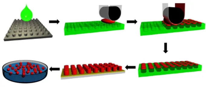
An easy to grasp analogy is to consider PRINT molds as ice-trays with
cubicles of defined size and shape that can be filled with solutions of one’s choosing. After PRINTing, the particles (ice cubes) can be popped out of the tray, resulting in a
homogenous population of monodisperse particles of defined chemical composition (Figure 1-4). Using PRINT, well-controlled experiments isolating the effects of
29
Figure 1-4. Schematic of the PRINT fabrication process.
Master templates (grey) are fabricated using advanced lithographic techniques to defined size and shape parameters. A liquid fluoropolymer (green) is poured on the
surface of the master template and photochemically cross linked to generate a precise mold having micro- or nanoscale cavities (green). A liquid solution
comprised of user-specified components (red) fills the cavities in the mold through capillary forces. Once the liquid in the mold cavities is converted to a solid, the array of particles (red) are removed from the mold (green) through contact with a
harvesting film (yellow). Free flowing particles or stable dispersions can be obtained by separating the harvesting film from the particles, often with water, thus enabling
30
To summarize this section, biological interactions with NMP fundamentally depend on the physical and chemical parameters of particles, though there are no
overarching conclusions that can be drawn as regards all NMP for a given parameter. This is perhaps unsurprising given that the body does not process information in discreet groups but instead tailors an appropriate response to the
individual stimulus, as discussed earlier in this chapter. Just as the body responds uniquely to various types of bacteria despite similarities in size, shape and genus, it
follows that the response to NMP will also be context dependent. For these reasons, a bulk of the work presented in chapters 2 and 3 of this dissertation pertains to careful analysis of the mammalian innate immune response to NMP
through controlled experiments using PRINT particles of strictly defined size, shape and composition. These fundamental studies are necessary in order to enable the
rational design of NMP therapies for a range of health ailments that afflict 21st
century society. As evidence of the potential for NMP to fundamentally change how diseases are treated through modulation of the immune system, an overview of
immunoengineering approaches using NMP will now be discussed.
1.8 IMMUNOENGINEERING: WHERE WE ARE
The basic principle of immunoengineering is to design NMP that deliver biological signals in a targeted manner in order to program immune responses. This
is commonly done through incorporating biological ligands and receptors into or on the surface of NMP that mimic endogenous immune cell interactions. The
31
through engineering approaches is rapidly approaching parity with the actual variety of immune interactions we know to occur in situ. This is in part due to the broad
range of commercially-available immune active molecules, such as cytokines and antibodies to immune receptors, which enable modular approaches to programming immunity. We now present a survey of published immunoengineering studies which
reflect the potential of this approach to treating human disease.
NMP have been designed to release cytokines in order to elicit specific
immune outcomes. Modeling NMP after mast cell granules, the Abraham group complexed an immunostimulatory cytokine, TNF-α, into synthetic granules
composed of heparin and chitosan in order to boost and direct the quality of
immune responses [195]. These synthetic granules drained to the lymph node of mice and were able to enhance adaptive immune responses and survival to
influenza more efficiently than soluble delivery of TNF-α. Furthermore, the group
showed that NMP delivery of IL-12 was capable of skewing the adaptive immune responses towards the Th1 class. This simple yet powerful approach indicates
artificial skewing of immune environments is feasible. Though the broad therapeutic potential of this approach remains largely unexplored, evidence suggests
appropriately designed NMP can skew adaptive immune responses between classes (i.e., Th2 -> Th1, Th17->Treg) [196,197].
In an in vitro system, the Fahmy group generated artificial antigen presenting
cells (aAPC) by anchoring peptide-MHC complexes and co-stimulatory ligands to the surface of MNP composed of the biodegradable polymer poly(lactide-co-glycolide
IL-32
2 and incubated in culture with T cells to assess their potential to act as dendritic cells (DC). The results show a size dependent activation of T cells, whereby
microparticles 8µm in diameter were more efficient at stimulating proliferation of T cells than NP with 130nm diameter. This was not due to differences in total amount of ligands or receptors present. The study also found that particle encapsulated IL-2
was 10x more efficient than soluble IL-2 in terms of T cell proliferation, suggesting high localized IL-2 concentrations between the aAPC and T cell mediated more
robust signaling akin to that which would occur between DC and T cells. This artificial system has relevance to ex vivo manipulation of T cells for clinical
interventions, but also reveals how scaling biological interactions to the appropriate
dimension (i.e., nano- and micrometer) confers broad enhancement of biological outcomes.
Next-Generation Vaccines
Perhaps the strongest illustration of the promise for immunoengineering with NMP is progress in vaccine development. This is ironic, given that vaccination is
one of the most successful and oldest examples of engineering immunity in human society. Prior to the advent of vaccination in the late 18th century, the average
lifespan was 35 years of age. It is now approaching 80 years, in large part due to the success of worldwide vaccination campaigns [121]. With recent elucidation of the molecular mechanisms behind the efficacy of vaccines, researchers are now
33
possible in large part through our understanding of innate immune activation by microbes through specific pattern recognition receptors (PRR).
By coupling PRR activation to delivery of an antigen, antigen-specific adaptive immune responses can be generated with control over the quality of the adaptive response through judicious design of NMP [74,201]. As many ligands for
PRRs are now known and commercially available, immunoengineers are rapidly incorporating these into NMP along with antigens of interest to improve the next
generation of vaccines. PRR ligands published with NMP include those for the majority of TLRs, as well as those which activate CLRs and the NLRs NOD1 and NOD2 [120,202-207]. The power of NMP from a vaccine perspective lies not only in
the modularity afforded through selective or combinatorial PRR agonist inclusion, but also the ability to co-deliver innate immune agonists along with the antigen one
desires an adaptive immune response towards. This mimics the spatiotemporal immune activation that occurs during natural infections and couples innate immune activation to a specific antigen of interest. In point of fact, NMP delivery of TLR
agonists to mouse and human DC has been shown to be 100 times more effective than soluble delivery [206].
The Pulendran group recently used PLGA NP containing TLR4 and TLR7/8 agonists to show a profound synergistic enhancement of antibody and cell-mediated adaptive responses to co-delivered antigens in both mice and non-human primates.
Combinatorial activation of these TLRs through NP delivery elicited antibody responses that were orders of magnitude greater than individual TLR ligand
34
line with others who have seen a profound enhancement in adaptive immune
responses to synergistic TLR agonism [200,205,207]. An as yet unexplored area is
the use of PRR ligands from multiple classes of PRR families for use in vaccines, NMP or otherwise. It is our belief that combinatorial activation of multiple PRR classes, such as TLRs, NLRs and RIG-I-Lie receptors (RLRs), will likely result in
even more profound enhancement of vaccine-induced immune responses as this can recapitulate the natural immune response to microbial infections which
themselves trigger multiple PRR classes.
Design Control of Nanomaterials
A great strength of NMP approaches to vaccine delivery is the control over
numerous biological parameters that can be designed into NMP and tailored to the clinical need at hand. For example, production of CD4+ T cell responses is required
for potent humoral immunity while anti-viral responses require CD8+ T cell effector responses. These two responses depend on presentation of antigen through either MHC II (CD4+) or MHC I (CD8+), and the pathway to presentation is different
between the two [208-210]. Class II peptides are acquired from the extracellular space as intact antigen and processed within the acidic environment of phagosomes
into peptides that are then presented on MHC II receptors. Conversely, Class I peptides are generally acquired from the cytosol of cells. Thus, NMP vaccines with the goal of eliciting antigen-specific CD8+ T cell responses, such as those against
35
Here, the materials science component of NMP design comes to play, with generation of polymers which are biologically responsive and thus can ensure
cargoes within NMP are delivered to the cytosol. One approach is the use of mulit-lamellar liposomal NP. These are highly sensitive to phagosomal phospholipases and thus rapidly degrade after endocytosis, releasing cargo (antigen) into the cytosol
and potently enhancing CD8+ adaptive immune response [211]. An alternative approach for ensuring cytosolic delivery of NMP cargo makes use of the pH drop as
phagosomes mature. In this case, NMP are made with pH-sensitive polymers such as polypropyl acrylic acid that display membranolytic activity during acidification of the phagosome. This disrupts the phagosome, releasing the NMP into the cytosol
where MHC I presentation of antigenic cargo can occur to drive potent CD8+ T cell responses [212].
A key requirement for the success of immunoengineering approaches is the ability to target specific cells or regions of the body with NMP . Because regional lymph nodes are the location of adaptive immune response generation, ensuring
NMP can get to these areas is crucial. NMP design affords a variety of approaches to this issue. Through size selection (500-2000nm diameter), one can passively
target antigen presenting cells, such as macrophages and dendritic cells, in the periphery where they then can traffic to regional lymphnodes. Conversely, smaller particles (<100nm) will drain to regional lymph nodes on their own, where they can
then be processed by resident immune cells [213]. For particles in the 200-500nm range, it is less clear whether they drain to lymph nodes or are passively trafficked
36
NMP can also be directly targeted to certain immune cell subsets through the use of surface conjugated ligands and antibodies. DEC-205 is a cell surface
receptor on a subclass of DC (CD8+) that are especially suited to priming CD8+ T cell responses. Thus, decorating MNP with antibodies to DEC-205 has been used to specifically target particles to this immune cell subset [215,216]. Interestingly, one
study found that varying the surface density of DEC-205 antibody on NP could modulate the ensuing innate immune response from targeted DC, with increasing
antibody density correlating with heightened production of the anti-inflammatory cytokine IL-10 due to enhanced cross-linking of the DEC-205 receptor [178].
Other studies targeting human DC through the DC-SIGN receptor showed
that both the distance between the conjugated antibody and NMP surface and the size of the particle were critical parameters in ensuring targeted cell-specific uptake
[217,218]. As evidence of how carefully coordinated the immune response is and how NMP design can both probe these basic mechanisms while offering
therapeutically relevant tools, the Figdor group assessed uptake and antigen
processing of NP targeted to either the carbohydrate recognition domain (CRD) or the neck region of the DC-SIGN receptor [219]. They found that NP which targeted
the neck region of DC-SIGN were taken up through a clathrin-independent mechanism that led to prolonged retention in endosomes and enhanced MHC I presentation of antigen as compared to the CRD-targeted NP. The
cross-presentation of antigen using this strategy was 1000 times more effective than soluble antigen alone, again pointing to the power of biological scale imparted by
37
Whereas most current vaccines are delivered via intramuscular or
subcutaneous injections, NMP afford a variety of alternative delivery routes, such as
inhalation, oral and transdermal microneedle. Studies addressing route of NMP delivery have seen an impact on the quality of immune responses, with Th1 response in particular being more sensitive to delivery route than Th2 responses
[220]. Importantly, it is increasingly appreciated that tissue-specific immune
responses are critical to controlling local infections [16]. This is perhaps best seen
as regards immunity of mucosal organs, such as the lungs and digestive tract. As the most common site of entry for infection, eliciting mucosal immunity via
vaccination is a critical parameter to success [221-223].
A powerful example of mucosal targeting is the induction of antigen-specific CD8+ T cells and production of a Th17 immune environment in the lungs following
intranasal vaccination with a NP designed to target DC and release antigen into the cytosol of these cells [224]. Such responses are crucial to protecting against
respiratory infections with bacteria and viruses, such as influenza. The paradigm of
inducing mucosal immunity also holds true for targeting mucosal tumors, whereby vaccination through the intranasal route is able to elicit anti-tumor CD8+ T cell
responses while intramuscular vaccination fails to do so in orthotopic head, neck and lung cancers derived from the human papillomavirus 16 (HPV16) E7–expressing
38 Cancer Immunotherapy
In addition to the use of NMP for microbial vaccination, immunoengineering
with NMP offers entirely new approaches to cancer treatment while avoiding the systemic toxicity associated with soluble delivery of cytokines or antibodies [136]. Similar to vaccines against microbes, a number of research groups are developing
NMP-based cancer vaccines which incorporate PRR agonists and antigens
associated with specific cancers [137,225-228]. While these approaches do show
promise, a difficulty will be identifying cancer-specific antigens that don’t result in autoimmune pathology in healthy tissues. Thus, modulating the immune
environment of cancer is a potentially more viable option.
As discussed earlier, cancers co-opt their microenvironment in order to hide from the immune system and divert nutrients towards their growth [130]. Therefore,
some nascent anti-cancer NMP approaches involve blocking the mechanisms by which cancer quells immune responses in the tumor microenvironment. One example is through inhibiting the function of STAT3, a transcription factor
upregulated in tumors that dampens production of Th1 cytokines while increasing secretion of immunosuppressive factors [229,230]. Designing NPs to carry siRNA
against STAT3 has shown promise in shifting the immunosuppressive tumor environment towards one that enables immune surveillance and tumor clearance [231,232]. Another approach coupled NP-mediated delivery of IL-2, a T-cell
activating cytokine, with a small molecule inhibitor of the immunosuppressive cytokine TGF-β. This multi-pathway approach inhibited tumor growth, provided
39
molecules, as either compound alone or delivered in soluble fashion failed to protect mice [233]. This reflects a growing understanding in the cancer field that targeting
multiple pathways at once will be required to avoid tumor resistance [234]. NMP are well suited to deliver multiple cargoes in this regard.
Due to clearance by innate immune cells as well as vasculature abnormalities
in the tumor bed, it can be difficult to ensure NMP reach cancer regions at sufficient levels to exert therapeutic benefit in vivo [235]. Thus, modification of immune cells ex vivo prior to implantation back into the host is a ‘trojan-horse’ approach that relies
on the intrinsic ability of immune cells to navigate to tumor sites in vivo. A creative use of this concept was reported by the Irvine group where they conjugated NP to
the surface of T cells ex vivo using free thiol groups present on the surface of T cell membranes. By loading these NP with IL-15 and IL-21, potent T cell activating
cytokines, these NP could in effect serve as paracrine signaling hitchhikers on T cells and greatly enhanced the T cell tumor killing capabilities in vivo [236].
Hitchhiking Particles and Inflammation
The ‘hitchhike’ tactic has been used by other groups in non-cancer
applications to deliver NMP to sites usually occluded from particle transport. For
example, by feeding macrophages NP with antiviral cargo, researchers have enabled delivery of anti-virals to the brain after these NP-laden macrophages navigated through the blood-brain barrier [237,238]. Access to the central nervous
40
therapeutic delivery of drugs or immune-modulators to this region have a tremendous amount of clinical potential [239,240].
Inflammation itself is a driver of numerous pathologies, wreaking havoc in the CNS after stroke for example, so approaches to limit inflammation are promising areas for NMP [34,96,241]. While encapsulating anti-inflammatory small molecules
like cyclosporine or glucocorticoids in NP is an effective means of dampening inflammation, more recent efforts make use of immune signaling pathways to
broaden the impact of NMP interventions [242,243]. One means of targeting inflammation is the development of reactive oxygen species-sensitive (ROS) polymers [244]. ROS production is upregulated by cells during inflammation
associated with cancer and infection, thus NMP designed with ROS-sensitive polymers may selectively degrade in targeted areas of inflammation and release
therapeutic payloads only in these sites.
A conceptually simple yet elegant approach to reduce inflammatory pathology is the passive targeting of monocytes with therapeutic NP that inhibit monocyte
recruitment to sites of inflammation where they perpetuate and worsen disease. This can be done through encapsulating siRNA to CCR2 in NP, as CCR2 is a cell
surface chemokine receptor required for recruitment of inflammatory monocytes. NP-delivery to these cells reduces upregulation of CCR2 and inhibits their ability to respond to inflammatory alarm signals because they are not able to sense it from
their environment in the absence of CCR2. In mouse studies, this approach attenuates the number of inflammatory monocytes in atherosclerotic plaques,