THE FORMATION AND FUNCTION OF THE HISTONE LOCUS BODY IN HISTONE mRNA BIOGENESIS
Kaitlin Pauline Koreski
A dissertation submitted to the faculty of the University of North Carolina at Chapel Hill in partial fulfillment of the requirements for the degree of Doctor of Philosophy in the Department
of Biology
Chapel Hill 2019
Approved by: Bob Duronio Bill Marzluff Jeff Sekelsky Brian Strahl
© 2019
ABSTRACT
Kaitlin Pauline Koreski: The formation and function of the Histone Locus Body in histone mRNA biogenesis
(Under the direction of Robert J. Duronio and William F. Marzluff)
The genome exerts spatial and temporal control of gene expression through the
compartmentalization of nuclear space into specialized substructures known as nuclear bodies (NBs). NBs are defined by light microscopy as the concentration of factors involved in specific biological reactions. In concentrating reaction factors and substrates in a distinct
of multivalent interactions in the formation of the HLB. This work provides insights into how the HLB forms and how this formation is related to the HLBs role in coordinating the steps in
ACKNOWLEDGEMENTS
I am incredibly thankful for everyone that has encourage and supported me on the path to this degree. Thank you will never be enough. I would first like to thank my mentors, Bill
Marzluff and Bob Duronio. By letting me be a member of both of their labs, I have had the amazing opportunity to work with two great men and become more knowledgeable and skilled in complementary areas of biology. They have let me explore my interests while keeping me from getting too far off the rails. More importantly, they have let me make mistakes, but they guided me in how I could fix them. They have helped me become a more confident scientist, a skill that is hard to learn. Bob, no one has ever challenged me the way you do, and I appreciate every moment of that. You have made me a stronger scientist. Bill, your knowledge of, everything, never ceases to amaze me and I have gained so much by just being around you. Thank you for always giving encouraging words when things got difficult with projects. I can honestly say that I do not know where I would be without your guidance, then and now.
Thank you to both the Marzluff and Duronio labs. My interaction with all of you over the years have helped make me into the scientist I am. You have supported me when graduate just got too hard.
PREFACE
Chapter 2: Histone locus regulation by the drosophila dosage compensation adaptor protein clamp. Leila E. Rieder*, Kaitlin P. Koreski*, et al. (2017). Genes Dev. Jul 15;31(14):1494-1508 *These authors contributed equally.
Most of this work in this chapter is a previously published research article(Rieder et al. 2017). This work was done with Dr. Leila Rieder and Dr. Erica Larschan who made the initial discovery of CLAMP at the histone locus and its localization to the HLB. All the
genomic/sequencing work was done by the Larschan lab in addition to the CLAMP mutants and embryo staining. I had previous shown (McKay et al. 2015), that HWT transgenes formed an HLB similar to the endogenous genes, with the exception of Mute (Fig. 2.1). This provided the basis of creating the GAGA mutant transgenes as we could assay their ability to form an HLB. These transgenes, and the deletion analysis in Supplemental 1 3, were created by a former
postdoc associate in the Duronio Lab Dr. Kara Boltz. I assisted her in making this histone array. I created the HWT transgene used in the paper and performed the retargeting experiments,
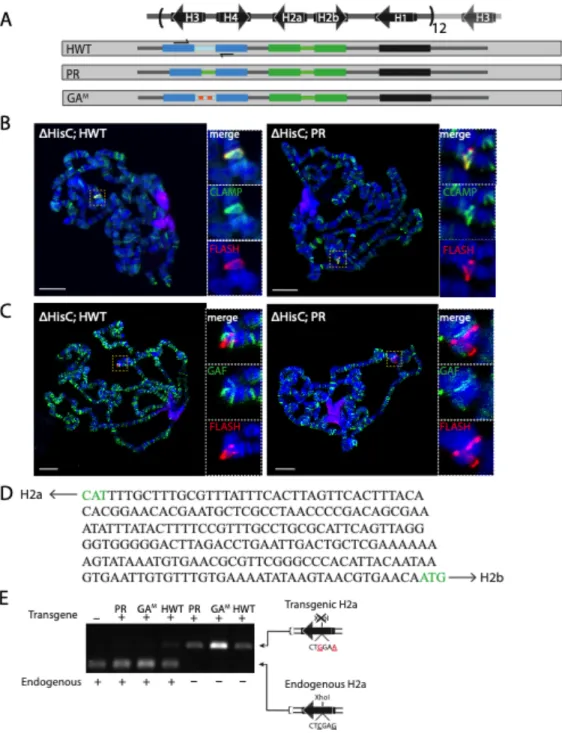
Figure 2.1
Figure 2.1. The transgenic histone gene locus assembles an HLB that accurately processes histone transcripts
. (B) Confocal images of blastoderm stage embryos stained for FLASH (green), Mxc (red), and Lamin (magenta) for the two indicated genotypes. (D) Confocal images of embryos at 2-4hrs, 4-6hrs, and 6-8hrs stained for FLASH (green), Mute (red) and Lamin (magenta) for wild type and 24× Rescue embryos. For B and D, the maximum projection of four 0.5-micron slices is shown. McKay, D. J., S. Klusza, T. J. Penke, M. P. Meers, K. P. Curry, S. L. McDaniel, P. Y. Malek, S. W. Cooper, D. C. Tatomer, J. D. Lieb, B. D. Strahl, R. J. Duronio and A. G. Matera (2015). "Interrogating the function of metazoan histones using engineered gene clusters." Dev Cell 32(3): 373-386
Chapter 3: Histone Locus Body Formation: Different ways to a common end.
the experiments with the exception of a few panels in Fig. 3.5C, of which my undergraduate, John Atwater, took a few images. My undergraduate, Lyndsey McLain, screened for transgenic fly lines and identified all 3 (HWT, PR, PR*) used in the study. I wrote the first draft of the manuscript which was then edited by Dr. Marzluff and later by Dr. Duronio.
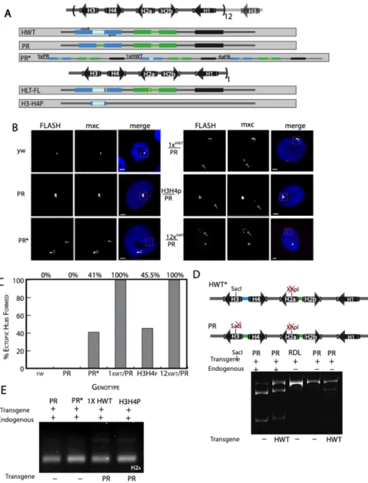
Figure 3.1
Figure 3.1 For this study I created a designer histone locus. This can be used to ask many questions regarding the regulation of histone gene expression, processing, and more. We designed it with unique restriction sites after each stop codon(red), after each histone
TABLE OF CONTENTS
LIST OF FIGURES ... xi
LIST OF ABBREVIATIONS ... xii
CHAPTER 1. INTRODUCTION ...1
Overview ...1
Nuclear Body Assembly ...2
Self-organization ...3
Seed and Grow ...4
Phase-separation ...5
Nuclear Body Function ...8
Histone Locus Body ...10
Histone mRNAs and Components ...10
Histone Locus Body (HLB) Assembly ...13
Dissertation Goals ...17
2. HISTONE LOCUS REGULATION BY THE DROSOPHILA DOSAGE COMPENSATION ADAPTOR PROTEIN CLAMP ...19
Introduction ...19
Drosophila strains ...23
Promoter Alignment ...24
FISH and immunofluorescence ...24
Western blotting ...25
MNase-seq ...26
Quantitative real-time PCR ...27
Embryo mRNA-seq ...27
Staged embryo ChIP-seq ...28
Analysis of H2a expression from the ectopic array ...28
Results ...29
CLAMP is recruited to the histone locus via the H3– H4p ...29
Conserved GA repeat cis elements in the H3–H4p are required for ectopic HLB formation ...32
CLAMP promotes recruitment of HLB components to an ectopic histone locus ...36
CLAMP regulates histone locus chromatin and histone gene expression ...38
CLAMP is specifically retained at the HLB in clamp nulls ...43
GAF localizes to the HLB when CLAMP is depleted ...44
Discussion ...46
Regulation of the histone locus ...47
A single transcription factor can mediate multiple
domains of coordinated gene activation ...49
The relationship between GA-binding factors at the histone locus ...51
Acknowledgements ...52
3. CHAPTER 3: HISTONE LOCUS BODY FORMATION: DIFFERENT WAYS TO A COMMON END ...54
Introduction ...54
Materials and methods ...57
Culture conditions and fly strains ...57
Locus Building ...57
Northern Analysis ...57
Histone Expression Analysis ...58
Immunofluorescence ...58
FISH-IF ...58
Results ...59
The H3-H4 promoter stimulates HLB formation ...60
A histone gene array lacking the H3-H4 promoter forms HLBs and is expressed in the absence of the endogenous genes. ...62
HLB assembly at the PR locus occurs at the normal time during embryogenesis ...67
Histone mRNA from the 12xPR transgenic locus is properly processed ...69
A WT-array can activate the 12xPR array when present in trans at the homologous locus ...69
4. CHAPTER 4: DISCUSSION ...77
The GAGA sequences in the H3/H4 promoter that triggers HLB assembly and biogenesis of histone mRNA. ...78
Things to Test ...79
Assembly of the HLB-looking for a "seed" ...81
Robustness of the PR HLB ...84
The HLB is built on DNA ...84
APPENDIX 1 ...87
APPENDIX 2 ...88
LIST OF FIGURES
Figure 1.1 Phase-separation schematic ...6
Figure 1.2 Drosophila Replication-Dependent Histone Gene Locus ...10
Figure 2.2 CLAMP localizes with makers of the HLB ...31
Figure 2.3 Localization of CLAMP at the HLB is conserved across drosophilids ...33
Figure 2.4 The GA repeats in the H3-H4p are required for HLB formation, and retargeting CLAMP recruits HLB factors ...35
Figure 2.5 CLAMP regulates histone gene transcription and histone locus chromatin accessibility ...39
Figure 2.6 CLAMP remains at the histone locus in CLAMP-depleted larvae and embryos ...42
Figure 2.7 GAF localizes to the HLB when CLAMP is depleted ...46
Figure 3.2 The H3-H4 bidirectional promoter promotes but is not required for HLB formation ...63
Figure 3.3 GA repeats are not required for CLAMP recruitment to the HLB ...66
Figure 3.4 HLB assembles at the PR locus occurs at the normal timing during embryogenesis ...68
Figure 3.5 Activating the PR via trans interactions ...71
Appendix 1 ...88
LIST OF ABBREVIATIONS
CB Cajal Body
CLAMP Chromatin linked adaptor for MSL proteins CPSF Cleave and polyadenylation specificity factor
EN Endogenous genes
FISH Fluorescent In Situ Hybridization FLASH FLICE-associate huge protein
GAF GAGA factor
HCC Histone Cleavage Complex HCC Histone Cleavage Complex HDE Histone Downstream Element HLB Histone Locus Body
HWT Histone Wild Type
HWT* Histone Wild Type that doesn’t have the H3 gene silently marked IDR Intrinsically disordered region
LacI Lac Repressor
LacO Lac Operator
MSL Male specific lethal
Mute Muscle wasted
Mxc Multi sex combs
NBs Nuclear bodies
ncRNA non coding RNA
nts. Nucleotides
PR Promoter Replacement
PR* Promoter Replacement with one wild type repeat RD Replication-dependent
RRM RNA Recognition Motif
RT-PCR Reverse Transcriptase- Polymerase Chain Reaction SLBP Stem-loop Binding Protein
CHAPTER 1: INTRODUCTION
OVERVIEW
Cells are faced with an important challenge: in the complex and crowded cellular
environment cells must both spatially and temporally regulate thousands of simultaneous yet
diverse molecular reactions to function properly. To accomplish this arduous task, cells display
a high degree of compartmentalization which is thought to help regulate biochemical reactions.
This compartmentalization is classically thought of as being achieved by numerous
membrane-bound organelles such as the nucleus that sequesters our genetic information or the richly-shaped
endoplasmic reticulum (ER). Membrane- bound organelles were beginning to be described in the
19th century. (Theory of Organelle Biogenesis: A Historical Perspective). Considering this, it is
no surprise membrane-bound compartments provide our textbook understanding of intracellular
organization. Despite this prevailing view of organization some of the first compartments to be
described did not follow this paradigm (Shin and Brangwynne 2017). The nucleolus was
formally described in 1898 (Pederson 2011) and the Cajal body was described in 1903 (Gall
2003) and we now know that these subcellular compartments are not surrounded by a membrane
but rather freely exchange components with the surrounding environment.
In the nucleus spatiotemporal control over biochemical reactions is partially achieved
through the formation of membraneless compartments known as nuclear bodies. Nuclear bodies
(NBs) are microscopically defined by the concentration of factors involved in biochemical
reactions, i.e transcription. By concentrating reaction factors, and excluding others, NBs are
associated biological reactions. There is some evidence for this, but the function of NBs, and
further why they form, remains largely unknown. To fully appreciate, and be able to test, the
function of NBs, there is a need to have a thorough understanding of how they form. For my
dissertation I sought to understand the relationship between NB formation and function.
To do this I have used a NB that forms at the replication-dependent histone genes as a
model. The replication-dependent histone mRNAs are the only eukaryotic cellular mRNAs to be
identified, even after multiple rounds of deep sequencing, that do not end in a poly (A+) tail, but
rather end in a conserved stem-loop structure. The unique 3’ end on the histone mRNA requires
a specialized suite of factors to be properly processed and many of them are localized in the
Histone Locus Body (HLB) (Liu et al. 2006). In this work, using the HLB, I addressed three
related questions: (1) What contributions do NBs make to their in vivo reactions? (2) How do
NBs specifically recognize their sites of function? (3) What cis acting elements contribute to NB
formation?
In this chapter I will introduce our current understanding of nuclear body assembly and
function. I will then discuss the Histone Locus Body, both the current ideas of formation and its
role in histone processing. Finally, I will discuss using the HLB as a model to understand how
NB formation is related to function.
Nuclear body assembly
In the nucleus, spatial and temporal organization of molecules and reactions is achieved
by numerous membraneless compartments. These compartments are defined by their
components and the reactions with which their components participate in, suggest a function.
bodies (PML), histone locus bodies (HLB), and several others. NBs perform a diverse set of
functions and are composed primarily of proteins and nucleic acids (RNA/DNA). NBs can be
detected in the light microscope by using FISH probes to know sequences or antibodies against
known components. It is not really understood how they concentrate components, control their
composition, or influence their associated biochemical activities (Banani et al. 2017). In recent
years there has been incredible strides made towards our understanding of these questions and
with this have come the realization that misregulation of these processes can lead to devastating
disease (Woulfe 2008).
It is attractive to look at the cell nucleus and observe many nonmembrane bound
compartments and study their dynamics, composition, or function but the first challenge in
understanding these compartments is defining what is required for the initial nucleation step that
leads to formation. How do these molecules initially come together to give rise to what we see in
the light microscope? Through years of biochemical and genetic analysis several major concepts
of NB assembly have been proposed
Self-organization
An important organizing principle in cell biology is dynamic self-assembly, also referred
to as molecular self-organization (Rajendra, Praveen, and Matera 2010). Put another way, this
property says that a macromolecular complex determines its own structure based on the
interactions between its components and further the interactions between its parts determine its
function (Misteli 2001). When thinking about nuclear organization, as well as cytoplasmic, this
property may seem familiar. Many components of NBs have the inherent ability to self-organize
One possibility is the stochastic, or random, assembly of components; the localization of one
component does not depend on the other. Support for this assembly model was demonstrated by
work from the Dundr Laboratory (Kaiser, Intine, and Dundr 2008) using a LacO/LacI tethering
system to immobilize LacI tagged Cajal Body (CB) components to a genomic encoded 256
repeat LacO array. Subsequently, by staining for endogenous CBs components and screening for
those that overlapped with the LacO array, they could determine the ability of CB components to
nucleate the formation of a CB de novo. They found that the CBs could form by stochastic
self-organization as assembly could be initiated by many CB components but only in the if CB
components coilin and SMN (survival of motor neurons protein) were both present. This
suggested that coilin and SMN are components that act cooperatively to facilitate CB formation.
Conversely, it has been proposed that self-organization of components follows a hierarchal
assembly pathway, with components associating in a defined sequence of steps (Dundr and
Misteli 2010).
Seed and grow
Alternatively, assembly can occur as a combination of the two models. A single, or
subset of components, may be required to initiate NB formation and continued assembly can
occur via random localization of components. A study done by the Duronio lab (White et al.
2011) highlighted this potential hybrid model of NB assembly. Using a high throughout
microscopy-based genome wide RNAi screen and genetic analysis, they identified Mxc and
FLASH as HLB components required for localization of other HLB factors but not the
vice-versa. This suggested stochastic self-organization does not tell the complete story of the HLB but
a “seed and grow” concept of assembly where an initial nonrandom nucleation event occurs
followed by stochastic self-organization assembles a NB. Many NBs form at the sites of
transcription, suggesting that RNA may serve as an important seed in assembly. This has been
shown for paraspeckle formation. Paraspeckles are NBs that contain long ncRNA species, Men
ε/β (NEAT1), and various proteins. Paraspeckles are important players in the control of gene
expression through nuclear retention of RNAs that have been subject to A-to I editing (Fox and
Lamond 2010). Using a LacO/LacI tethering assay, much like the one mentioned previously,
multiple paraspeckle proteins were able to recruit other paraspeckle proteins, however not very
efficiently, but none were able to recruit the RNA associated with paraspeckles. Previous studies
have shown that depletion of Men ε/β ncRNAs in cells disrupts paraspeckles indicating that these
RNAs act as important structural component. Using a MS2 system to directly visualize
transcription of Men ε/β ncRNAs and the recruitment of paraspeckle proteins it was found that
transcription of Men ε/β was able to efficiently recruit all the paraspeckle proteins examined.
This indicated that Men ε/β ncRNAs were the initial nucleating factor and provided a seed on to
which to recruit other paraspeckle proteins stochastically (Mao et al. 2011).
Phase-separation
Recently a number of studies have shown that there are common features of
membraneless compartments in both the nucleus and cytoplasm. For example, (1) there is a
dynamic exchange of components with the surrounding nucleoplasm, or cytoplasm, (2) their
structures are largely spherical, and (3) these structures can fuse and then relax into one spherical
structure. These properties suggest that membraneless compartments behave like liquids (like oil
2017). Phase transitions are commonplace in nature when one state switches to another. Liquid
phase separation can be seen in everyday life. For example, if two immiscible liquids are mixed,
such as oil and water, this will always “demix” and separate into two distinct “compartments”.
Another example that is easily recognized is water. Water can exist in liquid, solid, or gas
phases. In each of these phases the chemical composition of the water is the same, but the
molecular organization is drastically different (Shin and Brangwynne 2017).
Phase separation usually occurs in a concentration dependent manner where there is a
solubility limit, or threshold concentration, below which everything is mixed and once this limit
is passed two phases exists; one in which is small and highly concentrated in a set of molecules
and the other which is a low concentration dilute phase (Fig. 1.1) (Shin and Brangwynne 2017;
Patel et al. 2015). This has been long observed in the process of X-ray crystallography. In
supersaturated protein solutions, phase separation can occur resulting in two distinct phases with
widely different concentrations of protein. Due to this phase separation, in the high protein
concentration phase crystallization can occur much faster (Martin Muschol 1997).
Figure 1.1 Adapted from Taylor et. al 2016 (Taylor, Brown, and Cleveland 2016)
Figure 1.1 Schematic representing phase separation.
Proteins exist in two phases- a dense phase and a dilute
phase. On the left proteins are in a dilute phase. On the
right, protein within the dilute phase transition to a
Even though phase-separation seems to have been recognized by structural biologists for
some time it has only recently come to the stage as a common theme in organizing intracellular
space. Membraneless compartments have been studied for a very long time but forces driving
their formation have remained out of reach. The first step forward in this came with the
observation that RNA and protein-rich P-granules in Caenorhabditis elegans displayed liquid
like behavior. P-granules exist as a soluble phase and a condensed phase which is appeared to be
spherical, when these granules attached to the nucleus, they became nonspherical displaying an
appearance that resembled liquid droplets wetting a surface and moreover, P-granules
occasionally fused. In addition, P-granules are dissolved and rapidly condense in the posterior of
the embryos upon division. This suggested that P-granules may behave as a liquid and undergo
phase separation (Brangwynne et al. 2009). Then, not long after, nucleoli were shown to have
some of the same liquid- like properties (Brangwynne, Mitchison, and Hyman 2011). The
number of membraneless compartments that display liquid-like behavior is growing but
important questions still remain.
NBs are made up of many types of components (RNAs, DNA, proteins). In the bodies
where they have been analyzed a small number of these components are required for the integrity
of the body. These components are referred to as “scaffolds”. An important property of scaffolds
is the multivalent nature of the proteins, meaning that these proteins harbor multiple interaction
motifs that drive intra- or inter- molecular interactions(Banani et al. 2017). These proteins
include ones that harbor multiple modular interacting domains and proteins with stretches of
intrinsically disordered regions (IDRs) and these provide multiple weakly adhesive elements
which aid in the dynamic nature of NBs (Banani et al. 2016). Multivalent proteins are
phase separation behavior (Feric et al. 2016; Mitrea and Kriwacki 2016; Li et al. 2012; Hyman
and Simons 2012). Proteins are not the only thing that can provide multivalent interactions.
Nucleic acids (RNA and DNA) which are often found in membraneless compartments can also
contain multiple regions that bind to other nucleic acids and/or proteins. Together these scaffolds
with multivalent interactions provide a mechanism to regulate the formation of these
membraneless components.
Nuclear body function
Nuclear bodies form functionally distinct compartments within the 3D volume of the
nucleus. The biological function of many NBs is known. For example, the nucleolus is the site
of ribosome RNA biogenesis, CBs are involved in the assembly and modification of snRNPs
(Mao, Zhang, and Spector 2011), paraspeckles are important for nuclear retention of RNAs that
have been subject to A-to I editing (Bond and Fox 2009), and HLBs are involved in the
transcription and processing of histone mRNAs (Marzluff and Koreski 2017) but how these
bodies contribute to their in vivo reactions is not very well understood. By concentrating proteins
and RNAs involved in specific biological reactions, NBs create distinct microenvironments that
are postulated to increase the efficiency of these processes.
As mentioned, CBs play an important role in snRNP biogenesis but a detailed description
about how they do this is still lacking. The pathway of snRNP biogenesis ends with the
generation of a splicing competent tri-snRNP U4/U6•U5. In the first step of biogenesis U4 and
U6 snRNAs are brought together by the combinatorial action of SART3 and Lsm2-8 proteins.
SART targets U6 to the CBs allowing for annealing to occur in the CBs. After the U4/U6 duplex
associates with the U5 snRNP and becomes the mature U4/U6•U5 tri-snRNP. Since the early
discovery of coilin as a scaffolding protein required for formation of CB, the role CBs play in
snRNP metabolism has been a matter of debate (Staněk and Fox 2017). Mathematical modeling
and snRNP kinetic studies in suggested that snRNP assembly increased by a factor of 10 in CBs.
This suggests that CB provide a cellular advantage in snRNP assembly. However, coilin
depletion has been studied in Arabidopsis thaliana, Drosophila melanogaster, Danio rerio and
Mus musculus. Loss of function mutations in coilin in plants and fliesresulted in the dispersal of
CBs yet, surprisingly, no major defects in viability or fertility were observed. In contrast
depletion of coilin in zebrafish and mice had negative effects. In coilin -/- mice there was a
dramatic effect on viability and fertility and in zebrafish embryos depletion of coilin was lethal
within 24hrs and there was a reduction in snRNP levels and spliced mRNAs. This defect was
rescued by the injection of assembled snRNPs into the embryo suggesting that the main function
of coilin is to promote assembly of snRNPs (Machyna, Neugebauer, and Stanek 2015). These
results highlight that disruption of a NB doesn’t always have obvious impacts on the biochemical
Figure 1.2
Figure 1.2. Drosophila Replication Dependent histone locus. These genes are present at a single
locus as a tandemly arrayed 5kb repeat present in ~100 copies. Downstream of the histone
processing signals (stem-loop and HDE), on all five histone genes there are cryptic
polyadenylation signals (PAS). These are only used if the histone processing reaction doesn’t
occur efficiently and results in polyadenylated histone transcripts.
Histone locus body
Histone mRNAs and Components
Histone mRNAs are tightly regulated and present in high levels only in S-phase, to
provide the histone proteins necessary for packaging the newly replicated DNA. The high
demand for histone protein in S-phase, is met by the coordinated expression of multiple copies of
the replication dependent histone genes. In metazoans all five replication-dependent histone
genes have remained tightly clustered through evolution. This could reflect their presence in a
biogenesis. This is supported by the fact that in C. elegans the mechanism for 3’-end formation is
different than the U7 snRNP dependent mechanism. This resulted in loss of the tight linkage of
all the histone genes, and of the HLB
As mentioned above the replication-dependent histone genes are the only mRNAs that
are not polyadenylated but instead end in a conserved stem-loop structure that is critical for their
regulation (Fig. 1.2) (Pandey and Marzluff 1987). The stem-loop participates in all aspects of
histone metabolism and is bound by the Stem Loop Binding Protein (SLBP) which provides all
the functions of the polyadenylated tail. The stem-loop and SLBP complex function in the
processing (Lanzotti et al. 2002; Sullivan et al. 2001), transport(Sullivan et al. 2009), and
translation (Cakmakci et al. 2008) of histone mRNA (Marzluff and Koreski 2017).
In addition to SLBP the formation of the unique 3’ end of the histone message requires
additional factors to be properly processed. Formation of the 3’ end is mediated by two sites in
the RNA; the stemloop and the histone downstream element (HDE). The stemloop is bound by
SLBP and the HDE base pairs with U7 snRNP 3’ of the cleavage site. The U7 snRNP is
composed of U7 snRNA which is a small (<70 nt) RNA and a heptametric ring of Sm proteins
that surrounds the U7 snRNA. 5 of the Sm proteins are those found in spliceosomal snRNPs: B,
D3, E, F, and G and 2 proteins, Lsm10 and Lsm11, replace the spliceosomal proteins SmD1 and
SmD2. Lsm11 is much larger than other Sm proteins, 360 aa in mammals and 256 aa in
Drosophila, and the N-terminus of Lsm11 plays a critical role in histone pre-mRNA processing
(Burch et al. 2011). This complex makes up the core U7 snRNP which is required for processing
in Drosophila and mammals. The N-terminus of Lsm11 binds the N-terminus of another
essential processing factor, FLASH (Flice Associated Huge Protein). FLASH was first identified
processing (Yang et al. 2009). The N-terminus of FLASH interacts with the N-terminus of
Lsm11 and together form a platform on to which recruit the HCC (Histone Cleavage Complex).
The HCC is a complex of polyadenylation factors which includes CPSF73, the endonuclease that
performs a single cleavage between the HDE and stemloop to generate the mature histone
message and interesting enough. The identification of CPSF73 was a big surprise as this is the
endonuclease that also cleave polyadenylated mRNA (Yang et al. 2013) Some of the factors
discussed above, and others below, are found within the HLB.
However, initial studies identified this body as a specialized Cajal body. In mammalian
cells it was observed that U7 snRNA was localized near the histone genes, and that it colocalized
with the Cajal body marker protein, coilin, suggesting it was present in a subset of Cajal bodies
(Frey and Matera 1995). The first protein discovered to localize specifically to histone genes was
NPAT. NPAT was discovered as a cyclin E substrate that localized in a nuclear body near the
histone genes in mammalian cells, and like U7 snRNA, is not present in other Cajal bodies so it
was thought to be a specialized Cajal body (Zhao et al. 2000; Ma et al. 2000). It wasn’t until Joe
Gall’s lab observed U7 snRNA localized in a nuclear domain that was separate from U85 and U2
scaRNAs, which are unique to the Cajal body. This distinct body was named the histone locus
body (HLB) and was found to often lay close to the Cajal body. (Liu et al. 2006) Subsequently,
the Marzluff and Duronio labs identified Mxc (multi sex combs) as the Drosophila ortholog of
human NPAT(White et al. 2011).
There is an ever-expanding list of additional components of the HLB that have been and
continue to be identified. Below I will discuss some of the defining members of the HLB. As
mentioned above, NPAT and the Drosophila ortholog Mxc are scaffolding proteins in the HLB
is required for activation of histone expression(Wei, Jin, and Harper 2003; White et al. 2007).
FLASH, another defining member of the HLB, was originally identified as a factor required for
activation of caspase 8 in apoptosis(Imai et al. 1999) and further shown to be an essential
processing factor in mammals and Drosophila(Yang et al. 2009). Using an EMS-mutagenesis
screen to identify components required for myogenesis, Mute (muscle wasted) was identified as
a protein that caused progressive muscle loss in Drosophila embryogenesis. Mute was observed
to localize to the nucleus as a single prominent focus. This was shown to colocalize with known
HLB components Lsm10 and FLASH, so it was identified as a component of the HLB. Further,
it was shown that when Mute was knocked down there was increased levels of histone gene
transcripts. Based on this observation it has been proposed that Mute serves as a negative
regulator of histone gene transcription. (Bulchand et al. 2010) YARP (YY1 associated protein
related protein) is a homologue of Drosophila Mute, binds specifically to NPAT and likely acts
as a repressor as well. (Yang et al. 2014). The proteins outlined above are constitutive members
of the HLB and once assembled, do not require ongoing transcription as they are present in G1,
when histone genes are not active. There are many more proteins that have been identified in the
HLB, some of which are only present during S-phase (e.g transcription elongation factor Spt6, or
the scaffolding protein found in the HCC Symplekin).(Duronio and Marzluff 2017)
HLB assembly
As discussed above a critical component of nuclear bodies are scaffolding factors(Banani
et al. 2016). These proteins and nucleic acids are necessary for the coherence of the body. In the
HLB, genetic knockout studies or mutations(Terzo et al. 2015) in Drosophila have demonstrated
the histone genes themselves are also a scaffold. Mxc’s scaffolding role is exemplified by the
binding of both FLASH and Mute/YARP to different regions of Mxc/NPAT’s C-terminus (Yang
et al. 2014). The domains at the C-terminus of Mxc that bind FLASH, are essential to localize it
to the HLB. In addition, by knocking down Mxc in S2 cells there was a disruption in HLB
formation (White et al. 2011)
An important feature of NBs is that they are composed of several different types of
molecules and these must remain dynamic and in constant flux with the surrounding
nucleoplasm. This can come from multivalent weak interactions between molecules which are
necessary for formation/maintain the body. (Hyman, Weber, and Julicher 2014) Oligomerization
of scaffolding proteins are commonly seen in NBs. This has been observed for Paraspeckles.
Some paraspeckle proteins form homo-or hetero oligomers and associate, via their RRM, to
RNA and when these interacting motifs are deleted there is a loss of paraspeckles (Mao et al.
2011). Similarly, this has been shown for Mxc. In the N-terminus of Mxc, two domains mediate
self-interaction and have multivalent binding ability that could help oligomerize Mxc. This is
important for Mxc function as changing 3 amino acids render Mxc unable to support HLB
formation.
The studies on the mechanisms for HLB assembly have been done in both mammals and
Drosophila, using different approaches and this resulted in two non-mutually exclusive
pathways. Dundr and colleagues used a LacO/LacI system to study the ability of functionally
related RNAs to form major NBs (e.g. HLBs, paraspeckles, nuclear speckles, and nuclear stress
bodies) in mammalian cells. In this system a specific RNA was tagged with MS2 stemloops and
immobilized on 256 genomically encoded LacO repeats through LacI-MS2 coat protein’s
nucleate HLBs, H2b RNA tagged with MS2 stem loops was tethered to the LacO array. NPAT
and FLASH accumulated on the tethered histone RNA and this was dependent on the histone
stem-loop and HDE as deletion or mutation, respectively, abolished de novo HLB formation.
Additionally, tethering components involved in the expression (NPAT) or processing of histone
mRNA (FLASH, Lsm10 and 11, SLBP, CPSF73, CPSF100, and CPSF30) resulted in formation
of an HLB. This suggest that histone RNA or multiple factors involved in its expression or
processing can nucleate an HLB. (Shevtsov and Dundr 2011) This supports a stochastic model of
assembly wherein the order of assembly is of little matter.
Contrastingly, our laboratory has proposed a hybrid model of assembly (outlined above)
where Mxc/NPAT and FLASH provided the protein seed for assembly. Evidence for this comes
from: (1) when Mxc or FLASH was knocked down via RNAi HLB assembly was dramatically
affected, suggesting a scaffolding role, (2) using tightly timed embryo collections Mxc and
FLASH foci formed a cycle before histone transcription begins and the stochastic recruitment of
additional HLB components is visible only one cycle later, and finally (3) in mitosis the HLB
dissembles but small amounts of Mxc and FLASH remained chromosome bound supporting a
“bookmarking” role. These data suggest a model in which both hierarchal assembly followed by
stochastic self-organization builds the HLB (White et al. 2011), a “seed and grow” model.
Considering this and the disparity in methodology, the finding of Duronio and Dundr do not need
to be at odds but rather are complementary.
Continuing studies have identified how additional components in the HLB come together
and these have shown that the domains for localization to the HLB are not the same as the
domains for function. For example, the N-terminus of FLASH binds to the N-terminus of
required for proper processing (Burch et al. 2011; Tatomer et al. 2016a). Yet, the C-terminus of
FLASH is required for its localization to the HLB. Further, U7 snRNP localization to the HLB
requires the C-terminus of FLASH likely together with Mxc but does not require the region of
Lsm11 required for processing.
We are beginning to understand total of interactions between the proteins in the HLB and
how they come together and aid in assembly but the question regarding the initial nucleation or
“seeding” event is still one that is not fully understood. The HLB invariably associates with the
replication-dependent histone genes, in addition, as mentioned above, HLB components remain
associated with the chromosome in mitosis, and once assembled, the HLB is present in G1 cells
which do not have active histone transcription(White et al. 2007). This suggests that the histone
genes themselves serve as the seed to initiate HLB assembly, and they also act as a scaffold for
the HLB. To test this Duronio and colleagues have used a single histone repeat and various
mutant forms to determine what, if any, part of the locus was capable of nucleating HLB
components. A full-length histone repeat was capable of nucleating HLB and drive transcription
of the locus. When just the H3/H4 or H2a/H2b gene pair were used, only the H3/H4 gene pair
was capable of nucleating HLB components capable of driving histone transcription. This
suggested that there was something different about the H3/H4 gene pair so to probe further into
the requirements for HLB formation a promoter swap experiment was used. In this experiment,
the H3/H4 coding regions were driven by the H2a/H2b promoter and the H2a/H2b coding
regions were driven by the H3/H4 promoter. In this, the ability to nucleate HLB components and
drive transcription followed the H3/H3 promoter. To investigate this more and to determine if
any sequence in the histone pre-mRNA contributed to HLB assembly, just the H3/H4 promoter
drive transcription into the vector sequence. As I described above, the formation of Mxc and
FLASH foci appeared prior to histone transcription up which further recruitment of components
occurred(White et al. 2011). Considering this, to investigate if transcription from the H3/H4
promoter was required for full HLB assembly mutations in the TATA boxes in the H3/H4
promoter, preventing transcription, was assayed for HLB formation. This resulted in decreased
recruitment of HLB components and no detectable transcription from not only the H3/H4 gene
pair but also from the H2a/H2b gene pair. These data indicated that transcription from the H3/H4
promoter is necessary for not only full HLB formation but transcription from the H2a/H2b gene
pair. This study identified the ~300nt H3/H4 promoter as a potential “seed” for HLB assembly
and transcription from this is required for full HLB recruitment and expression for all
replication-dependent histone genes in the locus(Salzler et al. 2013).
Dissertation goals
In my thesis project I have used the replication-dependent histone genes and associated
Histone Locus Body as a model for nuclear body formation and function. It remains an open
question as to whether condensates (NB and cytoplasmic compartments) provide a function, if
any, to the cell. To fully understand this, rather than focusing solely on the output of the body
there needs to be an appreciation of how and what is necessary for them form. These are
important questions to understand as many cellular compartments have been implicated in
neurodegenerative disease. Understanding the more about mechanism of condensate formation
and how this formation is linked to function will provide a better understanding of what goes
In the 2nd chapter I discuss work I did, together with Dr. Leila Rieder at Brown
University, in defining the GAGA repeats found within the H3-H4 bidirectional promoter as
critical sequences in HLB formation and expression. We identified CLAMP as the DNA binding
protein that binds to the GA repeats within the promoter. Using the histone array technology
developed in the McKay, Duronio and Matera labs(McKay et al. 2015), we found that when the
GAGA sequences were mutated HLB formation and histone transcription were abolished. When
CLAMP was tethered to an ectopic locus it could recruit additional HLB components. These
results provided insight into how the HLB recognizes the histone locus
In the 3rd chapter I discuss my current work on the cis acting sequences that contribute to
HLB formation and function. Using a number of synthetic histone gene arrays created to assess
the contribution of the H3-H4 promoter to HLB formation and development, I found that the
requirement of the H3-H4 promoter for HLB formation changes depending on the presence of
the endogenous histone gene repeat. If the endogenous histone genes were deleted the H2a-H2b
promoter nucleated HLB components. My results suggest a model of assembly where the H3-H4
promoter and the GAGA repeats are a higher affinity binding site for critical HLB components
and sequester components away from the H2a-H2b promoter. This is consistent with HLB
CHAPTER 2: HISTONE LOCUS REGULATION BY THE DROSOPHILA DOSAGE COMPENSATION ADAPTOR PROTEIN CLAMP
INTRODUCTION
Within the complex environment of the nucleus, coordinated gene expression is facilitated
by membraneless structures known as nuclear bodies (NBs). NBs are critical for the precise spatial
and temporal regulation and processing of RNAs and include nucleoli, Cajal bodies, and histone
locus bodies (HLBs) (Mao, Zhang, and Spector 2011). NBs share properties and assembly
mechanisms with larger nuclear domains that regulate coordinated gene expression, such as the
dosage-compensated X chromosome (in mammals, the Barr body). NBs improve the efficiency
and coordination of nuclear processes, such as transcription and RNA processing, by concentrating
factors to promote interactions that would otherwise be stochastic(Matera et al. 2009; Mao, Zhang,
and Spector 2011; Tatomer et al. 2016a) . Despite their importance, our understanding of how
specific NBs are formed early during development remains incomplete.
The HLB is a highly conserved NB that assembles at the replication-dependent histone
genes(Liu et al. 2006), which are present in multiple clustered copies in most metazoans(Duronio
and Marzluff 2017). Humans have two histone gene clusters, a major cluster on chromosome 6
and a minor cluster on chromosome 1(Albig and Doenecke 1997; Marzluff et al. 2002), while most
Drosophila species have a single replication-dependent histone gene locus. In Drosophila
melanogaster, the histone locus resides on chromosome 2L and consists of a tandem array of ∼100
iLeila E. Rieder*, Kaitlin P. Koreski*, et al. (2017). Histone locus regulation by the Drosophila dosage compensation adaptor
protein CLAMP. Genes Dev. Jul 15;31(14):1494-1508 *These authors contributed equally
(Lifton et al. 1978; McKay et al. 2015). Coordinated expression of histone genes is necessary to
maintain nucleosome subunit stoichiometry, and this requirement is reflected in the arrangement
of the Drosophila histone genes that encode nucleosomal core proteins: In each 5-kb gene cluster,
H2A and H2B share a bidirectional promoter, as do H3 and H4. This same arrangement is present
in other species, such as budding yeast(Smith and Murray 1983; Eriksson et al. 2012). Histone
production is also tightly coordinated across the cell cycle, leading to a burst of histone mRNA
production at the beginning of each S phase(Marzluff, Wagner, and Duronio 2008). Many factors
involved in the cell cycle-regulated transcription and processing of histone transcripts are
concentrated in the HLB (Duronio and Marzluff 2017).
A common theme for NB assembly is that a “scaffolding” protein serves as a platform to
recruit other NB components. In Drosophila, HLB scaffolding is mediated by the multi-sex
combs (Mxc) protein, the ortholog of mammalian NPAT (nuclear protein of the ataxia
telangiectasia-mutated locus), a Cyclin E/Cdk2 substrate that is essential for both HLB assembly
and histone gene expression(Ma et al. 2000; Zhao et al. 2000; Ye et al. 2003; White et al. 2007;
Terzo et al. 2015). Early during Drosophila development, before the initiation of zygotic histone
gene expression, Mxc assembles into a “proto- HLB” along with FLASH (FLICE-associated
huge protein)(White et al. 2011; Salzler et al. 2013) , a protein necessary for endonucleolytic
cleavage to form mature histone mRNA(Yang et al. 2009; Burch et al. 2011; Tatomer et al.
2016a). Once Mxc and FLASH assemble into a proto-HLB, other factors involved in histone
mRNA biosynthesis are recruited to the HLB (White et al. 2011; Salzler et al. 2013) , including
the mRNA processing factor U7 snRNP (Strub and Birnstiel 1986; Mowry and Steitz 1987) and
YY1-associated protein (Bulchand et al. 2010; Yang et al. 2014). These data suggest that ordered
recruitment of factors contributes to HLB assembly.
How the process of scaffolding the HLB is initiated and functionally linked to regulation
of the histone locus chromatin and histone gene expression is not understood. Nucleation of
Mxc/FLASH proto-HLBs does not require expression of histone mRNA(Salzler et al. 2013).
Thus, one possibility is that a factor expressed during early development binds DNA at or near
the histone genes and initiates HLB assembly and histone gene activation, perhaps by interacting
with scaffolding factors such as Mxc/NPAT. Using engineered histone transgenes, Salzler et al.
(2013) determined previously that the ∼300-base- pair (bp) bidirectional promoter between the
Drosophila H3 and H4 genes (H3–H4p) is necessary and sufficient to recruit HLB factors,
including Mxc, FLASH, U7 snRNP, and Mute. Although transcription from the H3–H4p is
necessary for full recruitment of HLB factors, some Mxc and FLASH is recruited even in the
absence of an active H3–H4p (Salzler et al. 2013). In addition, once fully formed, HLBs do not
require ongoing transcription for maintenance, as they are present in G1 arrested cells that do not
express histone genes(Liu et al. 2006; White et al. 2007). Thus, some HLB component likely
recognizes a cis element in the DNA at the histone locus. The scaffolding protein Mxc contains
one AT-hook domain, but there is no evidence that Mxc or NPAT directly binds DNA (Miele et
al. 2005; Terzo et al. 2015; Wei, Jin, and Harper 2003).
The H3–H4p is highly conserved among 12 Drosophila species and contains two GA
repeat cis elements(Salzler et al. 2013). GA-rich cis elements have been implicated in a variety
of nuclear processes in Drosophila, including RNA polymerase II pausing (Tsai et al. 2016),
zygotic genome activation(Chen et al. 2013), three-dimensional genome organization(Quinn et
zinc finger transcription factors directly interact with GA repeats. The first, the well-studied
GAGA factor (GAF; trithorax-like [trl]), opens chromatin and modulates transcriptional pausing
at many genes(Guertin et al. 2012; Fuda et al. 2015). The second, chromatin-linked adaptor for
male-specific lethal (MSL) proteins (CLAMP), is a zinc finger DNA-binding protein that is
required for male X-chromosome dosage compensation (Larschan et al. 2012). CLAMP binds
throughout the genome but is enriched at evolutionarily conserved long GA repeats on the X
chromosome (Kuzu et al. 2016), where it recruits the MSL complex (Larschan et al. 2012;
Soruco et al. 2013). The MSL complex generates a chromosomal domain of coordinated gene
activation that increases transcript levels of male X-linked genes twofold, equalizing expression
between XY males and XX females(Belote and Lucchesi 1980; Hamada et al. 2005). While not
historically considered a NB, the male Drosophila X chromosome represents a distinct domain
of coordinated gene activation similar to the histone locus.
Using genetic, genomic, and biochemical approaches, we show that the conserved GA
repeats within the H3-H4p direct HLB formation. CLAMP, but not GAF, binds to these repeats
early during development, before zygotic genome activation and prior to formation of the mature
HLB. CLAMP is critical for histone gene expression and opening of chromatin at the histone
locus. Furthermore, tethering CLAMP to an ectopic histone locus is sufficient to recruit HLB
factors. Therefore, the presence of CLAMP and the absence of GAF at GA repeats at the HLB
and the male X chromosome (Soruco et al. 2013) are common properties shared by two different
MATERIALS AND METHODS
Drosophila strains
We used the MTD (Bloomington, #31777) and a stock expressing a shRNA against
clamp (Bloomington, #57008) made by the Transgenic RNAi Project (TRiP). For the H3–H4p
deletion experiments we inserted promoter sequences into the pMulti-BAC vector containing a
single histone repeat unit (McKay et al.2015) and inserted these transgenes into site 86Fb on
chromosome3 using ϕC31-mediated integration (Bestgene) (Groth et al. 2004). The full
sequences of engineered H3–H4p deletion sequencesare in the Supplemental Material. For the
LacO array experiments, we synthesized H3–H4ps (Genescript) and used restriction digest
cloning to insert the promoter containing LacO sequences in place of the wild-type promoter in
the single histone repeat unit. We built an array of 12 histone repeat units in pMulti-BAC for
each transgenic promoter and integrated each into site VK33 on chromosome 3 using
ϕC31-mediated integration (Model Systems Injections). The full sequences of engineered
H3–H4p with LacO sequences are in the Supplemental Material. We inserted CLAMPQ-LacI
and LacI into the pUbi-GFP (gift from Mark Peifer), in which we swapped LacI for GFP using
the LacI-HP1a vector (gift from Lori Wallrath). We amplified the CLAMP polyglutamine
domain for Gibson assembly (New England Biolabs) using the primers F
TAGGTCCTGTTCATTGAATGGAAGACCTTACCAAAAAC-3′) and R
(5′-GTTACTGGTTTCACCATAGCCACAATTTGCTGAAG-3′). We drove transcription of both
CLAMPQ-LacI and LacI genes using the ubiquitin promoter and integrated these transgenes into
site VK20 on chromosome 3 using ϕC31-mediated integration (Genetivision). To make
GFP-CLAMP, we cloned clamp cDNA into a vector containing the ubiquitin promoter (pUbi-GFP;
Promoter alignment
We obtained promoter sequences from D. simulans (DNA Data Bank of Japan [DDBJ]
accession no. AB055959) (Tsunemoto and Matsuo 2001), Drosophila erecta (DDBJ accession
no. AB073634) (Kakita et al. 2003), Drosophilia pseudoobscura (DDBJ accession no.
AB249651) (Nakashima et al. 2016), and D. virilis. We aligned sequences using T-Coffee
(Notredame et al. 2000) and formatted the alignment using BoxShade.
FISH and immunofluorescence
We used primary antibodies at the following concentrations: rabbit anti-CLAMP (1:1000;
Novus/SDIX) (Larschan et al. 2012), rabbit anti-CLAMP∗ (1:1000; custom antibody generated
by our laboratory through a contract to Abcam; both anti-CLAMP antibodies were raised against
the same N-terminal amino acids, CLAMP#22–121), guinea pig anti-Mxc (1:2000) (White et
al.2011), guinea pig anti-Mute (1:5000) (Bulchand et al. 2010), rabbit anti-C terminus FLASH
(1:2000) (Yang et al. 2009), rabbit anti-Lsm10 (1:1000), mouse anti-MPM-2 (1:100; Millipore),
rabbit anti-GAF (1:1000; gift from Giacomo Cavalli), mouse anti-LacI (1:1000; Millipore), and
chicken anti-GFP (1:400; Life Technologies). We used Alexa fluor secondary antibodies
(Thermo Fisher Scientific) at a concentration of 1:1000. We detected in situ probes using 15
μg/mL streptavidin-DyLight-488 (Vector Laboratories). To make the FISH probe, we made a
PCR product that spanned all five histone genes using a wild-type histone repeat in pMulti-BAC
as the template (primers F [AAAGGAGGTTGGTAGGCAGC] and R
[ACGCTAGCGCTTTATCTGCA]) (McKay et al. 2015).We made biotinylated FISH probes by
nick translation using the purified PCR product: 1 μg of purified PCR product was incubated for
2 h at 15°C in a total of 50 μL containing 1×DNAPolI buffer (Fisher Optizyme); 0.05mMeach
2-mercaptoethanol;0.004 U of DNaseI (Fisher Optizyme); and 10 U of DNAPol I (Fisher
Optizyme). The reaction was purified on a PCR purification column (Thermo Scientific) and
diluted in hybridization buffer (2× SSC,10%dextran sulfate, 50%formamide, 0.8 mg/mL salmon
sperm DNA) to a final volume of 220 μL. We performed FISH according to Grimaud et al.
(2005) except that we added hybridization mixture with the probe to the slide before heating. We
added a coverslip, sealed it with rubber cement, and heated the slide for 2 min on a 91°C heat
block. We obtained embryos (mixed sex) by mating virgin females,aged 3–4 d, of either
genotype (1) homozygous MTD or (2) MTD crossed to Bloomington #57008 for clamp RNAi
with w1118 males. Embryos were fixed in 3.7% formaldehyde in PBS with an equal volume of
n-heptane for 20 min, immunostained using the above antibody concentrations, mounted using
Prolong Diamond anti-fade reagent (Thermo Fisher), and imaged on a Zeiss laser scanning 510
or 800 confocal microscope equipped with a 63×/1.4 oil immersion plan apochromat objective
and Zen software. We performed polytene chromosome squashes from salivary glands of mixed
sex larvae. We passed glands through fix 1 (1.5% formaldehyde, 1% Triton X-100, in 1× PBS)
for 1 min, fix 2 (1.5% formaldehyde, 50% glacial acetic acid) for 2 min, and 1:2:3 solution (ratio
of lactic acid:water:glacial acetic acid) for 5 min prior to squashing and spreading. Slides were
immunostained using the antibody concentrations above and mounted using Prolong Diamond
anti-fade reagent (Thermo Fisher), and spreads were imaged on a Zeiss Imager.M1 using a
40×/0.75 plan neofluar objective and AxioVision software.
Western blotting
We conducted Western blotting as in Urban et al. (2017). We collected 2- to 4-h embryos
of the relevant genotypes (at least 150 per sample) on grape juice agar plates and washed them
and then washed them with several milliliters of PBS before transferring them to lysis buffer (50
mM Tris-HCl at pH 8.0, 150 mM NaCl, 1% SDS, 0.5× protease inhibitor). For salivary glands,
we dissected glands from third instar larvae (n = 10 per sample) in cold PBS and froze samples
in liquid nitrogen. We extracted total protein from samples by homogenizing the samples in cold
lysis buffer using a small pestle. We cleared the samples by centrifuging at 14,000g for 10 min at
room temperature. To blot for CLAMP and Actin, we ran 20 μg of total protein on a
Novex4%-12% Tris-glycine precast gradient gel (Life Technologies). We transferred proteins to PVDF
membranes using the iBlot transfer system (Thermo Fisher Scientific) and probed the
membranes for CLAMP (rabbit anti-CLAMP, 1:1000; rabbit anti-CLAMP∗, 1:1000) and Actin
(mouse anti-Actin, 1:400,000; Millipore ) using the Western Breeze kit, following the
manufacturer’s protocol (Thermo Fisher Scientific).
MNase-seq
We maintained S2 cells in standard Schneider’s medium (Gibco) with 10% heat
inactivated fetal bovine serum (Thermo Fisher Scientific). We performed RNAi as in Soruco et
al. (2013).We performed and analyzed MNase-seq data as in Mieczkowski et al. (2016). We
mapped reads to the custom histone locus genome (McKay et al. 2015) using Bowtie aligner
with the parameters “-M 5 -k 1 -I 50 -X 500 --solexa-quals --best –chunkmbs 256” (Langmead et
al. 2009). We identified genomic positions with abnormally high numbers of mapped reads
(Z-score = 7) and discarded tags mapped to such positions. We computed read frequencies in
100-bp nonoverlapping bins and normalized for the library size. We calculated MNase accessibility
(MACC) values for each bin by fitting linear regression on the normalized read frequencies
computed for each titration point (1.5-, 6.25-, 25-, and 100-U MNase concentrations). We used
correction to obtain the final accessibility scores (MACC values). The chromatin accessibility
data are available at NCBI Gene Expression Omnibus (GEO) with series number GSE99894.
Quantitative real-time PCR
We conducted qRT–PCR as described in Urban et al. (2017) using the embryo RNA
obtained for mRNA-seq (below) as well as RNA extracted from unfertilized oocytes laid by
unmated mothers and collected 0–2 h after egg lay. We used four biological replicates for each
genotype and time point. Primers for histone transcripts H3 and H4 and the normalization gene
rp49 are listed in Bulchand et al. (2010), while clamp and pka primers can be found in Urban et
al. (2017). We normalized histone transcript abundance against rp49 and clamp transcript
abundance against pka. We analyzed data using a Student’s t-test, comparing transcript
abundance between clamp RNAi embryos or oocytes and matched MTD control embryos or
oocytes.
Embryo mRNA-seq
We used embryo RNA collected for qRT–PCR (above). As in Wood et al. (2016), we
used 100 ng of total RNA as input for theOvation Universal RNA-seq kit with Drosophila rRNA
depletionmodule (NuGEN). We sequenced libraries on an Illumina HiSeq2500 in 1 × 50-bp
mode. We used at least four individually isolatedbiological replicates for each time point and
RNAi condition. Wemapped reads using TopHat version 2.0.13 with default parameters
(Trapnell et al. 2009) and counted fragments mapping tohistone gene exons (see Supplemental
Table S1 for all FPKM[fragments per kilobase per million mapped fragments] valuesfor histone
genes). See Supplemental Table S2 for a list of significantlyaffected genes. The mRNA-seq data
Staged embryo ChIP-seq
To obtain female embryos, we mated +; SD72/CyO females to19-3, yw, Rsp[s]
B[s]/Dp(2:y)CB25-4, y+, Rsp[s]B[s]; SPSD/CyOmales (both kind gifts from Cynthia Staber) to
obtain +/Dp(2:y)CB25-4, y+, Rsp[s]B[s]; SPSD/SD72 males, which we then matedto yw; attP2
PCNA-EGFP females (kind gift from Shelby Blythe).We performed 0- to 4-h timed lays and
collected and fixed embryosaccording to Blythe and Wieschaus (2015). We then handsorted
embryos using a Zeiss Discovery.V8 microscope underGFP excitation using an X-CITE 120Q
stereo light source. Wepooled 200 (NC 11–14) to 400 (NC < 11) embryos and performedChIP
as in Blythe and Wieschaus (2015) using 3 μL of rabbitanti-CLAMP antibody per sample. We
synthesized libraries usingthe NEBNext ChIP-seq kit (New England Biosystems) and sequenced
libraries on an Illumina HiSeq 2500 in 2×100-bp mode.We mapped CLAMP ChIP-seq reads to
the custom histone locusgenome (McKay et al. 2015), allowing only unique alignments byusing
Bowtie aligner (Langmead et al. 2009).
Analysis of H2a expression from the ectopic array
We isolated total RNA from larvae (n = 10) of the indicated genotypes by flash freezing
samples in liquid nitrogen and homogenizing them with a steel bead using a Retsch MM300
TissueLyser Mixer Mill. We then performed phenol/chloroform (Invitrogen) total RNA
extractions. We reverse-transcribed 1 μg of total RNA using the SuperScript II kit (Invitrogen)
using random primers according to the manufacturer’s instructions. We analyzed expression
from the transgenic array Histone2a genes via PCR and restriction digestion as in McKay et al.
RESULTS
CLAMP is recruited to the histone locus via the H3–H4p
While studying CLAMP in the context of male X-chromosome dosage compensation, we
noticed distinct CLAMP puncta in the nuclei of early D. melanogaster embryos. Costaining
revealed that these CLAMP puncta colocalized with markers of the HLB in embryos (Fig. 2.2 A)
and cultured cells (Supplemental Fig. S1A) and on the giant salivary gland polytene
chromosomes of third instar larvae (Fig. 2.2 B). A GFP-tagged full-length CLAMP also
colocalized with HLB markers on salivary gland polytene chromosomes (Supplemental Fig.
S1B). Both CLAMP and GAF recognize GA repeats throughout the genome, often at the same
loci(Kasinathan et al. 2014; Kuzu et al. 2016). However, we found that GAF was not present at
the HLB (Fig. 2.2 C). The endogenous Drosophila histone locus on chromosome 2L contains
∼100 tandem copies of a 5-kb gene cluster(Lifton et al. 1978; McKay et al. 2015), each
containing a single copy of the five replication-dependent histone genes (Fig. 2.2D). To
determine the exact location of CLAMP binding within the histone locus, we mapped existing
Drosophila cell culture CLAMP ChIP-seq (chromatin immunoprecipitation [ChIP] combined
with high- throughput sequencing) data from our laboratory(Soruco et al. 2013) and GAF
ChIP-seq data(Fuda et al. 2015) to a custom genome containing a single copy of the 5-kb gene
cluster(McKay et al. 2015). With this approach, the ChIP-seq signal represents an average
binding profile across all ∼100 gene clusters. We found that CLAMP localized precisely to the
H3–H4p in both male S2 and female Kc cultured cells (Fig. 2.2D), which is the same region of
the gene cluster that is minimally sufficient for recruitment of HLB components(Salzler et al.
that CLAMP provides a unique function at the histone locus, similar to our results on the
dosage-compensated X chromosome(Soruco et al. 2013; Kuzu et al. 2016).
A number of factors are present at the HLB constitutively throughout the cell cycle, while
others are present only during S phase, when the histone genes are transcriptionally
active(Duronio and Marzluff 2017). The scaffolding protein Mxc is present at the HLB
throughout the cell cycle but is phosphorylated only when Cyclin E/Cdk2 is active (e.g., during S
phase in cultured cells), creating a phosphoepitope recognized by the MPM-2 antibody(White et
al. 2011). Therefore, to characterize whether CLAMP localization to the HLB is cell
cycle-dependent, we used the MPM-2 antibody to label S-phase HLBs and the Mxc antibody to label
all HLBs. Unlike the MPM-2 epitope, CLAMP was present at the HLBs in all cultured cells
(Supplemental Fig. S1A), suggesting that CLAMP localizes to the HLB throughout the cell
cycle.
A “proto-HLB” composed of FLASH and Mxc forms before the onset of zygotic histone
gene transcription(White et al. 2011; Salzler et al. 2013). However, neither FLASH nor Mxc nor
any other previously known component of the HLB has been shown to bind DNA and target the
HLB to the histone locus. Our observations that CLAMP localizes to the H3–H4p (Fig. 2.2) and
is present at the HLB throughout the cell cycle (Supplemental Fig. S1A) led us to hypothesize
that CLAMP may be a factor that is recruited to the histone locus prior to activation of zygotic
histone gene expression (i.e., by embryonic nuclear cycle 10) and therefore may be a component
of the “proto-HLB.” To test this hypothesis, we performed CLAMP ChIP-seq from pools of
200-400 hand-sorted precisely staged embryos(Blythe and Wieschaus 2015). We identified pools of
embryos in cycle 10 and younger to obtain sufficient chromatin from such young embryos for
ChIP-seq analysis. We then mapped ChIP-seq reads to the histone gene cluster. CLAMP is
present at the embryonic H3– H4p, as observed in cultured cells (Fig. 2.2D), and was present at
all assayed nuclear cycles, including by nuclear cycle 10 (Fig. 2.2E). In contrast, other HLB
components (i.e., Mxc, FLASH, Mute, and U7 snRNP) are not detectable at nuclear foci prior to
cycle 10(White et al. 2007; Terzo et al. 2015). These observations demonstrate that CLAMP is
present at the embryonic histone locus prior to zygotic genome activation and suggest that it is
recruited to the histone locus before the mature HLB is formed(White et al. 2011; Salzler et al.
2013). Collectively, our observations led us to hypothesize that CLAMP regulates the histone
locus during development by recognizing critical cis elements within the H3–H4p.
Figure 2.2
Hoechst
Mute
CLAMP ~0.05 mm
Hoechst
Mxc
GAF 0.05 mm
Mxc
CLAMP
ChIP-seq En
richment
1,000bp 2,000bp 3,000bp 4,000bp 5,000bp
0bp
H2B H2A H4 H3 H1
S2 CLAMP ChIP
S2 GAF ChIP
Kc CLAMP ChIP
0 5 10 15 C D
1,000bp 2,000bp 3,000bp 4,000bp 5,000bp 0bp NC≤10 0.0 0.5 1.0 1.5 2.0 0 1 2 3 4 5 6 ChIP signal
1,000bp 2,000bp 3,000bp 4,000bp 5,000bp 0bp NC11 0 2 4 6 8
100bp 1,000bp 2,000bp 3,000bp 4,000bp 5,000bp NC12
0
5
10
15
200bp 1,000bp 2,000bp 3,000bp 4,000bp 5,000bp NC13 0 1 2 3 4
50bp 1,000bp 2,000bp 3,000bp 4,000bp 5,000bp NC14
E. Staged embryo CLAMP ChIP-seq B
A
Figure 2.2. CLAMP colocalizes with markers of the HLB. Embryos (A) and third instar larvae salivary gland polytene chromosomes (B,C) immunostained for CLAMP (green), HLB components (Mxc and Mute; red), and GAF (yellow). (A) CLAMP forms distinct puncta in the syncytial nuclei of wild-type Drosophila embryos that colocalize (arrowheads) with Mxc foci. (B) In salivary gland polytene chromosomes, CLAMP colocalizes with Mute at the histone locus near the chromocenter (yellow arrow). (C) GAF, another GA-binding factor, does not colocalize with Mxc. (D) We mapped our previous CLAMP ChIP-seq (chromatin immunoprecipitation [ChIP] combined with highthroughput sequencing) data from cultured S2 (male; green) and Kc (female; purple) cells (Soruco et al. 2013) and existing GAF ChIP-seq (yellow) from cultured S2 cells (Fuda et al. 2015) to the histone gene cluster. Shading represents 95% confidence intervals. ChIP-seq data were normalized to inputs. (E) We performed CLAMP ChIP-seq from precisely staged early embryos and mapped reads to the histone gene cluster. CLAMP is present at the H3–H4p as early as nuclear cycle 10, before zygotic genome activation. We normalized CLAMP ChIP-seq data to ChIP input, as in D.
Conserved GA repeat cis elements in the H3–H4p are required for ectopic HLB formation
We showed previously that a single transgenic copy of the ∼300-bp H3–H4p is necessary
and sufficient to recruit HLB components to an ectopic locus. We identified two conserved GA
repeat motifs within the H3–H4p as potential CLAMP-binding sites (Fig. 2.3A,B; (Salzler et al.
2013)). There is a substantial expansion of one GA repeat in D. melanogaster compared with
other drosophilids, including closely related species such as Drosophila simulans (Fig. 2.3B).
Because we reported recently that expanded GA repeats facilitate CLAMP-mediated X-
chromosome dosage compensation(Kuzu et al. 2016), we asked whether CLAMP localization to
the HLB was specific to D. melanogaster by staining polytene chromosomes from D. simulans
(Supplemental Fig. S2A,B) and Drosophila virilis (Fig. 2.3C,D), which diverged from D.
melanogaster >40 million years ago(Russo, Takezaki, and Nei 1995). The genome of D.
simulans contains a single histone locus near the chromocenter, while the genome of D. virilis
contains two histone loci in the middles of chromosome arms(Schienman, Lozovskaya, and
Strausbaugh 1998; Berloco et al. 2001). We found that CLAMP is present at the histone locus in
with HLB factors in other species. Therefore, CLAMP localization to the histone locus is not
specific to D. melanogaster and is not only due to the GA repeat expansion in the D.
melanogaster H3–H4p (Fig. 2.3B).
Our CLAMP ChIP-seq results (Fig. 2.2D,E) and the sequence conservation of the H3–
H4p (Fig. 2.3B) led us to hypothesize that the GA repeats may function to promote HLB
formation. To identify regions in the H3–H4p that are important for HLB factor recruitment, we
constructed four transgenes containing deletions in the ∼300-bp H3- H4p. In three of the four
constructs, either one or both of the GA repeats are deleted (Supplemental Fig. S3A). We found
that HLB factors were efficiently recruited only to the H3–H4p transgene constructs that
preserve both GA repeats (Supplemental Fig. S3B–D).