ELUCIDATING THE ROLE OF ROR2 WITHIN THE WNT PATHWAY AND ITS
CONTRIBUTIONS TO RENAL CELL CARCINOMA TUMORIGENESIS
Neal R Rasmussen
A dissertation submitted to the faculty of the University of North Carolina at Chapel
Hill in partial fulfillment of the requirements for the degree of Doctor of Philosophy in
the Curriculum in Genetics and Molecular Biology.
Chapel Hill
2013
Approved by:
W. Kimryn Rathmell, MD, PhD
Victoria Bautch, PhD
ii
©2013
Neal R Rasmussen
ALL RIGHTS RESERVED
iii
ABSTRACT
NEAL R RASMUSSEN: Elucidating the role of the receptor tyrosine kinase Ror2
within the Wnt pathway and its contributions to renal cell carcinoma tumorigenesis
(Under the direction of W. Kimryn Rathmell, MD, PhD)
Ror2, an important mediator of Wnt signaling cascades, has been shown to be
aberrantly expressed in RCC promoting cell migration, invasion, and tumor growth. In this
work, our goal was to elucidate the role of Ror2 as a Wnt receptor within its contextual
expression of RCC. Utilizing microarrays we examined gene expression in RCC tumors and
found Ror2 was significantly correlated with expression of several Wnt signaling genes
including the classical feedback target gene, Axin2. Subsequent analysis in RCC cells
showed that Ror2 expression results in a poised state for canonical Wnt signaling through
an increased signaling pool of β-catenin, leading to an enhancement of target genes
following Wnt3a stimulation. In addition, we utilized siRNA and dickkopf to inhibit LRP6 in
order to show that Ror2 stabilization of β-catenin was independent of LRP6 but required for
the downstream response to Wnt3a. Finally, we also saw that the Ror2 kinase domain is
required for stabilization of soluble β-catenin and enhancement of canonical signaling.
Due to Ror2’s correlation with aggressive disease in several cancers we have aimed
to examine its contributions to cell migration and tumor growth, and its potential as a
prognostic biomarker for RCC. Utilizing RCC cells and human tumors we have shown that
both MMP2 and SFRP2 exhibit a significant correlation with Ror2. We show that Ror2
expression results in increased cell migration that is dependent upon an intact Ror2 kinase
domain. We also examined the effects of Ror2 overexpression in xenografts and found that
iv
potential of Ror2 as a prognostic biomarker we first examined Ror2 expression in relation to
the ccA and ccB molecular subtypes in RCC tumors, finding that Ror2 expression was
significantly higher in ccB. Finally we assessed Ror2’s potential as an independent
prognostic biomarker for RCC; we show that high Ror2 expression correlates with increased
tumor growth and significant reduction in overall survival. These results exhibit Ror2’s
v
This dissertation is dedicated to my wife, Annie Atkin Rasmussen and my two
wonderful sons, Joseph and John, who have given me so much and helped me to
vi
ACKNOWLEDGEMENTS
I want to thank all those that have helped me to complete my dissertation and have
enriched during these years. In particular I would like to thank:
My advisor – Dr. W. Kimryn Rathmell, who has been a wonderful mentor and has
worked tirelessly in helping me to achieve my evolving goals for both my dissertation
research and future career path. I am grateful for her continued faith in me despite many
setbacks.
My committee – Drs. Victoria Bautch, Lee Graves, William Kim, and Mark Peifer for
their contributions and insightful suggestions.
My coworkers, past and present - Alex Arreola, Dr. Rose Brannon, Samira Brooks,
Dr. Shufen Chen, Dr. Lance Cowey, Dr. Zufan Debebe, Dr. Michelle DeSimone, Catherine
Fahey, Kate Hacker, Dr. Heather Horne, Leslie Kennedy, Dr. Caroline Martz Lee, Courtney
McGuire, Adam Sendor and Dr. Tricia Wright. These people provided scientific commentary,
friendship, and laughter over the years.
Collaborators – Jennifer Green for willingness to share her Ror2-GFP constructs
which has formed the basis of so much my work presented here and continuing within the
lab. Matt Walker for his help in navigating the Wnt world. Liz Lessey for her help in making
my beautiful Rho and Rac blots. My many good friends of the CICBDD; Bill Janzen, Emily
Hull-Ryde, Catherine Simpson, and Jacqueline Norris for their help in better understanding
vii
Department, faculty and staff – Patrick Brandt who has guided me to grow
professionally and personally. Finally, universities cannot run without administrative
assistants. Special thanks to Kathy Allen, Becky Muller, and Sausyty Hermreck.
Friends – From my time in graduate school I have gained so many wonderful friends
who have made this time so enjoyable. First and foremost, Rose Brannon,without whose
help I know I would never learned to navigate the life/work balance, and who has never
stopped believing in me. Luke Roode, for the many lunches together that helped to maintain
sanity for both us. Ryan Tanner for his quiet friendship over these years and my many other
friends with whom I have enjoyed many a good game night here at UNC.
Family – I would not be here without the loving support of my Mom and Dad. Thank
you for everything. In addition I have gained a wonderful father and mother-in-law who,
despite my moving their daughter across the country, have continued to support me in this
endeavor. I would also like to thank my wonderful siblings for all their love and support
through the years.
My wife, Annie Atkin Rasmussen who has been my greatest support through these
years and many late nights. You have always been there to listen and console me, despite
the fact that a fair amount of the time you had no idea what I was talking about, including
during your tenure as my editor for this dissertation.
While I am sure that I am forgetting to list someone here, please know that you
viii
PREFACE
Author Contributions
Neal R Rasmussen – Was responsible for the design of the experiments and
execution of all experiments not specifically noted below. Additionally was the primary
author for each manuscript.
Tricia M Wright – Responsible for the design of the V5-HisA hRor2 constructs and
development of the 786-0 hRor2 overexpression cell lines. She also initiated the xenografts
studies utilizing the 786-0 hRor2 overexpression cell lines.
Rose Brannon – Provided ccA and ccB assignment along with all survival analyses
of Ror2 in the ccRCC TCGA dataset.
Samira A Brooks – Was responsible for the RT-PCR utilizing the Human Wnt
Signaling RT2 Profiler PCR Array and our 786-0 cell panel.
Kathryn E Hacker – Responsible for SAM analysis of 95 human ccRCC tumors and
helped with the analysis of the Human Wnt Signaling RT2 Profiler PCR Array results.
Adam B Sendor – Helped with the in-vitro transwell migration assays.
Oishee Sen – Peformed all IHC staining for hRor2 overexpression xenografts.
Matthew P Walker & M Ben Major – Provided siLRP6, NFAT-Luciferase reporter,
and Tap-hRor2 construct or use in in-vitro assays.
Jennifer Green & Geoffrey M Wahl – Provided eGFP and hRor2-eGFP constructs for
use in in-vitro assays.
W. Kimryn Rathmell - Was responsible for helping in the design of experiments and
ix
TABLE OF CONTENTS
LIST OF TABLES ... xiii
LIST OF FIGURES ... xiv
LIST OF ABBREVIATIONS AND SYMBOLS ... xvii
Chapter One INTRODUCTION ... 1
Renal cell carcinoma ... 1
VHL/HIF signaling in clear cell renal cell carcinoma ... 2
RCC targeted therapeutic development ... 5
Potential therapeutic targets in VHL/HIF signaling in RCC ... 7
Developing targeted therapies for downstream HIF targets ... 11
PI3K/AKT/mTOR signaling in clear cell renal cell carcinoma ... 12
Developing PI3K/AKT/mTOR pathway targeted therapeutics ... 14
x
Receptor tyrosine kinase, Ror2 ... 17
Ror2 a developmental receptor tyrosine kinase ... 20
Ror2 in Cancer ... 21
Wnt Signaling ... 25
β
-catenin-dependent Wnt signaling ... 26
β
-catenin-independent Wnt signaling ... 27
Wnt antagonists ... 28
Wnt signaling in renal development and RCC ... 29
Significance ... 30
Chapter Two ROR2 EXPRESSION CREATES A POISED STATE OF WNT
SIGNALING IN RENAL CANCER ... 51
Introduction ... 51
Results ... 53
Expression of Ror2 in primary human RCC tumors correlates with
expression of Wnt signaling component gene ... 53
xi
Suppression of Ror2 results in decreased
β
-catenin mediated
transcription in RCC cells ... 57
Overexpression of Ror2 enhances
β
-catenin mediated transcription
in renal and RCC cells ... 58
Ror2 expression results in an increased pool of stable
β
-catenin
independent of exogenous Wnt stimulation ... 61
Ror2 dependent stabilization of
β
-catenin is independent of LRP6 ... 63
Intact Ror2 kinase domain is required for poised Wnt signaling state ... 65
Wnt/PCP and Wnt/Ca
2+Signaling is independent of Ror2 in RCC cells ... 68
Discussion ... 69
Materials and Methods ... 73
Chapter Three ROR2 IS A NOVEL PROGNOSTIC BIOMARKER FOR
RENAL CELL CARCINOMA ... 83
Introduction ... 83
Results ... 86
Determination of Ror2 correlated genes in RCC cells and tumors ... 86
Ror2 dependent MMP2 expression is enhanced by Wnt3a stimulation ... 88
xii
Intact Ror2 kinase domain is required for MMP2 expression and
migration in RCC cells ... 90
SFRP2 expression is Ror2 dependent in RCC cells ... 91
Expression of Ror2 promotes in vivo tumor growth & vascularity ... 92
Ror2 expression is higher in ccB tumors ... 93
Ror2 expression predicts poor clinical outcome in patients with RCC ... 95
Discussion ... 99
Materials and Methods ... 102
Chapter Four CONCLUSIONS AND DISCUSSIONS ... 110
Overall summary ... 110
Comparisons to previous work ... 112
Ror2 activities promoting cancer ... 118
Is Ror2 a pseudokinase? ... 121
Regulation and activation of Ror2 in RCC ... 123
Ror2 inhibition in cancer ... 125
xiii
LIST OF TABLES
Table 1.1: Stage of development for new targeted therapies for RCC………10
Table 1.2: Summary of the role of Ror2 in various cancers………..22
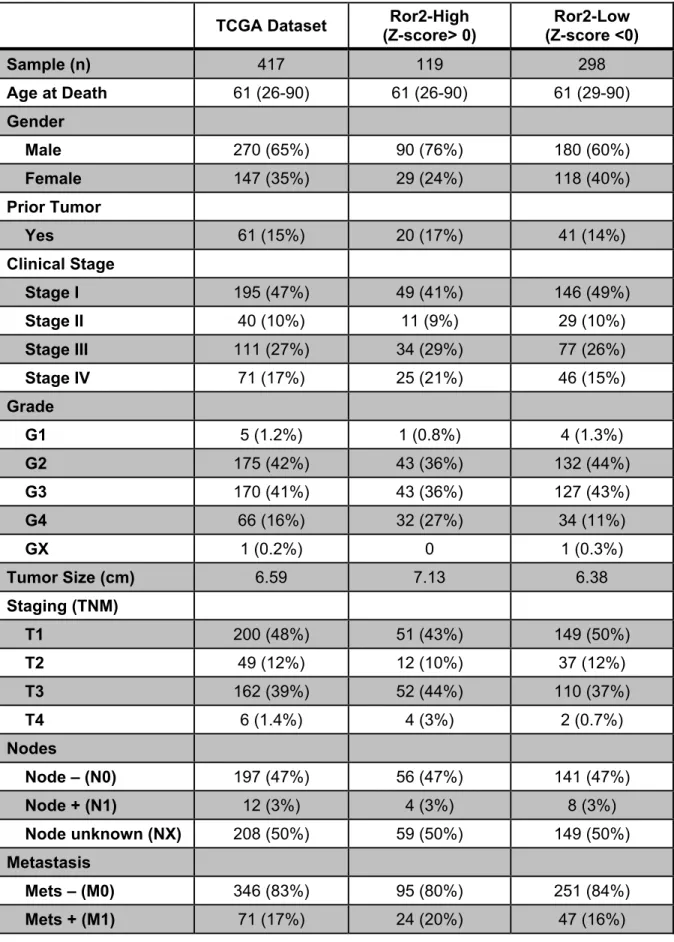
Table 3.1: Summary of clinical characteristics of patients in the
TCGA ccRCC dataset……….…….96
xiv
LIST OF FIGURES
Figure 1.1: Overview of the VHL/HIF pathway and dependent targets
in RCC……….….5
Figure 1.2: Overview of the PI3k/AKT/mTOR pathway and potential
targets in RCC ………...………..14
Figure 1.3: Structural representations of Ror orthologs …………..………..19
Figure 1.4: Overview of Wnt signaling pathways………28
Figure 2.1: Expression of Ror2 in primary human RCC tumors correlates
with expression of Wnt signaling component genes………...………55
Figure 2.2: Expression of Ror2 in RCC cells results in changes of
canonical Wnt regulators and target genes………..57
Figure 2.3: Suppression of Ror2 results in decreased
β
-catenin
mediated transcription in RCC cells………..58
Figure 2.4: Overexpression of Ror2 enhances
β
-catenin mediated
transcription in renal and RCC cells ………...60
Figure 2.5: Ror2 expression results in an increased pool of stable
xv
Figure 2.7: Intact Ror2 kinase domain is required for poised Wnt
signaling state………..….67
Figure 2.8: Wnt/PCP and Wnt/Ca2+ Signaling is independent of
Ror2 in RCC cells………...69
Figure 2.9: Ror2/Wnt3a signaling in RCC cells………..………….70
Figure 3.1: Determination of Ror2 correlated genes in RCC cells and
tumors………..…..88
Figure 3.2: Ror2 dependent MMP2 expression is enhanced by Wnt3a
stimulation……….………….89
Figure 3.3: Migration in RCC cells is enhanced by Ror2 expression…….……..……90
Figure 3.4: Intact Ror2 kinase domain is required for MMP2 expression
and migration in RCC cells……….……….………...91
Figure 3.5: SFRP2 expression is Ror2 dependent in RCC cells ……….…...……….92
Figure 3.6: Expression of Ror2 promotes
in vivo tumor
growth and
vascularity……….……….……93
Figure 3.7: Ror2 expression is higher in ccB tumors………..………...94
Figure 3.8: High Ror2 expression trends towards more aggressive
ccRCC………..……….….97
xvi
Figure 4.1: Kinase targets in clinical trials and the currently targeted
kinome………..111
Figure 4.2: Positions of induced Ror2 mutations within the kinase domain……….115
Figure 4.3: Sequence alignment of TKD domains from Ror1 and Ror2………122
xvii
LIST OF ABBREVIATIONS AND SYMBOLS
ANOVA
Analysis of variance
Ang1
Angiopoietin
1
Ang2
Angiopoietin
2
APC
adenomatous
polyposis
coli
aPKC
atypical
protein
kinase
C
CAIX
carbonic
anhydrase
IX
CCND1 cyclin
D1
ccRCC
clear cell renal cell carcinoma
CK1
α
casein
kinase
1
α
CRD
cysteine-rich
domain
DAAM1 Disheveled-Associated
Activator Of Morphogenesis 1
DKK1
dickkopf-1
DVL
dishevelled
DKK
dickkopf
DVL
Dishevelled
ECM
extracellular
matrix
EGFR
epidermal
growth
factor
receptor
EMT
epithelial-mesenchymal transition
xviii
GIST
gastrointestinal
stromal
tumor
Glut1
glucose
transporter
1
GFP
green
fluorescent
protein
GSK3-
β
glycogen synthase kinase beta
HIF
hypoxia
inducible
factor
HSP90
heat shock protein 90
IGFR
insulin-like growth factor receptor
IRP1
iron-regulatory
protein-1
JNK
c-Jun
N-terminal
kinase
LDA
lactate
dehydrogenase-A
LEF
lymphoid
enhancer-binding
factor
LMS
leiomyosarcoma
LRP
low-density
lipoprotein receptor-related protein
LRP5/6
lipoprotein receptor-related protein 5 or 6
MAPK
mitogen activation protein kinase
MuSK
muscle-specific
kinase
MMP
matrix
metalloprotease
MMP1
matrix
metalloprotease
1
MMP2
matrix
metalloprotease
2
MMP9
matrix
metalloprotease
9
MMP13
matrix metalloprotease 13
mTOR
mechanistic
target
of
rapamycin
xix
mTORC2
mTOR complex 2
NFAT
nuclear factor associated with T cells
NF
κ
B
nuclear factor kappa B
PCP
planar
cell
polarity
PDGF
platelet-derived
growth
factor
PDGFR
platelet derived growth factor receptor
PDK1
phosphoinositide
dependent protein kinase 1
PI3K
phosphatidylinositide
3-kinase
PIP3
phosphoinositol-3,4,5-triphosphate
PKA
phospho-glycerase
kinase
PTEN
phosphatase and tensin homolog
pRS
pRetroSuper
pVHL
von
Hippel-Lindau
protein
PYGO1 pygopus
1
Raptor regulatory-associated protein of mTOR
RCC
renal
cell
carcinoma
Rictor
Rapamycin-Insensitive companion of mTOR
ROCK1
Rho kinase 1
Ror2
receptor tyrosine kinase-like orphan receptor
RTK
receptor
tyrosine
kinase
RT-PCR
Reverse transcription polymerase chain reaction
SAM
Significance
Analysis of Microarrays
xx
SFRP
secreted
Frizzled-related
protein
shRNA
short hairpin RNA
siRNA
small
interfering
RNA
TCF
T
cell
factor
Trk
tropomyosin
receptor
kinase
VHL
Von Hippel-Lindau
VEGF
vascular endothelial growth factor
Chapter One
INTRODUCTION
Renal cell carcinoma
Renal cell carcinoma (RCC) is a common epithelial tumor that continues to rise in
prevalence in the face of recent advances in its treatment. It is the sixth most commonly
diagnosed cancer in men and the eighth most common in women. Though the cause of the
disparity of incidence rates between the genders is as yet undetermined, men are twice as
likely to develop RCC as women. It is estimated there will be over 65,000 new cases and
13,000 deaths in the United States in 2013, nearly one-third of these new cases presenting
with advanced RCC (1). For those patients with metastases upon diagnosis, the 5-year
survival rate is bleak at less than 10% (2). Moreover, advanced RCC is quite difficult to treat
because it is highly unresponsive to traditional single-agent or combination
chemotherapeutic strategies. While cytokine therapies have consistently shown promise for
a small subset of patients, the overall toxicity of current cytokine therapies interferes with
their widespread use(3).
RCC consists of several distinct histological subtypes including chromophobe,
papillary, and clear cell renal cell carcinoma (ccRCC). ccRCC accounts for 70-75% of RCC
cases and derives its name from its histologically characteristic clear cytoplasm, setting it
apart from the other subtypes (4). The clear cytoplasm is due to ccRCC’s highly glycolytic
2
and vascular nature, which results in a buildup of glycogens and lipids within the cytoplasm.
All subsequent work and discussion presented here will focus solely on ccRCC, being
referred to as either ccRCC or RCC.
VHL/HIF signaling in clear cell renal cell carcinoma
ccRCC is molecularly characterized by inactivation of the von Hippel-Lindau tumor
suppressor gene (VHL), leading to constitutive stabilization of the hypoxia-inducible factors
(HIF-1α and HIF-2α) (5). This feature is seen both in sporadic ccRCC and in patients with
VHL syndrome, as a result of either biallelic mutations or functional inactivation of the VHL
protein (pVHL) in 75-85% of ccRCC cases (6-9). Under normoxia or normal oxygen
conditions, pVHL serves as part of the E3 ubiquitin ligase complex that recognizes
hydroxylated prolyl residues of HIFα subunits mark them for proteasomal degradation.
However, under hypoxic conditions or when pVHL is inactivated, the HIFα subunits
accumulate and heterodimerize with HIF-1β (ARNT), translocating into the nucleus and
regulating transcription of target genes. The skewed VHL/HIF axis seen in ccRCC results in
increased expression of a variety of hypoxia-inducible genes shared by both HIF-1α and
HIF-2α including the pro-angiogenic factors platelet-derived growth factor (PDGF) and
vascular endothelial growth factor (VEGF), the extracellular matrix (ECM) remodeling protein
matrix metalloprotease 2 (MMP2), and glucose transporter 1 (Glut1) (Figure 1.1). Although
HIF-1α and HIF-2α are highly similar, with 48% overall amino acid identity, they exhibit
unique expression profiles and induction of distinct target genes (10). HIF-1α-targeted
induction of lactate dehydrogenase-A (LDA) and phospho-glycerase kinase (PKA) are
representative of the increased metabolic activity observed in RCC. However, HIF-2α
expression, can increase in-vivo tumor growth in RCC cells when acting alone, uniquely
targets erythropoietin, Cyclin D1 (CCND1), and Oct-4, an important factor in stem cell
3
In addition to its function as part of the E3 ubiquitin ligase complex regulating HIF,
pVHL has been shown to regulate cell cycle, apoptosis, fibronectin binding, and extracellular
matrix assembly, independent of HIF (13-18). The established hallmarks of cancer include
the escape of programmed cell death and cell cycle checkpoints leading to sustained cell
proliferation (19). As previously noted, traditional therapeutics have had limited efficacy in
RCC because of its resistance to radiotherapy and cytotoxic treatments. These features of
RCC may be partially attributed to increased anti-apoptotic signaling within the tumor. The
contribution of loss of pVHL to these tumorigenic signaling networks is well illustrated by its
regulation of several proteins known to be central to these cellular processes.
The tumor suppressor p53 serves as a key regulator of cell cycle and has been
observed to be mutated in a significant portion of solid tumors. While many studies have
noted the distinct lack of p53 mutations in RCC (6), pVHL has been shown to directly
associate with p53, inhibiting Mdm-2 ubiquitination and the promotion of its transactivation.
The loss of pVHL and proper regulation of p53 in RCC cells results in attenuated apoptosis
or abnormal cell cycle arrest upon DNA damage (15). pVHL also serves as a negative
regulator of the transcription factor nuclear factor κB (NFκB) through inhibitory
phosphorylation of the NFκB agonist Card9 (20,21). Thus the loss of pVHL drives RCC
tumorigenesis through the resulting increased anti-apoptotic signaling and improper cell
cycle control. Finally, pVHL has been found to bind and inhibit atypical protein kinase C
(aPKC) which is associated with cell proliferation, cellular survival, and in establishment and
maintenance of cell-cell junctions (22,23).
Another hallmark of cancer is the ability of tumor cells to migrate and invade,
allowing the cancer to metastasize throughout the body (19). Critical to cells’ ability to do
this is the loss of cell-cell adhesion, along with the remodeling of the surrounding ECM,
which normally acts as a structural support for cells. pVHL has been shown to directly bind
4
requires the ECM to mediate cell signaling,as removal of the ECM substrate in-vitro, results
in a reduction of epithelial differentiation and growth arrest at high cell density (16).
Correlating with these changes in epithelial differentiation, pVHL loss in RCC cells results in
an epithelial-mesenchymal transition (EMT) marked by decreased expression of epithelial
markers that promote cellular adhesion (e.g. E-cadherin and γ-catenin) and accompanying
increased expression of N-cadherin, vimentin, integrins, MMP2, and MMP9 characteristic of
motile mesenchymal cells (21,22). Reintroduction of VHL expression or inhibition of NFκB in
VHL null cells is also capable of reverting expression of these EMT markers in RCC cells
(21). Taken together, these findings coalesce into a clearer understanding of how loss or
inactivation of VHL drives many aspects of RCC tumor biology including resistance to
5
RCC targeted therapeutic development
Better understanding of the molecular underpinnings of RCC has led to the advent of
the use of targeted therapies such as the anti-angiogenic VEGF and PDGF receptor
tyrosine kinase inhibitors sorafenib (Nexavar, Bayer/Onyx), pazopanib (Votrient, Glaxo
6
(Avastin, Genentech) (24-28). Each of these compounds has contributed significantly to
improving the progression-free survival of patients with RCC, thus advancing the standard of
care for the disease. Two mTOR inhibitors, temsirolimus (Torisel, Wyeth), and everolimus
(Afinitor, Novartis) have also shown clinical efficacy in the care of advanced RCC, with
temsirolimus extending overall survival for patients with extremely poor-risk disease (29,30).
Despite the progress seen with the introduction of these targeted therapies, there
remains considerable room for improvement, considering that no available targeted
therapies are currently capable of inducing remission, and de novo or acquired resistance to
these targeted therapies is an on-going challenge facing clinicians. Further complicating
matters, several of the currently approved therapies exhibit undesirable and dose-limiting
side effects due to toxicity, most likely on account of both on- and off-target effects. Given
the limitations of current targeted therapies there is a great need in the field for more targets
for development of therapeutics for use as first-line agents or in combination with available
therapies.
Because loss of VHL is one of the defining molecular characteristics of RCC, its
interactions in the disease have been well characterized. As mentioned above, upon loss or
inactivation of pVHL the HIF subunits accumulate and lead to the transcriptional induction of
various target genes including VEGF and PDGF (Figure 1.1) (31). The molecular targeted
therapies described above were designed to take advantage of this aspect of VHL
deficiency, and are thought to primarily affect the VEGF and PDGF receptor tyrosine
kinases expressed on tumor-supporting endothelial cells and pericytes. The efficacy of
these drugs in inducing disease response and stabilization implicates this pathway as one
important to the maintenance of renal tumors. Promising new targets exist at an array of
points all along the VHL-HIF axis which could possibly address the problems of side effects
7
Potential therapeutic targets in VHL/HIF signaling in RCC
The most immediate potential targets for therapeutics are the HIF transcription
factors themselves, with HIF-2α expression being sufficient to induce in-vivo tumor growth
(12). Various efforts have focused on targeting one or both HIF factors, with initial strategies
directed toward the first of these factors to be characterized, HIF-1α. Transcription factors
are notoriously difficult to inhibit compared to targets with enzymatic activity. Even so,
strategies aimed at reducing the production or stability of these factors can be effective.
HIF-1α is regulated by a variety of mechanisms in addition to the E3 ubiquitination provided via
it’s interaction with pVHL. These include the phosphatidylinositide 3-kinase (PI3K)/mTOR
pathway, mitogen activation protein kinase (MAPK) signaling, and other autocrine circuitry
(32). Agents intersecting all of these signaling pathways are presently in development for
RCC and other tumor types and need to be evaluated in the context of ccRCC.
Several unique approaches have been undertaken to therapeutically reduce HIF-1α
levels. EZN-2968, has highlighted the potential of antisense oligos, as it is able to effectively
reduce both HIF-1α and target gene expression in-vitro and in-vivo (33) (Table 1.1).
Oxygen-independent stablization of HIF-1α is also mediated by heat shock protein 90
(HSP90), providing an alternate target which has shown significant promise in reducing
HIF-1 levels in the setting of VHL loss, and is currently being evaluated in clinical trials (34-36).
YC-1 (3-(5’-hydroxymethyl-2’-furyl)-1-benzylindazole), originally identified as agent-targeting
cyclic GMP, has been found to exhibit activity in repressing HIF-1α and to inhibit tumor
growth (37). The small molecule inhibitor PX-478
(S-2-amino-3-(4’-N,N,-bis(2-chloroethyl)amino) phenyl propionic acid N-oxide dihydrochloride) has been shown to inhibit
translation of HIF-1α and to exhibit antitumor activity (38,39). Other mechanisms also
modulate HIF-1α production, but they are not as well understood. For example, one group
8
tumor cell HIF-1α levels (40). These demonstrate some unique and upcoming inhibitors to
HIF-1α; a more comprehensive review of all potential inhibitors can be found here (41).
Because several reports have suggested a primary role for HIF-2α in promoting
ccRCC tumorigenesis, HIF-2α has begun to set itself apart as the more promising
therapeutic target for pharmaceutical development. HIF-2α may in fact provide an essential
function in ccRCC, and thus emerge as the key target. Recent studies inhibiting HIF-2α in
VHL-null cells resulted in inhibition of cell growth (42,43). In vitro and in vivo experiments
have further shown that stable expression of HIF-2α can drive tumor growth even in the
presence of pVHL (12,44,45). Finally, examination of ccRCC tumors reveals that these
tumors either express HIF-2α exclusively (H2 tumors) or HIF-2α along with HIF-1α (H1H2
tumors), leaving HIF-2α expression as the common denominator of VHL-mutated cancers
(46). Thus, the evidence that HIF-2α is possibly a driving force in RCC tumorigenesis sets it
apart as a promising target.
Less is understood about the regulation of HIF-2α, but efforts to target this factor are
rapidly emerging from in vitro studies. One intriguing compound, emetine, is a protein
synthesis inhibitor that has been shown to specifically inhibit HIF-2α protein stability in the
setting of VHL loss, although the effect of sustained treatment with emetine in functional
tumor assays remains unknown (47) (Table 1.1). An alternate class of inhibitors has shed
light on the transcriptional regulation of HIF-2α. In a screen for small molecules to blunt
HIF-2α transcription, the identified compounds enhance the binding of iron-regulatory protein-1
(IRP1) to an iron-responsive element in the HIF-2α 5’-untranslated region, effectively
suppressing transcription of HIF-2α (48). This novel approach provides an effective strategy
to limit HIF-2α accumulation, but remains to be studied in in vitro or in vivo assays of
tumorigenesis.
These opportunities to effectively reduce the HIF transcriptional signal might be
9
the seeming linearity of these events. However, preliminary evidence suggests that
removing the powerful stimulus of HIF transcriptional signals on a broad array of
pro-angiogenic and other factors may enhance the activity of VEGF pathway-directed agents.
This interesting pre-clinical observation heralds important considerations for ccRCC clinical
10
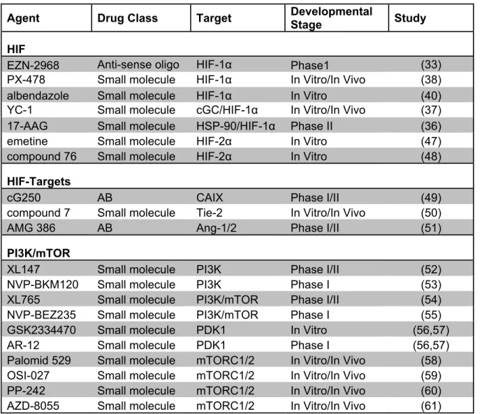
Table 1.1 Stage of development for new targeted therapies for RCC
Agent Drug Class Target Developmental
Stage Study
HIF
EZN-2968 Anti-sense oligo HIF-1α Phase1 (33)
PX-478 Small molecule HIF-1α In Vitro/In Vivo (38)
albendazole Small molecule HIF-1α In Vitro (40)
YC-1 Small molecule cGC/HIF-1α In Vitro/In Vivo (37)
17-AAG Small molecule HSP-90/HIF-1α Phase II (36)
emetine Small molecule HIF-2α In Vitro (47)
compound 76 Small molecule HIF-2α In Vitro (48)
HIF-Targets
cG250 AB CAIX Phase I/II (49)
compound 7 Small molecule Tie-2 In Vitro/In Vivo (50)
AMG 386 AB Ang-1/2 Phase I/II (51)
PI3K/mTOR
XL147 Small molecule PI3K Phase I/II (52)
NVP-BKM120 Small molecule PI3K Phase I (53)
XL765 Small molecule PI3K/mTOR Phase I/II (54)
NVP-BEZ235 Small molecule PI3K/mTOR Phase I (55)
GSK2334470 Small molecule PDK1 In Vitro (56,57)
AR-12 Small molecule PDK1 Phase I (56,57)
Palomid 529 Small molecule mTORC1/2 In Vitro/In Vivo (58)
OSI-027 Small molecule mTORC1/2 In Vitro/In Vivo (59)
PP-242 Small molecule mTORC1/2 In Vitro/In Vivo (60)
AZD-8055 Small molecule mTORC1/2 In Vitro/In Vivo (61)
HIF, hypoxia-inducible factor; cGC, cyclic GMP; HSP-90, heat shock protein-90; AB,
antibody; CAIX, carbonic andhydrase; Ang, Angipoietin; PI3K, phosphatidylinositol 3-kinase;
mTOR, mammalian target of rapamycin; PDK1, phosphoinositide dependent protein kinase
11
Developing targeted therapies for downstream HIF targets
In addition to targeting the HIF transcription family members themselves, HIF factors
promote the transcription of over 100 immediate targets and innumerable secondary targets.
In terms of broad classification, this spectrum of targets provides the opportunity to
selectively inhibit angiogenesis with current or new putative targets, interfering with
HIF-mediated cell growth and apoptotic resistance, impacting cell migration and extracellular
matrix regulation, and finally identifying novel ways to harness altered cellular metabolism. A
few selected targets downstream of HIF stabilization have already begun to present
particularly intriguing possibilities for therapeutic development in RCC (Figure 1.1).
Carbonic anhydrase IX (CAIX) is one of many hypoxia-inducible target genes, but is
unique in that it is found to be highly expressed in many ccRCC tumors (62). Due to these
high expression levels, CAIX in RCC is now being pursued as a prognostic indicator,
diagnostic tool, and prospective target for chemotherapeutics. CAIX serves a regulator for
maintaining the pH in the cell and the microenvironment surrounding the cell (63,64).
Strategies have emerged utilizing anti-CAIX antibody (also known as cG250) to identify
tumors which are highly expressive of this moiety using molecular directed nuclear imaging
(49). As a highly expressed cell surface marker in RCC, this antibody has also been
investigated as a therapeutic modality to target agents more effectively to the tumor or as an
agent with direct antitumor activity in the context of a large placebo-controlled adjuvant
study (Table 1.1).
Anti-angiogenic therapies have become the standard of care for RCC due to its
highly vascular nature and its resistance to traditional chemotherapeutic approaches, but
resistance is common. Because this resistance appears to occur as a result of angiogenic
escape mechanisms, expanding the repertoire of anti-angiogenic factors related to ccRCC is
highly desirable. Additional angiogenic targets have been identified in RCC including the
12
targets of HIF transcriptional activity. Like other targeted angiogenic kinases, Tie2
expression is mostly restricted to endothelial cells, but has been shown to correlate with
angiopotietin-2 expression in RCC tumors (65). Being an enzymatically active kinase,
conventional small molecule inhibitor screens have been effective in producing lead
compounds (50) (Table 1.1). These have been shown in preclinical models to be effective at
reducing aberrant tumor vessel growth. Several Tie2 inhibitors are being seriously
considered for clinical trials as stand-alone anti-angiogenics, or as therapeutic options to
combat VEGF receptor inhibitor resistance. Similarly, the inhibition of angiopoietins
themselves provides another tractable anti-angiogenic strategy. Angiopoietins 1 and 2
(Ang1, Ang2) are both targets and mediators of HIF expression (66). These molecules
therefore present an interesting opportunity to dampen HIF expression while having a direct
effect on tumor angiogenesis. One drug, a neutralizing peptibody, AMG 386 (Amgen) is
currently being evaluated in clinical trials (51) (Table 1.1). These targets not only present
possible new therapeutic options for the care of RCC patients but a unique opportunity to
better understand the underlying biology of these tumors. Sequential or combinatorial
therapies involving targeting Ang1, Ang2, and Tie2 also would serve to better understand
the mechanisms of resistance in RCC, if it is through compensatory upregulation of HIF or
simply engagement of untargeted angiogenic receptors among the endothelial cells. In
addition, as Tie2 expression is limited solely to endothelial cells, as is thought to be the case
for the angiogenic VEGF receptor, this presents an opportunity to examine the interplay
between the tumor and the supporting microenvironment.
PI3K/AKT/mTOR signaling in clear cell renal cell carcinoma
VHL loss alone in mice has been unable to completely recapitulate the clinically
observed pathology of ccRCC , therefore additional driver mutations and signaling cascades
must play a role as well (67-69). The collaborative efforts found in the Cancer Genome Atlas
13
exhibit significantly high rates of mutation. Of note, two members of the mechanistic target
rapamycin (mTOR) signaling cascade were included; mechanistic target of rapamycin
(mTOR) and phosphatase and tensin homolog (PTEN) were among the eight most
frequently mutated members (6).
The mTOR signaling cascade serves as a major hub for signals from the
extracellular milieu, regulating a variety of intracellular processes including metabolism,
survival, protein and lipid synthesis, and autophagy (70). Mutations and misregulation within
the mTOR signaling pathway are implicated in a wide spectrum of cancers. Initiation of the
mTOR signaling cascade occurs through multiple receptor tyrosine kinases (RTK) including
epidermal growth factor receptor (EGFR), insulin-like growth factor receptor (IGFR), and
platelet-derived growth factor receptor (PDGFR) leading to activation of PI3K, which
produces the secondary messenger phosphoinositol-3,4,5-triphosphate (PIP3) and is tightly
regulated by the tumor suppressor PTEN. The resulting PIP3 recruits AKT (protein kinase B,
PKB) to the membrane, where AKT is phosphorylated by phosphoinositide dependent
protein kinase 1 (PDK1) to its fully activated form. The signaling cascade continues with
activated AKT directly activating mTOR by phosphorylation and indirectly activating MTOR
through inhibition of tuberous sclerosis 1 or 2 (TSC1/2), negative regulators of mTOR.
Subsequent formation of either mTOR complex 1 (mTORC1) or mTOR complex 2
(mTORC2), dependent upon the inclusion of either regulatory-associated protein of mTOR
(Raptor) or Rapamycin-Insensitive companion of mTOR (Rictor) respectively, regulates a
wide variety of cellular effects including cell growth, survival and angiogenesis (71) (Figure
14
Developing PI3K/AKT/mTOR pathway targeted therapeutics
The PI3K/AKT/mTOR pathway represents a prime target for the development of
therapeutics since it is one of the most common aberrant pathways activated in cancer,
15
migration (72,73). Capitalizing on the known effects of the mTOR pathway, mTORC1
complex inhibitors have become an additional tool for treatment of advanced RCC (29,30)
(Figure 1.2). Despite the initial successes of the mTOR inhibitors their efficacy has been
limited, highlighting the need for additional therapeutic targets.
Because PI3K serves as a bottleneck in response to several RTKs and is the most
proximal element, it has become another attractive target for RCC to be used alone or
concert with current mTOR inhibitors. Two prospective PI3K inhibitors, LY294002 and
wortmannin, have been shown to decrease AKT activation and significantly reduce cell
growth in vitro through induction of cell apoptosis. Use of the small molecule inhibitor
LY294002 in vivo was able to induce tumor regression (74). Although LY294002 and
wortmannin exhibit limited selectivity and high toxicity, they demonstrate the possibilities
associated with inhibiting PI3K in RCC. Ongoing studies using PI3K and dual PI3K/mTOR
inhibitors modified for clinical use are ongoing (52-55) (Table 1.1).
Similar to PI3K, PDK1, a key mediator of AKT activation, is poised to respond to
targeted inhibition by blockade of AKT signaling. Both highly specific inhibitors such as
AR-12 (Arno) and inhibitors with dual function on PDK1/PI3K or PDK1/AKT are in development
(56,57) (Table 1.1). These targets present another possibly robust way to render the AKT
pathway completely inhibited, mitigating the confounding issues of inhibition of each of the
AKT family members.
Upon phosphorylation AKT is known to interact with a large set of substrates,
impacting many key cellular processes such as cell cycle progression and apoptosis, both of
which execute a vital function in oncogenesis (75). In addition, AKT is constitutively
activated in RCC cell lines (74). Recent work has suggested mTORC2 provides feedback to
AKT, leading to its compensatory activation (76,77). AKT targeting via direct or indirect
16
expression of activated AKT, and as coordinate therapy with current mTOR inhibitors
curbing possible refractory responses.
mTOR performs a vital role in regulating critical cellular processes including cell
growth, proliferation, transcription, and protein synthesis (78). In addition to the
aforementioned effects of mTOR activation, mTOR plays an important part in regulating
HIF-1α expression. Targeting mTOR is already an FDA-approved strategy for the treatment of
RCC but both rapalogs temsirolimus and everolimus suffer from the drawbacks of seeming
to be solely selective for inhibiting mTORC1 and, as previously mentioned, can lead to
upstream activation of AKT through mTORC2 (76,77). This highlights the need for targeted
agents for mTORC2 or dual inhibitors of both mTORC1 and mTORC2. Inhibitors in these
categories are also being developed for clinical use (58-61) (Table 1.1).
Overcoming resistance in RCC
A continuing impediment to the treatment of RCC in the clinic is resistance to
established targeted therapies. The driving forces behind initial or developed resistance in
RCC are not yet fully understood. However, RCC xenografts submitted to a daily regimen of
the VEGF receptor inhibitor sorafenib showed a reduction in supporting tumor vasculature
that was reestablished prior to the onset of resistance (79). These findings suggest a
potential mechanism of resistance where, through either compensatory activity of the VEGF
or alternative angiogenic pathways, the supporting vasculature to the tumor is reestablished
(80). Several of the targets discussed above lend themselves as potential tools in
overcoming and better understanding these mechanisms of resistance in RCC.
As previously discussed, RCC is characterized by loss or inactivation of VHL, leading
to constitutive stabilization of HIFα. A plausible mechanism for this observed resistance is
through upregulation of HIF transcription factors and targeted pro-angiogenic genes VEGF
and PDGF. If a compensatory increase in HIF levels is truly driving the development of
17
PI3K/AKT/mTOR pathway may lead to renewed potentiation of VEGF-receptor targeted
therapies. This approach was examined in colon cancer xenografts using the VEGF
receptor inhibitor, sunitinib, with and without disruption of HIF-1α and HIF-2α, showing an
additive effect on angiogenesis and tumor cell proliferation (81). Additionally, the use of
mTOR inhibitors temsirolimus and everolimus has been shown to lead to a compensatory
activation of AKT which could spur a resurgence of vasculature through an upregulation of
HIF (76,77). Vertical combination approaches targeting PI3K, AKT, and PDK1 or use of
inhibitors that more fully target both mTORC complexes pose an intriguing possibility that
calls for further research. Another avenue for escape from treatment with the established
anti-angiogenic therapies lies in additional angiogenic pathways independent of VEGF. One
of these VEGF independent pathways, receptor Tie2 and ligands Ang1/2, has been
discussed previously here as a new target for RCC. Inhibition of the kinase Tie2 and
blocking of the angiopoietins or other adjunctive angiogenic signals offers a plausible route
to more fully restrict the reestablishment of the supporting tumor vasculature. Further
research using these strategies either in sequential or combination with available targeted
therapies is needed to establish their efficacy in reducing resistance in RCC.
Receptor tyrosine kinase, Ror2
An exciting, novel RCC therapeutic target recently identified is the developmentally
regulated receptor tyrosine kinase-like orphan receptor 2 (Ror2) which has been shown to
be expressed in a VHL- and HIF-dependent manner in RCC (82,83). Ror2 is a member of
the Ror family of receptor tyrosine kinases (RTK) which also includes Ror1. Ror1 and Ror2
are both characterized by a conserved series of domains, including an Immunoglobin-like
domain, a Frizzled-like cysteine-rich domain (CRD) allowing Ror RTK to serve as Wnt
18
RTK includes a tyrosine kinase domain, which shows a high degree of amino acid identity
with both muscle-specific kinase (MuSK) and the tropomyosin receptor kinase (Trk),
kinases, which served as the basis of the original identification of hRor1 and hRor2 genes in
humans by PCR screen (84). Both the mouse and human orthologs of Ror RTK also include
a series of domains following the tyrosine kinase domain, consisting of a proline-rich domain
flanked on both sides by serine/threonine-rich domains. These serine/threonine-rich
domains have been shown to be necessary for activity and mediating protein interactions
(85-89). This general structure of domains is well conserved with a few deviations across
species within the Ror othologs identified in A. californica (Apror) (90), C. elegans (CAM-1) ,
D. melanogaster (dRor) (91), X. laevis (xRor2) (92), into the split of two genes in M.
musculus (mRor1, mRor2) (93), and humans (hRor1, hRor2) (84) (Figure 1.3). Although
Ror1 and Ror2 share this overall conserved structure of domains they diverge in that overall
they share only 58% amino acid identity. However, there is a 92% amino acid identity
between mRor2 and hRor2, allowing studies in both to be reasonably used to inform one
20
Ror2 a developmental receptor tyrosine kinase
The role of Ror2 as a developmental kinase was first evident in CAM-1 mutants,
which exhibit improper cell migration, defects in asymmetric cell divisions, and axon
outgrowth in C. elegans (94-96). These mutational analyses also highlight the function of
Ror2 as a Wnt receptor, since overexpression of EGL-20 (a Wnt) phenocopies mutant
CAM-1 migration defects (97). Several independent knockout studies of mouse mRorCAM-1 and mRor2
have further elucidated their expression patterns and roles in development. Ror2 expression
is largely restricted to embryogenesis, with expression seen in the heart, brain, nervous
system, lungs, midgut, developing limb buds, and kidney (83,93,98-105) although some
limited expression of Ror2 has been observed in the adult within the cycling uterus, colon,
and small intestine (106,107).
Knockout of Ror2 results in perinatal lethality, most likely due to respiratory defects.
Loss of Ror2 also results in widespread skeletal abnormalities, ventricular septal defects in
the heart, and decreased branching of sympathetic neuron axons (93,98-102). Loss of
mRor1, however, exhibits none of the developmental defects seen in mRor2, though
mRor1/mRor2-null mice experience an increased severity of these developmental defects,
suggesting some level of redundancy or interaction within Ror RTKs (100-102). The skeletal
defects are the result of loss of Ror2 in proliferating chrondocytes, leading to reduced
endochondral ossification (98,99). Many of the developmental defects observed in the
mRor2 knockout are phenocopied in Wnt5a-null mice (108). In addition the spatiotemporal
expression of Wnt5a and Ror2 during development overlaps in many tissues, including the
facial primordia, limb mesenchyme, neural crest-derived tissues, and the genital tubercle
(100,101,108). Together, these observations suggest that Ror2 may function as a receptor
for Wnt5a during development.
Due to the widespread skeletal defects observed in mRor2-null mice, it is
21
developmental disorders: recessive Robinow syndrome and autosomal dominant
Brachydactyly type B (109-112). Robinow syndrome is characterized by skeletal dysplasia,
limb foreshortening, brachydactyly, craniofacial malformations, and genital hypoplasia.
These features are strikingly similar to the phenotypes observed in Ror2 knockout mice,
suggesting that Robinow syndrome is indeed due to loss of Ror2. However, Brachydactyly
type B effects are limited to hypoplasia of the distal phalanges and nails. The differences in
Robinow syndrome and Brachydactyly type B phenotypes is reflected by the mutational
profile of Ror2 for each. Robinow Syndrome is characterized by missense, nonsense, and
frameshift mutations within the CRD, Kringle, and tyrosine kinase domains, resulting in loss
of Ror2 function. However, the Ror2 mutations resulting in Brachydactyly type B are due to
truncation of the protein within or after the kinase domain (112).
Ror2 in Cancer
In addition to its contribution during development, expression of Ror2 has been
observed in an increasing array of cancers. Ror2 expression was first identified in SH-SY5Y
cells derived from a neuroblastoma (84). The aberrant expression of Ror2 in primary human
RCC tumors was initially shown in 2009, where its expression in RCC cells was found to be
VHL- and HIF-dependent (82,83). Expression of Ror2 in RCC cells was shown to promote
cell migration, anchorage-independent growth and in-vivo growth in xenografts (83). Since
that time Ror2 expression has been observed in several other cancers including
osteosarcoma, melanoma, prostate cancer, gastric cancer, gastrointestinal stromal tumor
(GIST), leiomyosarcoma (LMS), and squamous cell carcinoma of the head and neck
22
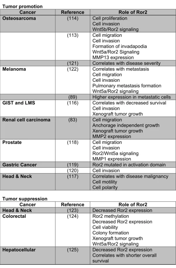
Table 1.2: Summary of the role of Ror2 in various cancers
Tumor promotion
Cancer Reference Role of Ror2
Osteosarcoma (114) Cell proliferation
Cell invasion
Wnt5b/Ror2 signaling
(113) Cell migration
Cell invasion
Formation of invadapodia Wnt5a/Ror2 Signaling MMP13 expression
(121) Correlates with disease severity
Melanoma (122) Correlates with metastasis
Cell migration Cell invasion
Pulmonary metastasis formation Wnt5a/Ror2 signaling
(89) Higher expression in metastatic cells
GIST and LMS (116) Correlates with decreased survival
Cell invasion
Xenograft tumor growth
Renal cell carcinoma (83) Cell migration
Anchorage independent growth Xenograft tumor growth
MMP2 expression
Prostate (118) Cell migration
Cell invasion
Ror2/Wnt5a signaling MMP1 expression
Gastric Cancer (119) Ror2 mutated in activation domain
(120) Cell invasion
Head & Neck (117) Correlates with disease malignancy
Cell motility Cell polarity
Tumor suppression
Cancer Reference Role of Ror2
Head & Neck (123) Decreased Ror2 expression
Colorectal
(124) Ror2 methylation
Decreased Ror2 expression Cell viability
Colony formation Xenograft tumor growth Wnt5a/Ror2 signaling
Hepatocellular (125) Decreased Ror2 expression
23
Since changes in Ror2 expression result in drastic defects in skeletal development, it
is fairly unsurprising to find the reexpression of Ror2 in osteosarcoma, a malignant
adolescent bone cancer. In-vitro analysis shows aberrant expression of Ror2 in
osteosarcoma increases cell proliferation, migration, invasion, and formation of invadapodia
(113,114). These invasive and migratory characteristics are mediated by Ror2-dependent
expression of the extracellular matrix remodeling protease, matrix metalloprotease 13
MMP13 (113,114,126). Surprisingly, either Wnt5a or Wnt5b in connection with Ror2 were
able to mediate these aggressive features in osteosarcoma cells. An analysis of primary
osteosarcoma tumors through IHC for Wnt5a and ROR2 revealed a positive correlation
between their high degree of expression in over 70% of samples. In addition, both Wnt5a
and Ror2 correlated with Enneking surgical stage and tumor metastasis. These findings
further confirm the role of Ror2 in tumor progression in osteosarcoma and its potential as
prognostic biomarker (121).
Previous works elucidating the process of tumor metastasis in melanoma have
shown Wnt5a to be overexpressed in metastatic melanoma tissues and correlated with
decreased survival in melanoma patients (127,128). In addition to Wnt5a’s role in promoting
tumor progression and metastasis in melanoma, it also serves in regulating Ror2 expression
in melanoma cells. Utilizing in-vitro and in-vivo approaches, suppression of Wnt5a/Ror2
signaling resulted in decreased cell invasion, migration, and metastasis (122). Reflective of
the shared role Wnt5a and Ror2 have exhibited in melanoma, Ror2 is similarly correlated
with metastasis (89,122).
Soft-tissue sarcomas are heterogeneous malignant tumors that currently have limited
therapeutic options and prognostic biomarkers. Edris et al. undertook an analysis using a
library of sarcomas to uncover novel RTK targets or biomarkers (116). Ror2 expression was
24
Knockdown of Ror2 reduced cell invasion in both GIST and LMS cells. Similar to our
previous finding in RCC cells (83), suppression of Ror2 in LMS significantly reduced in-vivo
tumor growth. Finally, Edris et al. showed that Ror2 can serve as an independent prognostic
factor for both LMS and GIST, with Ror2 expression predicting poorer clinical outcome
(116).
For most prostate cancer patients the rate of response to treatment is high if the
disease is caught in its early stages. However for the 20-30% of patients that experience
recurrence the picture is much more grim highlighting the need for identification of novel
biomarkers to predict recurrence and identify new therapeutic targets. Yamamoto et al.
showed that Wnt5a expression correlates with high Gleason scores, which are linked to
increased risk of recurrence. Suppression of Ror2 resulted in decreased Wnt5a-dependent
cell migration and invasion in prostate cancer-derived cells. These invasive characteristics
are partially mediated by MMP1 in a Wnt5a/Ror2 dependent signaling manner (118).
An analysis undertaken to identify additional RTK targets expressed in gastric cancer
screened all RTKs for mutations within the kinase domain in a series of gastric cancer cell
lines and 52 microdissected primary gastric cancer tumors. Ror2 was identified among other
RTKs to be expressed in poorly-differentiated invasive gastric cancers and to be a frequent
target of non-synonymous mutagenesis. The identified D644N mutation is found within the
kinase domain of Ror2, likely altering activity of the RTK Ror2. The indication that Ror2 is a
potential driving factor in the development and/or progression of RCC is strengthened by the
combined weight of these studies with similar suggestive correlations between increased
invasive malignancy and Ror2, marking it as a promising therapeutic target. These studies
also emphasize the potential of Ror2 as a prognostic and/or diagnostic biomarker.
Conflicting reports on whether Ror2 is more highly expressed in squamous cell
carcinoma have been published. Kobayashi states that Ror2 is more highly expressed in
25
(117), but Liu et al. states that Ror2 expression is downregulated in comparison to adjacent
corresponding normal tissues (123). Seeing that both studies rely upon very small sample
sizes, any firm conclusions cannot be made at this time and further studies are required to
clarify this discrepancy.
This ambiguity is further reflected in Ror2’s purported tumor-suppressive capacity in
both colorectal cancer and hepatocellular carcinoma. Epigenetic silencing of Ror2 through
hypermethylation was observed in both colorectal cancer-derived cell lines and in primary
tumors, with rescue of Ror2 inhibiting in-vitro and in-vivo tumor growth (129). Similarly
reduced Ror2 expression levels were seen in 63% of hepatocellular patients in comparison
to adjacent paired normal tissue samples and correlated with a significantly reduced overall
survival rate (125). Though these results are seemingly inconsistent with previous findings
regarding Ror2’s tumor-promoting capacity, it is important to note that reintroduction of Ror2
in colorectal cancer cells resulted in a decrease in β-catenin-dependent signaling,
constitutive activation of which serves as the driving force in colorectal cancer (129). These
results suggest Ror2’s identity as a tumor promoter or suppressor is dependent upon the
context of expression and the state of Wnt/β-catenin-dependent signaling within the tumor.
A precedent for this scenario may be observed in several of the Wnt antagonists including
dickkopf -1 (DKK1) and secreted Frizzled-related proteins 1-3 (SFRP1-3) which have shown
the ability to serve in both roles of tumor promoter and suppressor in various cancer
contexts (130,131).
Wnt Signaling
Wnt signaling is a highly conserved network effecting a diverse set of processes
within adult tissue homeostasis and embryonic development, including migration, cell/tissue
26
(132,133). Considering that Wnt signaling contributes to such a varied array of cellular
functions it is unsurprising that disruptions in the activity and/or regulation of Wnt signaling
have been linked to various developmental defects including skeletal diseases (134),
polycystic kidney disease (135-137), and many forms of cancer (134,138). The large range
of processes and signaling networks regulated by Wnt signaling can be partially attributed to
the multitude of ligand/receptor combinations possible between the 19 members of the Wnt
family, 10 Frizzled (Fzd) receptors, and additional Wnt receptors found in mice and humans.
In addition the tight regulation of the spatiotemporal expression of these Wnt ligands and
receptors during development allows for crosstalk between the Wnt signaling pathways.
Following the original discovery of the Wnt-1 (int-1) ligand, members were originally
divided into two classes: canonical (β-catenin-dependent) and non-canonical (β
-catenin-independent), according to their ability to transform C57mg mammary epithelial cells and
activate β-catenin-mediated transcription (139-141). This classification placed Wnt1, 3, 3a,
and 7a as canonical Wnts, with Wnt2, 4, 5a, 5b, 6, 7b and 11 as non-canonical Wnts.
However, this classification of Wnt ligands may be artifactual or incomplete since crossover
between the β-catenin-dependent and independent Wnt signaling has been well
documented as a result of receptor context. The most extensively used ‘non-canonical’
ligand, Wnt5a, has been shown to be able to either stimulate or repress β-catenin
dependent signaling both in vitro and in vivo depending upon the context of the
spatiotemporal expression of various Wnt receptors (142-144). Thus future examinations of
Wnt signaling cascades need to be viewed through cellular and receptor context.
β
-catenin-dependent Wnt signaling
β-catenin-dependent Wnt signaling is the most well-characterized cascade and is
triggered by Wnt ligand engaging Fzd and low density lipoprotein receptor-related protein 5
or 6 (LRP5/6) heterodimers. This signal is transduced through activation of dishevelled
27
of casein kinase 1α (CK1α), glycogen synthase kinase-β (GSK3-β), adenomatous polyposis
coli (APC), and Axin. The resulting accumulating stabilized Β-catenin is translocated into the
nucleus, binding to transcription factors including T cell factor (TCF) and lymphoid
enhancer-binding factor (LEF), regulating expression of target genes such as the negative feedback
regulator Axin2. However, under conditions where no Wnt ligand is bound, β-catenin is
sequentially phosphorylated by CK1α and GSK3-β within the destruction complex. β-catenin
is then polyubiquitinated following recruitment of the β-TrCP-containing E3 ubiquitin ligase,
marking it for proteasomal degradation (134,145) (Figure 1.4A).
β
-catenin-independent Wnt signaling
β-catenin-independent Wnt signaling does not utilize the coreceptor LRP5/6 or β
-catenin and is mediated solely through Fzd or other non-canonical Wnt receptors. The first
of the β-catenin-independent pathways is referred to as the planar cell polarity pathway
(PCP). Wnt/PCP cascade is initiated through Fzd receptor engagement of the Wnt ligand
resulting in activation of DVL, then Disheveled-Associated Activator of Morphogenesis 1
(DAAM1) and/or the GTPases Rac1 and RhoA. The signal is further transduced with the
activation of the kinases c-Jun N-terminal kinase (JNK) and Rho kinase 1 (ROCK1),
resulting in changes in cell polarity, movement, and inhibition of β-catenin-dependent
signaling (Figure 1.4B).
The second of the β-catenin-independent pathways is the Wnt/Ca2+ pathway which,
akin to the Wnt/PCP pathway, is mediated solely through the Fzd receptor. Upon
engagement with the Wnt ligand the Fzd receptor leads to the activation of DVL. The
cascade continues through activation of phospholipase C (PLC) resulting in a release of
intracellular Ca2+ and activation of calcineurin, calcium-calmodulin kinase II (CamKII) and
protein kinase C (PKC). Activated calcineurin rapidly dephosphorylates NFAT resulting in its
28
Wnt antagonists
An additional level of regulation of these Wnt signaling pathways is through several
families of secreted proteins referred to as Wnt antagonists. These factors have been
classified into two broad groups based upon their mechanisms of action. The first class of
secreted antagonist factors comprised of the SFRPs, Wnt inhibitory factor 1 (WIF-1), and
Cerberus bind directly to Wnt ligands allowing for inhibition of any the Wnt signaling
cascades. The SFRP family is comprised of 5 members, SFRP1-5, which were originally
identified in mammals for their regulatory contribution in embryonic development (146,147).
All members of the SFRP family consist of two domains, a CRD domain, allowing for binding
of Wnt and Fzd receptors or Tolloid metalloproteases; and a Netrin domain, which has also
been shown to participate in binding of Wnts (148-152). Although WIF-1 and Ceberus both
lack a CRD domain they have been shown to be able to bind Wnts and serve as antagonists
29
Wnt coreceptors LRP5/6. The DKK family is comprised of four members, DKK1-4, which are
characterized by two CRD domains (155,156). DKKs mediate their inhibition of β
-catenin-dependent signaling by binding LRP5/6 and initiate either the internalization or disruption of
its heterodimer with Fzd (157-159). DKK1-2’s inhibition of LRP5/6 requires the concomitant
binding of the transmembrane protein Kremen.
As with the other members, many of these Wnt signaling members are able to serve
in multiple capacities dependent upon the context of their expression. Although the SFRP
family has been classified as Wnt antagonists, further research has shown their part to be
much more complicated as they are capable of inhibiting and enhancing Wnt signaling in a
context-dependent manner (160). DKK2 is further set apart from its family members
because it is also capable of acting as LRP agonist (161).
Wnt signaling in renal development and RCC
As previously described, Wnt signaling plays an extensive role in development, but
of particular note for this work, previous studies have highlighted the function of Wnt
signaling in nephrogenesis. The loss of either Wnt9b or Wnt4 results in hypoplasia within the
developing kidney, due to their regulation of the epithelial differentiation of metanephric
mesenchymal progenitor cells (162,163). Wnt11 expression is restricted to the tip of the
ureteric bud, with its loss causing a mild form of hypoplasia within the kidney (164).
Expression of both SFRP1 and SFRP2 is present during nephrogenesis in tightly-regulated
spatiotemporal patterns within the developing mesenchyme, which complement or overlap
the expression of aforementioned Wnt4 (165-167). SFRP1 binds Wnt4, inhibiting β
-catenin-dependent signaling, resulting in decreased tubular differentiation and bud branching.
SFRP2 competes with SFRP1 for the binding of Wnt4, allowing a dynamic balance to
achieve proper differentiation and tubule formation within the kidney (165,166). In addition to
these secreted factors, several Wnt receptors including Ror2 and Fzd are also expressed in