TYROSINE KINASE MODULATION OF TRAFFICKING AND BIOLOGICAL FUNCTIONS OF THE ATYPICAL RHO GTPASE, WRCH-1
Jamie K. Alan
A dissertation submitted to the faculty of the University of North Carolina at Chapel Hill in partial fulfillment of the requirements for the degree of Doctor of Philosophy in the
Curriculum in Pharmacology
Chapel Hill 2010
Approved by
Abstract Jamie K. Alan
Tyrosine kinase modulation of trafficking and biological functions of the atypical Rho GTPase, Wrch-1.
(Under the direction of Dr. Adrienne D. Cox)
DEDICATION
To the two most important women in my life, my grandmother Mary Kathryn Farmer, and my mother Jacqueline Sue Farmer. You taught me that I can do anything, and with your love and support, I did. I am blessed to have two women like you in my life, and though
one is not here on this earthly planet, she will live in my heart and in this work for all times. God blessed me when he placed both of you in my life.
ACKNOWLEDGMENTS
My advisor,
Dr. Adrienne Cox-thank you for you support and faith in me. I may never write anything without typos, but you helped me become a good scientist, and I am grateful.
My committee members,
Dr. Channing Der, Dr. Gary Johnson, Dr. Michael Schaller, and Dr. Steve Rogers- thank you for your insight and guidance.
Past and present members of the Cox lab,
Dr. Vanessa Gonzalez-Perez, Dr. Donita Brady, Dr. Anastacia Berzat, Dr. James Fiordalisi, James Madigan, Rhonda Wilder, Dr. Antonio Baines, Dr. Jared Snider, Dr. Trey Kidd, Lauren Parker, Dr. Heather Hodges-Loaiza, Dr. Patricia Keller, Kelly Gewain, Meredith Ward, Ethan Suttles, Brad Sanders, Sarah Grabowski, Sarah Quinn, and Alex Berger- Thank you all for a wonderful environment to work in, and for all the fun times I will never forget. I want to give special thanks to Dr. Vanessa Gonzalez-Perez, for being the best friend and lab mate a girl could ever hope for. Your love got me through the days. Also to Dr. Donita Brady, who always listened and always had sound advice for me. Finally a special thanks to Dr. Anastacia Berzat, for introducing me to Wrch-1, and to Dr. James Fiordalisi, it's been fun working on the same project with you.
My collaborators,
Dr. Lee Graves and Dr. Brian Dewer My classmates,
Aurelie Gresset, Steve Cappell, Katy Cappell, and John Bauman. Thank you for helping me through everything. I am glad we met and went through this experience together.
You made it all worthwhile. My family members,
TABLE OF CONTENTS
LIST OF FIGURES ...x
LIST OF ABBREVIATIONS ... xii
CHAPTER I: INTRODUCTION ... 1
1.1 The Ras superfamily of small GTPases ... 1
1.2 The Rho family of small GTPases... 5
1.3 The Cdc42 subfamily of small GTPases ... 16
1.4 Wrch-1, an atypical Rho family member... 18
1.5 Cdc42 related GTPases and trafficking... 30
1.6 C-terminal Ser/Thr phosphorylation of Ras and Rho proteins ... 32
1.7 Src family kinases ... 33
1.8 EGFR family members... 35
1.9 Rho GTPases and cancer... 37
1.10 Evaluating the role of C-terminal tyrosine phosphorylation in regulating the localization and function of Wrch-1 ... 40
CHAPTER II: REGULATION OF THE RHO FAMILY SMALL GTPASE WRCH-1 BY SRC-MEDIATED PHOSPHORYLATION OF ITS C-TERMINAL MEMBRANE TARGETING DOMAIN ... 42
2.1 Abstract ... 42
2.2 Introduction... 43
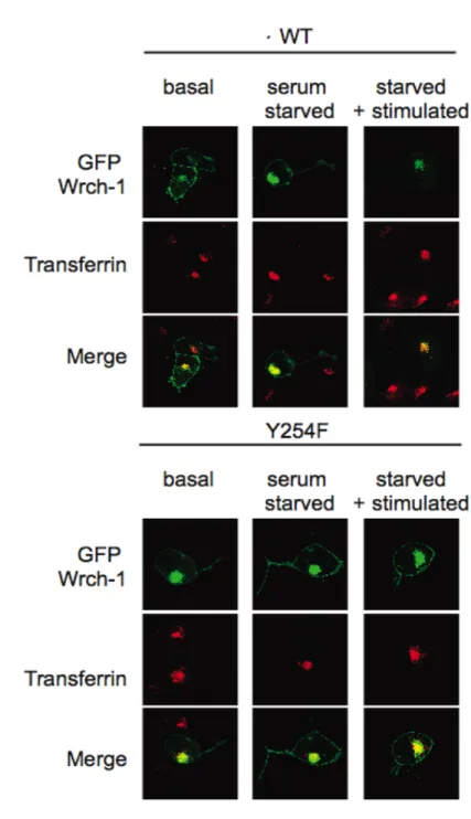
2.3.1 Wrch-1 rapidly relocalizes from the plasma membrane
to endosomal compartments in response to serum stimulation ... 47
2.3.2 Relocalization is dependent on the presence of a tyrosine at position 254 in the Wrch-1 C-terminal membrane targeting domain ... 47
2.3.3 Src can phosphorylate Wrch-1, and Src tyrosine kinase activity is required for both tyrosine phosphorylation and serum-stimulated relocalization of Wrch-1... 49
2.3.4 Phosphorylatable residue Y254 regulates Wrch-1 mediated transformation... 51
2.3.5 Phosphorylatable residue Y254 regulates Wrch-1-mediated epithelial morphogenesis ... 52
2.3.6 Wrch-1 interacts with its effector PAK at the plasma membrane, where it is GTP-bound and active, but not at endosomes, where it is GDP-bound and inactive ... 53
2.3.7 Serum-stimulated tyrosine phosphorylation and relocalization of Wrch-1 decreases its activation of downstream effectors in a Y254-dependent manner ... 56
2.4 Discussion ... 57
2.5 Materials and methods... 62
2.5.1Molecular constructs ... 62
2.5.2 Cell culture, transfections, and retroviral infection ... 63
2.5.3 Fluorescence, immunofluorescence, confocal microscopy and localization assays ... 64
2.5.4 Antibodies and western blot analysis ... 64
2.5.5 Immunoprecipitation... 65
2.5.6 In vitro tyrosine kinase assay ... 65
2.5.7 Anchorage-independent growth transformation assay... 66
2.5.8 Epithelial morphogenesis cyst formation assay ... 66
CHAPTER III: REGULATION OF MEMBRANE TRAFFICKING AND
BIOLOGICAL ACTIVITIES OF THE RHO FAMILY SMALL GTPASE WRCH-1,
BY THE TYROSINE KINASES EGFR AND SRC ... 82
3.1 Abstract ... 82
3.2 Introduction... 83
3.3 Results... 87
3.3.1 Wrch-1 is tyrosine phosphorylated in response to stimulation with EGF and PDGF but not bradykinin or LPA ... 87
3.3.2 Wrch-1 tyrosine phosphorylation in response to EGF is Src-dependent... 88
3.3.3 EGF stimulation causes Wrch-1 to traffick to endosomal compartments ... 88
3.3.4 EGF stimulation causes Wrch-1 to traffick to early endosomes, recycling endosomes and lysosomes... 89
3.3.5 EGF stimulation causes Wrch-1 to traffick to endosomal compartments with Src ... 90
3.3.6 Tyrosine phosphorylation of Wrch-1 on Y254 negatively regulates migration and invasion... 90
3.4 Discussion ... 91
3.5 Materials and methods... 94
3.5.1Molecular constructs ... 94
3.5.2 Cell culture and transfections ... 94
3.5.3 Fluorescence, immunofluorescence, confocal microscopy and localization assays ... 94
3.5.4 Antibodies and western blot analysis ... 95
3.5.5 Immunoprecipitation... 96
3.5.6 Scratch assays for cell motility ... 96
CHAPTER IV: SUMMARY AND FUTURE DIRECTIONS ...105 4.1 Summary ...105 4.2 Future Direction I: Determine how PDGFR phosphorylation of Wrch-1
alters its localization and function ...107
4.3 Future Direction II: Determine if Wrch-1 is necessary
for EGFR and PDGFR mediated biological functions...112 4.4 Future Direction III: Determine how Wrch-1 C-terminal ubiquitination
contributes to biological function ...118 4.5 Future Direction IV: Determine how Wrch-1 is
negatively regulated at endosomes ...127
LIST OF FIGURES
Figure 1.1 The Ras superfamily of small GTPases ... 3
Figure 1.2 Small GTPases are regulated by GDP/GTP binding ... 4
Figure 1.3 The Rho family of small GTPases... 6
Figure 1.4 Rho GDIs regulate Rho GTPases ... 7
Figure 1.5 Rho GTPases are modified post-translationally by lipid modifications ... 13
Figure 1.6 A second signal is required for proper Rho protein membrane localization ... 14
Figure 1.7 Cdc42-family members share sequence similarities ... 15
Figure 1.8 Wrch-1 contains N- and C-terminal extensions when compared to Cdc42 ... 24
Figure 1.9 Wrch-1 is modified by a palmitate fatty acid on a CFV motif ... 25
Figure 1.10 Wrch-1 is evolutionarily conserved... 26
Figure 2.1 Wrch-1 rapidly relocalizes to endosomal compartments upon serum stimulation... 68
Figure 2.2 Wrch-1 is tyrosine phosphorylated on Y254 in response to serum, and this phosphorylation is required for serum-stimulated relocalization... 69
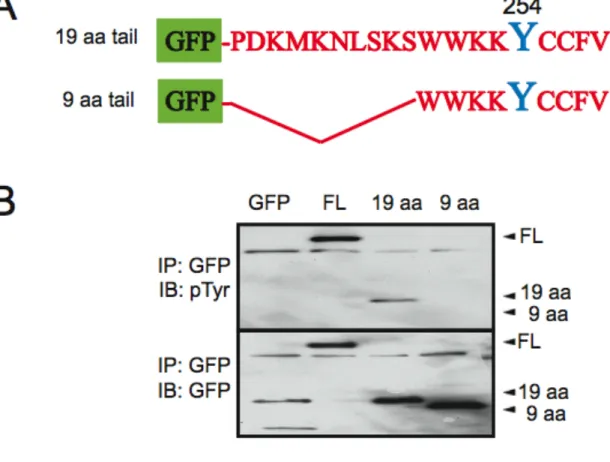
Figure 2.3 The C-terminal 19 amino acids of Wrch-1 are sufficient to become tyrosine phosphorylated and to relocalize in response to serum... 71
Figure 2.4 Src activity is required in vivo for tyrosine phosphorylation of Wrch-1 ... 73
Figure 2.5 Phosphorylation at Y254 negatively regulates Wrch-1 mediated biological functions ... 76
Figure 2.6 Wrch-1 is GTP-bound and active, and recruits its effector PAK1, at plasma membrane but not at endosomes ... 78
Figure 3.1 Wrch-1 is tyrosine phosphorylated in response to EGF and PDGF
But not LPA or bradykinin ... 98
Figure 3.2 Wrch-1 tyrosine phosphorylation in response to EGF is Src dependent...100 Figure 3.3 EGF stimulation causes Wrch-1 to traffic to endosomal compartments...101
Figure 3.4 EGF stimulation causes Wrch-1 to traffic to early endosomes,
recycling endosomes, and lysosomes ...102 Figure 3.5 EGF stimulation causes Wrch-1 to traffic
to endosomal compartments with Src. ...103
Figure 3.6 Tyrosine phosphorylation of Wrch-1 on Y254
negatively regulates migration and invasion ...104
Figure 4.1 Schematic of dimerization of PDGF and PDGFR ...110 Figure 4.2 Schematic of PDGR signaling pathways...111 Figure 4.3 Cdc42 sequesters c-Cbl through binding to p85-Cool-1/β-pix, resulting in decreased degradation of EGFR and increased expression levels of EGFR. ...117 Figure 4.4 Wrch-1 incorporates ubiquitin molecules. ...121
Figure 4.5 Ubiquitin is covalently attached to proteins as either a single monomer
(mono-ubiquitination) or a long chain (polyubiquitination). ...122
Figure 4.6 Wrch-1 is ubiquitinated
in the C-terminal membrane targeting region...123
Figure 4.7 Wrch-1 is ubiquitinated at one of three lysine residues
in the C-terminal membrane targeting region...124
Figure 4.8 Potentially ubiquitinated lysines cannot be completely substituted with arginines to support Wrch-1-mediated anchorage-independent growth ...125
LIST ABBREVIATIONS
a.a. amino acid
CAAX cysteine-aliphatic-aliphatic-unconserved amino acid Cdc42 cell division cycle 42
Chp Cdc42 homologous protein C-terminus Carboxyl-terminus Dbl diffuse B-cell lymphoma DH Dbl homology
DMEM-H high glucose Dulbecco’s modified Eagle medium EGF epidermal growth factor
EGFP enhanced green fluorescent protein EGFR epidermal growth factor receptor F farnesyl group
FITC fluorescein isothiocyanate FPP farnesyl diphosphate FTase farnesyltransferase GAP GTPase activating protein GDP guanine diphosphate
GEF guanine nucleotide exchange factor GFP green fluorescent protein
GG geranylgeranyl group
GTP guanine triphosphate HA hemagglutinin
H-Ras Harvey-rat sarcoma HV hypervariable
ICMT isoprenylcysteine carboxyl methyltransferase K-Ras Kirsten-rat sarcoma
NLS nuclear localization signal N-terminus amino-terminus
N-WASP neural Wiskott-Aldrich syndrome protein PAK p21-activated kinase
Par partitioning defective PAT protein S-acyltransferase PDGF platelet-derived growth factor PH Pleckstrin homology
PI3-K phosphatidylinositol 3-kinase 2-BP 2-bromopalmitate
TC10 teratocarcinoma 10 TCL TC10-like
TKB tyrosine kinase binding TNF tumor necrosis factor
TRITC Texas Red isothiocyanate
UNC-CH University of North Carolina at Chapel Hill WASP Wiskott-Aldrich syndrome protein
Wnt Wingless/Int
CHAPTER 1 INTRODUCTION
1.1 The Ras superfamily of small GTPases. The Ras (Rat sarcoma) superfamily of small GTPases consists of proteins that function to transmit intracellular signals initiated from
extracellular stimuli (Figure 1.1A). Ras small GTPases are involved in many divergent
cellular functions including cytoskeletal reorganization, cell survival and proliferation,
transformation and vesicular trafficking (Wennerberg et al., 2005) (Figure 1.1B). These
small GTPases function as tightly regulated molecular switches; when they are GTP-bound
they undergo a conformational change, can engage effectors, and are active. Conversely,
when they are GDP-bound, they cannot engage effectors and are inactive (Figure 1.2A).
Two types of proteins regulate the GDP/GTP cycle of these small GTPases. Guanine
nucleotide exchange factors (GEFs) are positive modulators; they function to exchange
GDP for GTP and consequently they activate small GTPases. GTPase accelerating proteins
(GAPs) work by hydrolyzing the terminal phosphate of GTP, leaving the protein GDP-bound
and inactive (Figure 1.2A). Oncogenically activating mutations that occur in cancer render
GTPases GAP-insensitive, and therefore constitutively GTP-bound and active (Figure 1.2B).
In Ras, these mutations typically occur at position 12, 13 or 61 (Figure 1.2C). These
mutations can also be utilized in the laboratory to study GTPase function. The Ras
superfamily is divided into 5 main subfamilies: Ras, Rho, Rab, Arf and Ran. Ras subfamily
proteins are well known for their role in driving cell proliferation. The Rho subfamily of
proteins are primarily known for regulating cytoskeletal dynamics. The Arf and Rab families
2
1.2 The Rho family of small GTPases. The Rho family of GTPases comprises a major subgroup of the Ras superfamily of small GTPases. Structurally, Rho GTPases are defined
by a Rho insert domain between the fifth β strand and the fourth α helix in the GTPase
domain. Rho family members are best known for their ability to control the actin cytoskeleton
by regulating structures such as actin stress fibers, lamellipodia, and filopodia (Wennerberg
and Der, 2004; Wherlock and Mellor, 2002). Within the Rho family, the core GTPase
domain contains a Rho insert domain is highly conserved between family members,
whereas they are divergent in their N-terminal extensions and in their C-terminal
hypervariable regions. In humans there are 23 members of the Rho family, which are
divided into 5 major families: Rho, Rac (Ras-related C3 substrate), Cdc42 (cell division cycle
42), Rnd (round), and RhoBTB (Rho Broad-Complex, Tramtrack and Bric à brac) (Figure
1.3). The best-studied members are RhoA, Rac1, and Cdc42. The Rho and Rac subgroups
are best known for their roles in cytoskeletal rearrangement, driving formation of actin stress
fibers and lamellipodia, respectively. Cdc42 family members are also involved in regulating
the actin cytoskeleton by inducing filopodia formation, but additionally they play a major role
in controlling cell polarity. The Rnd subgroup of Rho GTPases also plays an important role
in controlling the actin cytoskeleton (Nobes et al., 1998), and Rnd3 has also been shown to
be important in cell-cycle progression and transformation (Hansen et al., 2000; Villalonga et
al., 2004). Although less is known about the other family members, RhoBTB and Miro
proteins are being investigated for their potentially distinct roles in actin cytoskeleton control,
oncogenesis, and mitochondrial functions (Aspenstrom et al., 2004; Fransson et al., 2003;
Like Ras small GTPases, Rho GTPases also function as molecular switches that cycle
between a GTP-bound active state and a GDP-bound inactive state. Rho proteins are also
modulated by GEFs, which exchange GDP for GTP, and GAPs which hydrolyze the terminal
phosphate of GTP, rendering the protein GDP-bound. The first RhoGEF was isolated from a
gene that was transforming in human diffuse B-lymphoma cells (Dbl) (Eva et al., 1988; Ron
et al., 1988). Dbl is now known to be an exchange factor for Cdc42 (Hart et al., 1991). The
domain in Dbl responsible for exchange was subsequently found in many other RhoGEFs
and was termed the Dbl-homology (DH) domain. DH domain-containing GEFs are found in
many organisms, H. sapiensD. melanogaster, C. elegans, and S. cerevisiae to name a few.
DH domain-containing RhoGEFs also contain a pleckstrin homology (PH) domain, which is
usually immediately adjacent and C-terminal to the DH domain (Rossman et al., 2005).
Usually, both the DH and PH domain are minimally required to catalyze the exchange of
GDP for GTP. RhoGEFs work by promoting the dissociation of GDP, the stabilizing Mg2+
ion, and promoting a nucleotide-free intermediate. (Cherfils and Chardin, 1999). Because of
the high concentration of GTP relative to GDP in the cell, after GDP is released, GTP can
replace GDP in the nucleotide-binding region of the small GTPase. Remarkably, the number
of RhoGEFs outnumber target GTPases by a factor of 3, which suggests that multiple GEFs
can be capable of activating the same GTPase (Rossman et al., 2005; Schmidt and Hall,
2002). For example, it is known that the RhoGEF Trio is capable of activating both Rac1 and
RhoG in mammalian cells (Blangy et al., 2000). However, some GEFs are specific for a
particular Rho GTPase. For example, p115RhoGEF activates RhoA exclusively (Kozasa et
al., 1998). In general, RhoGEFs are regulated by their negative regulatory N-terminus; the
N-terminus sterically blocks the DH domain, and prevents exchange activity.
Phosphorylation of some RhoGEFs relieves the N-terminus/DH domain interaction, allowing
the GEFs to become active. Vav2 is a well-characterized example of a RhoGEF that is
(Han et al., 1997) relieves the N-terminal inhibition of Vav2, opening up the DH domain,
which can then interact with Rho GTPases (Aghazadeh et al., 2000; Crespo et al., 1997).
As previously mentioned, Rho proteins are also negatively regulated by RhoGAPs. Rho
proteins contain an intrinsic ability to hydrolyze GTP to GDP, however this process is very
slow, unless catalyzed by a GAP. Classically, RhoGAPs contain an "arginine finger" that is
the catalytically active subunit of the GAP. This "arginine finger" functions to accelerate the
hydrolysis of GTP to GDP by stabilizing the transition state of the substrate-binding site of
the small GTPase (Rittinger et al., 1997). There are currently approximately 170 proteins
that are predicted to possess GAP activity against various members of the Ras superfamily
and about 70 of these proteins contain a predicted Rho/Rac GAP domain (Lander et al.,
2001). In vitro, many of the GAPs that contain the Rho/Rac GAP domain interact with
several members of the Rho subfamily of small GTPases; however, some GAPs are specific
to a particular Rho GTPase. For example, p50RhoGAP can regulate RhoA, Rac1 (Lancaster
et al., 1994), and Cdc42 (Barfod et al., 1993), whereas p190RhoGAP (Ridley et al., 1993)
and RhoGAP6 (Prakash et al., 2000) are both specific for Rho. In addition to the GAP
domain, RhoGAPs also contain many putative protein and lipid interaction motifs that can
modify the localization, activity and/or substrate specificity of these proteins (Ligeti et al.,
2004).
As a third level of regulation, in addition to GEFs and GAPs, Rho proteins are also regulated
by guanine nucleotide dissociation inhibitors (GDIs). Three human RhoGDIs have been
identified: RhoGDI-1/RhoGDI-α, which is ubiquitously expressed (Ueda et al., 1990),
RhoGDI-2/RhoGDI-β, which is expressed in hematopoietic cells (Lelias et al., 1993; Scherle
et al., 1993), and RhoGDI-3/RhoGDI-γ, which is expressed only in lung, brain, and testes
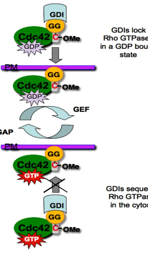
GDP dissociation and also sequester Rho proteins in the cytosol, rendering them inactive
(DerMardirossian and Bokoch, 2005) (Figure 1.4). When GDIs bind to Rho proteins, they
interact with the effector-binding domain with high affinity (low nanomolar range
(Nomanbhoy, 1996 #313)), which is comparable to or better than the affinity for the GTPase
for most effectors (Nomanbhoy et al., 1999). Additionally, RhoGDIs have a hydrophobic
pocket that can accommodate the geranylgeranyl or farnesyl groups attached to the CAAX
boxes of most Rho family proteins (Hoffman et al., 2000).
In addition to regulation by GEFs, GAPs, and GDIs, Rho proteins are regulated by their
subcellular localization. Typically Rho proteins are modified by the irreversible addition of a
prenyl group to a C-terminal CAAX motif (Adamson et al., 1992; Casey and Seabra, 1996).
These isoprenoid lipids are either a C15 farnesyl group or a C20 geranylgeranyl group
(Figure 1.5A). It is important to note that canonical CAAX motifs are not present in Wrch-1,
Chp/Wrch-2, RhoBTB1, or RhoBTB2 (Roberts et al., 2008). These lipids are added by the
enzymes, farnesyltransferase (FTase) or geranylgeranyltransferase I (GGTase I),
respectively. Following the addition of an isoprenoid group to the C-terminal cysteine, the
AAX group is proteolytically cleaved by a CAAX-specific protease, Rce1 (Boyartchuk et al.,
1997; Kim et al., 1999; Winter-Vann and Casey, 2005). Subsequently, the prenylated
cysteine is then methylated by isoprenylcysteine carboxyl methyltransferase (ICMT) (Clarke
et al., 1988; Winter-Vann and Casey, 2005) (Figure 1.5B). After the completion of these
steps, the post-translationally processed Rho protein translocates from the cytosol to the
plasma membrane (Figure 1.5B). Studies have shown that a cysteine to serine mutation in
the CAAX motif (CAAX>SAAX), which render the proteins unprenylated, results in
mislocalization of Rho proteins to the cytosol, resulting in their inactivity (Winter-Vann and
Casey, 2005). For example, mutation of RhoA CLVL>SLVL and RhoB CKVL>SKVL causes
in Rac1 (CLLL>SLLL), renders it insensitive to GGTase I modification, and results in a
mislocalization of Rac1 (Joyce and Cox, 2003). Furthermore, isoprenylation alone is not
sufficient to correctly target many Rho GTPases to their correct subcellular localization.
Proper posttranslational processing by both Rce1 and ICMT are both required for correct
localization of many Rho GTPases (Roberts et al., 2008). Rce1 activity is required for
proper localization of RhoB, Rnd1, Rnd2, Rnd3, TC10, TCL, RhoD, Rif and RhoH (Roberts
et al., 2008). ICMT activity is required for proper localization of RhoA, Rnd1, Rnd2, Rnd3,
TC10, TCL, Rif, and RhoH (Roberts et al., 2008). In addition to the prenyl group attached to
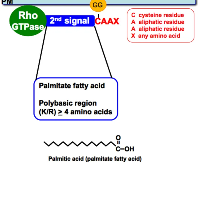
the C-terminal CAAX motif, Rho proteins are typically modified by a "second signal"
upstream of the prenyl modification. This "second signal" is required for proper plasma
membrane localization. There are two classic "second signals" that have been described
and characterized, first for Ras proteins, and then for Rho proteins: the addition of a
palmitate fatty acid, or the presence of a polybasic region upstream of the prenylation site
(Hancock et al., 1990) (Figure 1.6). For example, both RhoB and TC10 require a palmitoyl
modification upstream of the isoprenyl group for proper localization (Michaelson et al.,
2001), whereas Cdc42 relies on an upstream polybasic region (Williams, 2003). There is
data to show that "second signals" in the hypervariable domains can drive Rho proteins to
distinct subcellular compartments. RhoB, which is palmitoylated, is targeted to internal
membranes, Golgi, and peri-Golgi vesicles (Michaelson et al., 2001). TC10, which
incorporates both a palmitoylation site and a polybasic region, localizes to both the plasma
membrane and internal membranes (Michaelson et al., 2001). Rac1, which has a strong
polybasic region, localizes primarily to the plasma membrane. In contrast, both Cdc42 and
Rac2, which both have a weak polybasic region when compared to Rac1, localize primarily
to internal membranes, with a small proportion of the protein localizing to the plasma
membrane (Michaelson et al., 2001). Additionally, other additional flanking sequences in the
(Willumsen et al., 1996). Additional posttranslational modifications, such as phosphorylation,
can also modulate the localization of small GTPases, and this is the major focus of my
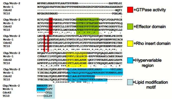
Figure 1.7 Cdc42- family members share sequence similarities.
1.3 The Cdc42 subfamily of Rho GTPases. Cdc42 is the founding and most well studied member of the Cdc42 subfamily. Cdc42 was originally identified in yeast where it functions
as a regulator of cell polarity and cellular division (Ziman et al., 1993). In mammalian cells,
Cdc42 regulates cytoskeletal organization, especially filopodial structures. Additionally,
roles for Cdc42 have been defined in normal cell growth and polarity, gene expression, cell
cycle progression, and in tumorigenesis when aberrantly regulated (Aspenstrom et al., 2004;
Wennerberg and Der, 2004). The Cdc42 subfamily of small GTPases consists of 5 proteins:
Cdc42, TC10 (teratocarcinoma 10), TCL (TC10-like) and the Wrch proteins, Wrch-1 (Wnt-
regulated Cdc42 homolog-1) and Chp (Cdc42 homologous protein)/Wrch-2. These proteins
share high sequence identity within the GTP- and effector-binding regions, and differ the
most in their N- and C-terminal extensions (Figure 1.7). As with all small GTPases, the
GTPase domain undergoes a conformational switch when it is GTP-bound, allowing these
proteins to interact with effectors through the effector-binding domain. Because the Cdc42
subfamily shares high sequence similarity within the effector-binding domain, it is easy to
think that these proteins utilize similar effectors to elicit biological function and are therefore
redundant. However, the presence of unique sequence elements within the effector-binding
domain, and divergent hypervariable regions (important for localization) within the subfamily
confer enough divergence to promote unique functions within the Cdc42 subfamily. Most
Cdc42 effectors contain the Cdc42/Rac-interactive binding (CRIB) domain that is required
for interactions with the effector-binding domain of Cdc42 (Cotteret and Chernoff, 2002).
Exceptions to this include phosphotidylinositol 3-kinase (PI3K), CIP4 and IQGAP, which
interact with a region in the effector domain that is distinct from the region that interacts with
the CRIB domains (Bishop and Hall, 2000; Cotteret and Chernoff, 2002). One of the major
effectors of Cdc42 is the serine/threonine kinase, p21-activated kinase (PAK), which binds
through its CRIB domain to Cdc42. When Cdc42 is GTP-bound, it can then bind and
et al., 1995). PAK activity downstream of Cdc42 has been shown to be important in many
Cdc42-driven biological activities, including transformation, morphogenesis and cell cycle
progression.
Cdc42 and related proteins reduce stress fibers and focal adhesions, and promote the
formation of filopodia. These cytoskeletal changes are thought to be induced by signaling
through PAK as well as other Cdc42 effectors. Another major class of Cdc42 effectors
includes the Wiskott-Aldrich syndrome protein (WASP) family members, which bind through
their CRIB domains to Cdc42 (Li et al., 1999). Downstream of Cdc42, WASP proteins
facilitate the formation of filopodia. Cdc42 also controls epithelial cell polarity, which is
mediated by the formation of distinct apical and basolateral membrane regions, which are
separated by tight junctions. Establishment of polarity is required for directional movement
of cells, and if polarity is disrupted it can contribute to more motile and invasive phenotypes.
Cdc42 controls tight junction formation by its interactions with two proteins; partitioning
defective 6 (Par6) and atypical protein kinase Cs (aPKCs). This dissertation will focus on
Wrch-1 (Wnt-regulated Cdc42 homolog-1), a more recent addition to the Cdc42 subgroup.
Wrch-1 shares some effectors with Cdc42 to elicit its cellular effects, and but also utilizes
other effectors, that are not associated with Cdc42.
Like Cdc42, Wrch-1 can bind to and stimulate autophosphorylation of PAK1 (Tao et al.,
2001) and, as discussed later in this dissertation, activation of PAK1 by Wrch-1 is regulated
by Wrch-1 phosphorylation status. Wrch-1 also binds to the Par6 and PKCζ complex (Saras
et al., 2004) to control epithelial cell morphogenesis (Brady et al., 2009). Unlike Cdc42,
Wrch-1 does not bind to WASP family proteins such as CIP4 or Spec1, as shown by a yeast
2-hybrid analysis (Aspenstrom et al., 2004). Taken together, this information suggests that
divergent from Cdc42 because it can utilize different effectors.
1.4 Wrch-1, an atypical Rho family member. Wrch-1 (Wnt-regulated Cdc42 homolog-1) and Chp/Wrch-2 (Cdc42-homologous protein), the most recently identified Rho family
members, share 57% and 52% sequence identity with Cdc42, respectively, and 61%
sequence identity with each other (Aronheim et al., 1998; Tao et al., 2001). Canonical Rho
GTPases include RhoA, RhoB, RhoC, Rac1, Rac2, Rac3, and Cdc42. These members of
the Rho family of small GTPases are considered canonical because they are tightly
regulated by GDP/GTP-binding, through regulation by GEFs, GAPs, and GDIs. Additionally,
they undergo typical post-translational modifications, such as an isoprenyl group on their
C-terminal CAAX motif, and an additional second signal such as a polybasic region or an
upstream palmitate, which will be discussed in more detail below. Atypical Rho proteins
vary in either their regulation of GTP/GDP-binding, the presence of other domains besides a
Rho insert domain, and variances in their N- and C-termini and/or posttranslational
modifications. For example, the Rnd subfamily of proteins are constitutively
GTPase-deficient and they are not stable in either a GDP-bound or a nucleotide-free form (Nobes et
al., 1998). Rnd3, also known as RhoE and Rho8, is constitutively GTP-bound in vivo
(Foster, 1996 #4;Nobes, 1998 #231). RhoD and Rif contain additional N-terminal
sequences when compared to the canonical Rho GTPases (Ellis and Mellor, 2000; Murphy
et al., 1996). RhoH, also known as TTF (translocated three four), is expressed specifically in
hematopoietic cells (Dallery et al., 1995), and plays a role in both non-Hodgkins lymphoma
and multiple myeloma (Preudhomme et al., 2000). In RhoH, similar to the Rnd proteins, the
residues analogous to G12 and Q61 in other Rho proteins are not conserved, and RhoH is
likely to also be GTPase-deficient (Li et al., 2002). When compared to the other members of
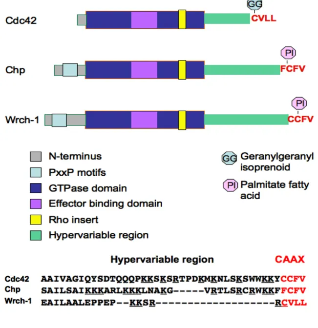
the Cdc42 family, Wrch-1 has elongated N-terminal and C-terminal extensions (Figure 1.8).
increases biological activity (Berzat et al., 2005b; Shutes et al., 2006; Shutes et al., 2004).
The N-termini of both Wrch-1 and Chp contain PxxP motifs, which may mediate interactions
with proteins containing SH3 domains, such as the adaptor proteins Grb2 and Nck. The
RhoBTB family consists of three proteins in humans: RhoBTB1, RhoBTB2, and RhoBTB3.
The RhoBTB subfamily proteins are also structurally different from the canonical Rho
GTPases, in that they contain a tandem repeat of BTB subdomains and lack C-terminal
CAAX motifs (Ramos et al., 2002). Additionally, their GTPase domains are altered when
compared to the canonical Rho GTPases. RhoBTB1 and 2 contains a Rho insert domain
that is longer than usual, contains many charged residues, and also contains insertions and
deletions in the GTPase domain. One of the deleted residues is analogous to Q61 in Ras,
and also the G12 position is substituted by an asparagine in RhoBTB1 and 2. These
proteins would be predicted to display impaired enzyme activity. Consistent with this
hypothesis, in vitro RhoBTB2 does not bind GTP (Chang et al., 2006). Furthermore,
RhoBTB3 has an extremely truncated GTPase domain, to the point where it is almost
unrecognizable as a GTPase domain, and does not bind GTP (Berthold et al., 2008) but
instead is an ATPase (Espinosa et al., 2009). However, like canonical Rho GTPases,
RhoBTB3 does have a prenylation motif, and is farnesylated (Berthold et al., 2008). Finally,
the Miro subfamily contains not only one, but two Rho GTPase domains (the N-terminal Rho
GTPase domain is most similar to other Rho GTPase domains) and two EF-hand motifs
(Fransson et al., 2003).
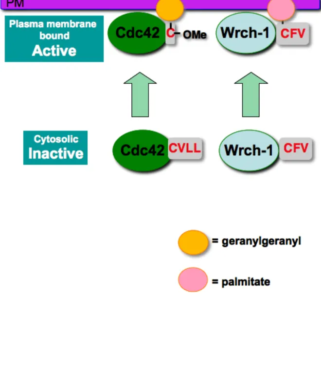
Wrch-1 is also divergent in the C-terminus, when compared to other members of the Cdc42
family. Like Chp (Chenette et al., 2006), instead of being irreversibly prenylated on a CAAX
motif, Wrch-1 is reversibly palmitoylated on a CFV motif (Berzat et al., 2005b) (Figure 1.9).
Although palmitoylation as a primary lipid modification has not been described for Rho
that terminate in CXX motifs, and their localization and function depend on the addition of a
palmitate fatty acid, rather than a prenyl group, on this motif (Ivanchenko et al., 2000; Lavy
et al., 2002). There are eleven Rho-related proteins in Arabidopsis, typically referred to as
Rops (Rho in plants) (Zheng and Yang, 2000) or RACs (Winge et al., 2000). Of the eleven
Rho proteins in Arabidopsis, eight are prenylated on CAAX motifs and three rely on
palmitoylation of a CXX motif for association with plasma membrane and proper elongation
of the root hair (Lavy et al., 2002). Additionally, there are two ROP proteins in maize, both of
which terminate in CXX motifs (Ivanchenko et al., 2000), similar to those found in
Arabidopsis, and also similar to Wrch-1 and Chp in humans.
The palmitoylation/depalmitoylation of proteins is a rapid and reversible cycle that utilizes
specific protein S-acyltransferases (PATs) for transfer of palmitoyl fatty acids onto cysteines,
and palmitoyl protein thioesterases (PPTs) for removal of the palmitates (Resh, 1999).
Although protein palmitoylation was discovered more than 20 years ago, the enzymes
responsible for this modification have only been recently described (Linder and Deschenes,
2003; Linder and Deschenes, 2004; Lobo et al., 2002; Roth et al., 2002) and their identities
and modes of action are still under active exploration. Although the addition of a palmitate
can occur spontaneously in vitro (Bharadwaj and Bizzozero, 1995; Duncan and Gilman,
1996; Quesnel and Silvius, 1994), two classes of protein palmitoylating enzymes have been
discovered (Lobo et al., 2002; Roth et al., 2002). Members of the first class of PATs share
a DHHC domain, which is a cysteine-rich domain with a conserved Asp-His-His-Cis motif,
suggested to be the domain responsible for palmitoyl transfer (Roth et al., 2002). Examples
of proteins that contain the DHHC domain are Erf2/Erf4 in yeast, which have been shown to
palmitoylate Ras, and Akr1 which is the related PAT for yeast casein kinase II. Members of
the second class of PATs act on proteins in the lumen of the secretory pathway (Chamoun
skinny hedgehog (also called Rasp, Central missing, and Sightless) is required for
palmitoylation of Hedgehog (Chamoun et al., 2001) and Spitz (Miura et al., 2006).
Additionally, another D. melanogaster protein, porcupine, is required for palmitoylation of
Wnt proteins (Zhai et al., 2004).
Palmitoyl protein thioesterases (PPTs) are required for the removal of palmitoyl
modifications. Although no direct work has been done to demonstrate the activity of PPTs
on Rho proteins, pulse-chase experiments were first done to demonstrate the turnover of
palmitate on H- and N-Ras (Magee et al., 1987), the transferrin receptor (Jing and
Trowbridge, 1987), and ankyrin (Staufenbiel, 1987), to name a few. Additionally, it has been
shown that there is a requirement for Ras to be in the native conformation, along with a
requirement for the presence of Mg2+ for PPT1 to remove the palmitate from H-Ras (Camp
and Hofmann, 1993). Other in vitro work has shown that PPT1 and acyl protein thioestrase
(APT1) can both remove palmitate modifications from Ras proteins (Duncan and Gilman,
1998). Importantly, the palmitoylation/depalmitoylation cycle has been shown to be dynamic
and reversible (Rocks et al., 2005; Zhang and Rock, 2008), and may serve as a dynamic
mechanism to regulate small GTPase localization and function. In support of this notion,
palmitoylation is required for proper membrane localization and biological function of Wrch-1
(Berzat et al., 2005b). A mutant that cannot be palmitoylated (Wrch-1 SSFV) is mislocalized
and causes a decrease in anchorage-independent growth (Berzat et al., 2005b). As
mentioned before, many Rho proteins utilize a "second signal", such as an upstream
palmitate or poly-basic region to properly regulate their localization to the plasma
membrane. Currently, there is no known "second signal" for Wrch-1 plasma membrane
localization; there is no identifiable polybasic region, and there is no upstream cysteine
Wrch-1 was initially discovered as a Wnt-responsive gene, whose expression (measured by
mRNA) was increased in response to Wnt-1 signaling in transformed cells,
Wnt-1-transgene-induced mouse mammary tumors, and Wnt-1 retrovirus-infected cells (Taneyhill
and Pennica, 2004; Tao et al., 2001). When Wrch-1 is mutationally activated (Q107L,
analogous to a Ras Q61L mutation) it phenocopied Wnt-1-mediated morphological
transformation of mammary cells (Tao et al., 2001). The Wnt family of secreted proteins are
critical mediators of cell-cell signaling in development. The first Wnt gene, Wnt1, was
discovered as a proto-oncogene that was activated by integration of a mouse mammary
tumor virus into mammary cells. Wnt proteins have been extensively characterized in D.
melanogaster and C. elegans. When cells are not exposed to Wnt, β-catenin is degraded
through interactions with Axin, APC and GSK-3. When Wnt ligands interact with the
transmembrane receptor Frizzled they work by inhibiting the Axin/APC/GSK-3 complex to
induce elevation of cytoplasmic β-catenin, which can then interact with TCF to control
transcription (Klingensmith and Nusse, 1994; Klingensmith et al., 1994; Peifer et al., 1994;
Siegfried et al., 1994; Theisen et al., 1994; Zeng et al., 1997). Normally Wnt signaling is
present in the developing organism, and is in place to promote normal development.
However in adults, mis-regulation of the Wnt signaling pathway can lead to a variety of
abnormalities and disease. Therefore, regulation of Wrch-1 in response to Wnt simulation
could be hypothesized to mediate both normal development, and disease driven by aberrant
Wnt signaling in adults.
Chp/Wrch-2, a protein highly related to Wrch-1, was identified in a screen that was designed
to look for proteins that interact with p21-activated kinase (Pak1) (Aronheim et al., 1998).
Both Wrch-1 and Chp/Wrch-2 are expressed ubiquitously at very low levels, with a higher
level of expression in the brain and placental tissue (Aronheim et al., 1998; Tao et al., 2001).
however they are expressed in different spatial and temporal patters during chick
embryogenesis, suggesting distinct roles in this process (Notarnicola, 2008 #434). Like
Wrch-1, Chp also contains and C-terminal extensions when compared to Cdc42. The
N-terminus of both Wrch-1 and Chp functions as an auto-inhibitory domain (Chenette et al.,
2005; Shutes et al., 2004). When the N-terminus of either Wrch-1 or Chp is deleted, both
proteins display enhanced biological functions such as increased transformation (Chenette
et al., 2005; Shutes et al., 2004). Additionally, both Wrch-1 and Chp terminate in a CFV
motif, and both are palmitoylated on the cysteine of this motif (Berzat et al., 2005b; Chenette
et al., 2005), a modification that is necessary for their membrane association and function
(Berzat et al., 2005b; Chenette et al., 2005). Because both Wrch-1 and Chp are
palmitoylated instead of being prenylated, neither protein interacts with RhoGDIs (Berzat et
al., 2005b; Chenette et al., 2006). Unlike Wrch-1, but similar to Cdc42, Chp requires the
presence of a stretch of basic amino acids in the C-terminal membrane-targeting motif
(Chenette et al., 2006). Chp also requires the presence of a conserved tryptophan in the
C-terminal membrane-targeting region (Chenette et al., 2006), and although Wrch-1
possesses a tryptophan residue in the 9aa minimal targeting sequence (Berzat et al.,
2005b), it has not been determined whether this residue is important for proper localization
of full-length Wrch-1. Interestingly, Chp has been shown to be activated at internal
membranes in response to stimulation by TNFα (Chenette et al., 2006), by a
yet-undetermined mechanism; however, Wrch-1 activation in response to TNFα has not been
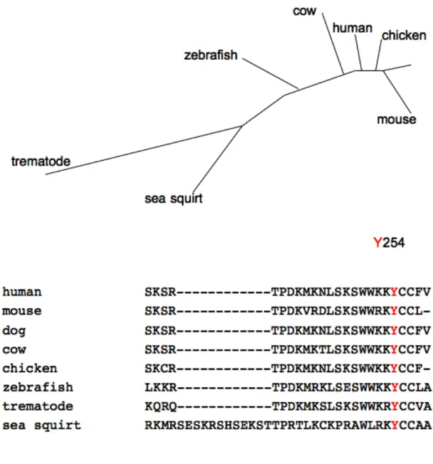
Wrch-1 homologs or orthologs have been found in many organisms, including human,
mouse, dog, chicken, zebrafish, sea squirt and trematodes, demonstrating evolutionary
conservation of Wrch-1 (Figure 1.9). Interestingly Wrch-1 is not found in some other
important model organisms, such as fruit flies, however there is a Wrch-1/Chp hybrid found
in C. elegans, named Chw-1. Currently there are no identified GEFs, GAPs, or GDIs known
to interact with and regulate Wrch-1. In vitro, Wrch-1 has a very high rate of GDP/GTP
exchange, suggesting that Wrch-1 may be constitutively GTP-bound in cells (Shutes et al.,
2006). However, a putative dominant negative of Wrch-1, T63N, does not behave like wild
type (Ruusala and Aspenstrom, 2008), supporting the idea that at least one GEF may still
be important to activate Wrch-1. In addition, in biological assays, a GTPase-insensitive
mutant of Wrch-1 (Q107L) shows significantly higher activity than wild-type Wrch-1 (Berzat
et al., 2005b; Brady et al., 2009; Brazier et al., 2009; Chuang et al., 2007; Ory et al., 2007;
Ruusala and Aspenstrom, 2008; Saras et al., 2004), suggesting that there is at least one
GAP that inactivates Wrch-1 which has yet to be identified.
Although Wrch-1 is a fairly recent addition to the Cdc42 subfamily, several effectors have
been identified. Wrch-1 shares some of the same effectors as Cdc42, including PAK1 and
the Par6/PKCζ polarity complex (Brady et al., 2009; Saras et al., 2004; Tao et al., 2001).
Wrch-1 also has effectors that it does not share with Cdc42, including the non-receptor
tyrosine kinase Pyk2 (Ruusala and Aspenstrom, 2008). Several important biological
functions have been ascribed to Wrch-1 including regulation of the actin cytoskeleton (Brady
et al., 2009; Ruusala and Aspenstrom, 2008; Saras et al., 2004; Tao et al., 2001),
anchorage-independent growth (Berzat et al., 2005b; Brady et al., 2009), focal adhesion
formation (Chuang et al., 2007 Ory, 2007 #124), cell migration (Brazier et al., 2009; Chuang
et al., 2007; Ory et al., 2007), osteoclastogenesis (Brazier et al., 2009; Ory et al., 2007) and
screen designed to identify genes that were up-regulated in a murine mammary epithelial
cells that stably express Wnt1 (Tao et al., 2001). In the same study, Wrch-1 was found to
activate both PAK-1 and C-Jun using in vitro kinase assays (Tao et al., 2001). Wrch-1 also
induced filopodia formation and stress fiber dissolution in Swiss 3T3 cells, and also
stimulated these cells to re-enter the cell cycle (Tao et al., 2001). Expression of
constitutively activated Wrch-1 caused morphological transformation of mouse mammary
epithelial cells, similar to Wnt-mediated morphological transformation (Taneyhill and
Pennica, 2004; Tao et al., 2001). Taken together, this data suggested that Wrch-1 may be
important in Wnt-mediated transformation of cells.
Wrch-1 was also identified in another screen designed to identify mRNA that was
upregulated during the differentiation of macrophages into osteoclasts (Brazier et al., 2006).
Following this observation, Wrch-1 was shown to localize to the podosome belt and is
associated with Src-induced podosomes in osteoclasts (Ory et al., 2007). Wrch-1 was also
shown to localize to focal adhesions, where it reduces overall adherence (Brazier et al.,
2009) and the number of focal adhesions in osteoclasts (Ory et al., 2007) and HeLa cells
(Chuang et al., 2007). Because focal adhesions and podosomes are important structures in
migration and invasion respectively, it was thought that Wrch-1 activity within these
structures may regulate migration and/or invasion. Indeed, Wrch-1 overexpression
increased migration in both osteoclasts and HeLa cells (Brazier et al., 2009; Chuang et al.,
2007; Ory et al., 2007). Conversely, Wrch-1 depletion led to an increase in focal adhesions,
by regulating myosin light chain, and a decrease in migration (Chuang et al., 2007). Wrch-1
mediated migration is thought to be accomplished through activation of Akt and JNK
because Wrch-1 is necessary for wound-healing mediated by Akt and JNK activation
Wrch-1 expression reduced osteoclast adhesion onto vitronectin but not fibronectin and also
increased osteoclast precursor aggregation (Brazier et al., 2009). Increased aggregation
and decreased adhesion in osteoclasts was mediated through Wrch-1 binding to the
cytoplasmic domain of integrin β3, and interference with adhesion induced Pyk2 and paxillin
phosphorylation (Brazier et al., 2009). Wrch-1 also binds to Pyk2 in PAE/PDGFRβ cells,
where activation of Wrch-1 results in increased Pyk2 phosphorylation and activation
(Ruusala and Aspenstrom, 2008). In these cells, Wrch-1 requires both Pyk2 and Src
expression and activity for the formation of filopodia (Ruusala and Aspenstrom, 2008).
Wrch-1 has also been shown to promote anchorage-independent growth in several cell
types (Berzat et al., 2005b; Brady et al., 2009). Wrch-1-mediated anchorage-independent
growth is dependent on the presence of a palmitate fatty acid on its CFV motif. A mutant of
Wrch-1 that cannot be palmitoylated (Wrch-1 SSFV) decreased the ability of Wrch-1 to form
colonies (Brady et al., 2009). Wrch-1-mediated anchorage-independent growth is also
regulated by its N-terminus; the removal of the N-terminal auto-inhibitory domain of Wrch-1
increases anchorage-independent growth (Brady et al., 2009). In addition to FTase and
GGTase I, another prenyl transferase called geranylgeranyl transferase II (GGTase II) has
been described, which modifies Rab GTPases (Seabra et al., 1992). Instead of recognizing
a CAAX motif, GGTase II recognizes Rab proteins which terminate in CCXX, CC, or CXC
motif (Khosravi-Far et al., 1992). Although Wrch-1 ends in an apparent CCXX motif, it is not
a target for GGTase II (Berzat et al., 2005b).
Most recently, we found that Wrch-1 negatively regulates the kinetics of tight junction
assembly through binding to the cell polarity protein Par6 in a GTP-dependent manner
(Brady et al., 2009). Polarity is important for the proper development and function of
(Wodarz and Nathke, 2007). Proper cell polarity is needed for the formation of apical and
basolateral membranes during morphogenesis, determination of cell fate during asymmetric
cell division, and proper development and migration of neuronal axons and dendrites
(Nelson, 2003). Although Wrch-1 had no detectable effect on overall cell polarity in a
confluent monolayer, it had a dramatic impact on the cytoskeleton and multilayering in cells
grown in three-dimensional culture (Brady et al., 2009). A precisely regulated level of
Wrch-1 seems to be required, because overexpression of activated Wrch-Wrch-1 or knockdown of
Wrch-1 disrupted epithelial cell morphogenesis (Brady et al., 2009).
Wrch-1 localizes to the plasma membrane and to internal membranes including endosomes
and lysosomes, potentially suggesting a role for Wrch-1 in intracellular trafficking. As stated
earlier, there is no known "second signal" for Wrch-1 plasma membrane localization; there is
no identifiable polybasic region, and there is no upstream cysteine available in the
C-terminus for palmitoylation. However there are many residues in the C-terminal
hypervariable region that could be substrates for post-translational modification, including
several lysines (ubiquitination), serines, threonines, and tyrosines (phosphorylation). This
dissertation will focus on the role of C-terminal tyrosine phosphorylation in regulating the
localization and function of Wrch-1. Chapters II and III will focus on the upstream kinases,
and the mechanism by which C-terminal tyrosine phosphorylation regulates Wrch-1.
1.5 Cdc42-related GTPases and trafficking. There are several examples of Cdc42 family members that are involved intracellular trafficking. Cdc42 shares high sequence similarity
with two other members of the Cdc42 family in addition to Wrch-1 and Chp, TC10 and TCL
(Figure 1.10). Similar to Cdc42, TC10 promotes the formation of filopodia, activates JNK,
and promotes SRF- and NF-κB-mediated transcription (Murphy et al., 1999). TCL promotes
2000). Interestingly, TC10 and TCL share some overlapping localizations with Wrch-1.
TC10 localizes mainly to the plasma membrane and intracellular membranes, whereas TCL
localizes to endosomes. TC10 localization and biological function are dependent on both
palmitoyl fatty acid modification of carboxyl terminal cysteine residues upstream of the
CAAX motif, and on isoprenylation of its CAAX motif (Murphy et al., 2001; Murphy et al.,
1999). Additionally, TC10 is phosphorylated by CDK5 on Thr197. Palmitoylation and
phosphorylation also regulate TC10 mediated biological functions. For example, TC10
regulation of GLUT-4 translocation to the plasma membrane requires palmitoyl-assisted
localization of TC10 to lipid rafts (Murphy et al., 2001; Murphy et al., 1999). Additionally,
phosphorylation at Thr197 by CDK mediates the association of TC10 with lipid rafts, a
requirement for its ability to modulate insulin-stimulated GLUT4 translocation (Okada et al.,
2008). TCL has been previously shown to associate with early endosomes, and regulate the
transfer of transferrin from early endosomes to recycling endosomes (de Toledo et al.,
2003). A proper balance of TCL activity is required for this function, because both loss of
TCL and constitutive activation of TCL restricts transferrin to early endosomes (de Toledo et
al., 2003). Furthermore, the C-terminal tail and proper localization were crucial for TCL
function, because the fusion of C-terminal sequences of TCL to either Cdc42 or TC10 had a
similar effect on transferrin trafficking (de Toledo et al., 2003). This further highlights the
importance of localization in regulating the function of Rho family proteins. Wrch-1 localizes
to the plasma membrane and internal membranes, and as discussed in this dissertation, its
localization is regulated by C-terminal terminal phosphorylation. The mechanisms of Wrch-1
1.6 C-terminal Ser/Thr phosphorylation of Ras and Rho proteins. There is increasing evidence that phosphorylation of small GTPases is required for both their localization and
specific functions. In one of the first studies of small GTPase phosphorylation, it was found
that K-Ras was phosphorylated by PKC (Ballester et al., 1987). In addition to being modified
by a prenyl group on its C-terminal CAAX motif, K-Ras also possesses an upstream
polybasic region that is required for proper membrane targeting (Hancock et al., 1990).
Because of these modifications, K-Ras falls into a broad class of proteins that are also
anchored into the plasma membrane via a lipid modification and a polybasic region, one of
which is the myristoylated alanine-rich C kinase substrate (MARCKS). The lipid
modifications are thought to insert into the lipid bilayer of the plasma membrane, and the
basic residues are thought to interact with the positively charged head groups of the
phospholipids (Leventis and Silvius, 1998). Previously, it was shown that MARCKS is a
substrate for PKC phosphorylation, which causes MARCKS to dissociate from the plasma
membrane by a process known as the "myristoyl-electrostatic switch" (McLaughlin and
Aderem, 1995). Subsequently it was found that K-Ras also underwent a similar process,
where it was phosphorylated and translocated from this plasma membrane, and this
translocation was then termed a "farnesyl-electrostatic switch" (Bivona et al., 2006). In
response to PKC-mediated phosphorylation of Ser181 in its C-terminal membrane-targeting
domain, K-Ras4B translocates from the plasma membrane to the mitochondria, where it
then promotes apoptosis instead of proliferation (Bivona et al., 2006). Similarly, RalA is a
target of Aurora-A kinase-mediated phosphorylation at Ser194 and PP2A Aβ-mediated
dephosphorylation; phosphorylation of this site depletes it from the plasma membrane (Lim
KH, 2009; Sablina et al., 2007; Wu et al., 2005). Furthermore, AuroraA phosphorylation of
RalA not only causes translocation from the plasma membrane, but also promotes activation
of RalA and its effector RalBP1 (Lim KH, 2009 #172). Other members of the Ras
phosphorylated on Ser180 by protein kinase A (Quilliam et al., 1991). Rho family proteins
whose localization and biological activity are regulated by phosphorylation include RhoA,
Rnd3/RhoE and TC10. RhoA localization and modulation of cell spreading and migration is
regulated by PKA-mediated phosphorylation on Ser188, which promotes its binding to
RhoGDI (Forget et al., 2002; Lang et al., 1996). Rnd3/RhoE is phosphorylated after
stimulation of PKCα, which results in translocation from the plasma membrane to internal
membranes, and PKC-mediated phosphorylation is required for Rnd3 to modulate the
Rho/ROCK pathway (Madigan, 2009 #435). Furthermore, Rnd3 is also phosphorylated by
ROCK-1 on multiple sites in its N- and C-termini, which then increases the stability of Rnd3
and alters its subcellular localization (Riento et al., 2005). The mechanism by which ROCK
phosphorylation of Rnd3 regulates its activity is thought to be mediated by promoting
binding of Rnd3 to the αG helix of ROCK-1, which then positions the N-and C-termini of
Rnd3 in close proximity to the kinase domain of ROCK-1 (Komander et al., 2008). TC10,
which is closely related to Wrch-1, is phosphorylated by CDK5 on Thr197, which regulates
its association with lipid rafts, a requirement for its ability to modulate insulin-stimulated
GLUT4 translocation (Okada et al., 2008). Thus, there is significant evidence for the
functional importance of phosphorylation of small GTPases.
1.7 Src family kinases V-Src, the first identified oncogene, is the transforming product of the Rous Sarcoma virus (Stehelin et al., 1977). Its cellular counterpart, c-Src, is part of a
nine-member family of non-receptor tyrosine kinases. Generally the Src family is divided
into 2 classes; those with a wide range of expression (Src, Yes, and Fyn) and those that
have restricted expression (Lyn, Fgr, Hck, Lck, Blk, and Yrc). In general, Src family kinases
are involved in cell adhesion assembly and turnover, motility, cell polarity, epithelial cell
morphogenesis, cell proliferation and survival (Thomas and Brugge, 1997). Src family
membrane), a unique domain, SH2 and SH3 domains (for interactions with other proteins), a
kinase domain (containing an autophosphorylation site), and a C-terminal regulatory
domain. Src is activated by undergoing a conformational change from a "closed" state to an
"open" state. Normally the SH2 domain of Src binds to a phospho group at Y527; when this
happens, Src adopts a "closed" and inactive conformation. De-phosphorylation at Y527
relieves this auto-inhibition, and Src is then in an "open" conformation (Cooper et al., 1986;
Cooper and King, 1986). After Src is in an "open" conformation, it can auto-phosphorylate
on Y416 within the kinase domain, rendering the kinase fully active (Kmiecik et al., 1988;
Kmiecik and Shalloway, 1987).
There are several well characterized effectors that Src utilizes to promote biological
functions, the most well studied effector being FAK (focal adhesion kinase) (Lipfert et al.,
1992). As the name suggests, FAK is a non-receptor tyrosine kinase that is localized to and
involved in focal adhesion formation and turnover. In primary chicken embryo fibroblasts
(CEFs), v-Src completely disrupts all focal adhesions over a 16-24 hour period, which
results in marked cell detachment (Fincham et al., 1995). Subsequently, it was shown that
v-Src induced tyrosine phosphorylation of FAK which mediated focal adhesion loss that
accompanies transformation (Fincham and Frame, 1998; Fincham et al., 1995). Cas
(Crk-and Src-associated substrate) is another Src effector that is involved in focal adhesion
formation and turnover, most notably downstream of integrin signaling (Sakai et al., 1994).
In addition to modulating focal adhesions, Src has also been implicated in regulating cell
proliferation through the transcription factor STAT3; the levels of STAT3 are elevated in
v-Src transformed cells, and dominant-negative STAT3 inhibits v-v-Src mediated transformation
(Bromberg et al., 1998).
peri-nuclear region, endosomes, and focal adhesions (Kaplan et al., 1992). Most active Src is
found either at the plasma membrane or at focal adhesions (Yamamoto et al., 2002). Src
can traffick to and from endosomes within the cell after extracellular stimuli (Donepudi and
Resh, 2008; Sandilands and Frame, 2008). Studies have shown that upon growth factor
stimulation, inactive c-Src at the peri-nuclear region can translocate via recycling
endosomes to the plasma membrane where it is active (Sandilands and Frame, 2008).
Additionally, Src can traffick with RTKs such as EGFR, VEGFR, and FGFR.
1.8 EGFR family members Epidermal growth factor receptors (EGFRs) are members of the family of receptor tyrosine kinases (RTKs). The members of this family include
EGFR/ErbB1/HER1, ErbB2/Neu/HER2, ErbB3/HER3, and ErbB4/HER4 (Hynes and Lane,
2005). All the members of this family have an extracellular ligand-binding domain, a
transmembrane domain, and a cytoplasmic protein-kinase domain. EGFR family members
are well known for their roles in cell proliferation, angiogenesis, adhesion, migration, and
invasion (Hynes and Lane, 2005). EGFR utilizes several effector pathways to promote
biological effects, the most well characterized being the Ras/Raf/MEK/ERK pathway (Gupta
and Davis, 1994; Hill et al., 1993; Sasaoka et al., 1994). Activation of the Ras/Raf/MEK/ERK
pathway by EGFR results in activation of c-myc and c-jun transcription factors, which leads
to increased transcription of genes involved in cell proliferation and survival (Hill et al.,
1993). EGFR also activates PI3K (phosphoinositide 3-kinase), a lipid kinase that
phosphorylates the 3' position hydroxyl group of the inositol ring of phosphatidylinositol
(PtdIns) (Laffargue et al., 1999). PI3K drives several biological outputs including cell growth,
proliferation, survival, differentiation, and motility (Kurosu et al., 1997; Roche et al., 1998).
Ligand binding to the EGFR family of receptors induces the formation of different homo- and
The kinase domain then autophosphorylates the receptor, creating binding sites for SH2
domains in other proteins, subsequently leading to the activation of various intracellular
signaling pathways (Ullrich and Schlessinger, 1990). The duration and intensity of
intracellular signaling initiated by EGFR activation is tightly regulated in cells. For example,
protein phosphatases can interfere with the amplitude and duration of EGFR signaling
responses (Samuels et al., 1993). Additionally, signaling initiated by there receptors is
attenuated by internalization and subsequent degradation of these receptors. Following
ligand binding, EGFRs are rapidly internalized via multiple different pathways (Dikic and
Giordano, 2003). After internalization, these receptors are initially delivered to early
endosomes, which then mature into late endosomes and multi-vesicular bodies. In the
multi-vesicular bodies, the receptors are either recycled back to the plasma membrane, or
directed to the lysosomes for destruction (Lai et al., 1989a; Lai et al., 1989b; Vieira et al.,
1996).
There are two main pathways that EGFR utilizes for receptor internalization following ligand
stimulation; ubiquitin-dependent and ubiquitin-independent pathways (Dikic, 2003; Dikic and
Giordano, 2003). It widely accepted that the Cbl family of E3 ligases is a major contributor
to ubiquitin-dependent EGFR endocytosis. E3 ligases are a class of proteins that, in
conjunction with E1 and E2 ubiquitin conjugating ligases, allow attachment of a ubiquitin
molecule onto a lysine in a target protein. These ubiquitin groups can be added as a single
monomer, or in poly-ubiquitin chains. Typically mono-ubiquitination acts as a signal for
intracellular trafficking, and poly-ubiquitin signals for destruction by the proteosome (Pickart
and Eddins, 2004). As previously stated, after EGF stimulation, EGFR family members
form dimers, and subsequently become autophosphorylated. These autophosphorylation
sites serve as docking sites for the Cbl family of E3 ligases (Y1045 in EGFR) (Schmidt and
EGFR, the receptor trafficks to endosomes, where it can then either be sorted to lysosomes
for destruction, or to recycling endosomes for recycling back to the plasma membrane
(Dikic, 2003).
Cbl can also mediate EGFR internalization through a ubiquitin-independent pathway,
through its association with CIN85 (Petrelli et al., 2002). The SH3 domain of Cbl binds to a
PxxP-motif in CIN85 (where x can be any amino acid). Proline-rich motifs in CIN85 mediate
its association with SH3 domains of endophilins, a family of proteins that are able to regulate
curvature of the plasma membrane during the early steps of endocytosis (Schmidt et al.,
1999). EGFR can utilize interaction with the Cbl-CIN85-endophilin complex, to traffick to
endosomal compartments. Inhibition of the Cbl-CIN85-endophilin complex was sufficient to
block EGFR endocytosis, independent of the E3 ligase activity of Cbl (Petrelli et al., 2002).
1.9 Rho GTPases in cancer The process of carcinogenesis involves self-sufficiency in growth signal, insensitivity to anti-growth signals, the evasion of apoptosis, sustained
angiogenesis, limitless replicative potential and eventually the initiation of migration and
invasion into nearby and distant sites (Hanahan and Weinberg, 2000). Rho GTPases are
known to regulate several of these processes, which suggests that, like Ras GTPases, Rho
GTPases may be aberrantly regulated in human cancers. Ras proteins are mutated in 30%
of all human cancer; however, there have been no identified naturally occurring mutations in
Rho GTPases, with the notable exception of RhoH/TTF (Pasqualucci, 2001 #39;
Preudhomme, 2000 #237). Although Rho GTPases are not mutationally activated in human
cancers, there are documented examples of overexpression of the GTPase itself or of their
positive regulators, or loss of negative regulators. Indeed, misregulation of Rho GTPases
leads to increased proliferation, dedifferentiation, invasion and metastasis (Ellerbroek et al.,
As previously mentioned, RhoH is the only Rho GTPase that is reported to be genetically
altered in human cancers (Pasqualucci et al., 2001; Preudhomme et al., 2000). RhoH, also
known as TTF (translocated three four), is expressed specifically in hematopoietic cells
(Dallery et al., 1995), and plays a role in both non-Hodgkins lymphoma and multiple
myeloma (Preudhomme et al., 2000). The rearrangement is caused by a
t(3;4)(q27;p11-13) chromosomal gene translocation, which results in the expression of a BCL3/LAZ3 fusion
protein (Dallery et al., 1995). Additionally, RhoH, similar to the Rnd proteins, lacks the
conserved residue analogous to G12 and Q61 residues in other Rho proteins, and is likely
to be GTPase-deficient (Li et al., 2002). Although there are no other known examples of
genetic modifications of Rho proteins, several Rho GTPases have been reported to have
altered expression in human cancers, including RhoA, RhoB, RhoC, Rac1, Rac1b, Rac2,
Rac3, RhoG, Cdc42, RhoH/TTF, Rnd3/RhoE (Ellenbroek and Collard, 2007), and in some
cases Wrch-1 (Kirikoshi and Katoh, 2002). Specifically RhoA is overexpressed in tumors of
the colon, breast, and lung (Fritz et al., 1999), as well as testicular germ cells (Kamai et al.,
2001) and head and neck squamous cell carcinoma (Abraham et al., 2001). RhoC
overexpression is found in breast cancer, is involved in driving the phenotype of
inflammatory breast cancer (van Golen et al., 2000) and has been shown to play a role in
promoting metastasis of these tumors (Wu et al., 2004).
In addition to Rho GTPase overexpression, aberrant RhoGEF regulation, such as
overexpression or constitutive activation, has been implicated in human cancers. For
example, chromosomal rearrangements in leukemia can result in a MLL-LARG chimera that
contributes to Rho family activation and tumorigenesis (Kourlas et al., 2000). Additionally,
downregulation of negative regulators of Rho GTPase activity has also been documented.