Recurrent adamantinoma of the tibia and lymph
node metastasis
Sunil B Gudaganatti, Meena N Jadhav, Rashmi K Patil, Shreekant K Kittur Department of Pathology, Belgaum Institute of Medical Sciences, Belgaum, Karnataka
Abstract
Adamantinoma is a rare bone tumor arising in the long bones. Metastasis has been reported in less than 10% of cases. Here we are presenting a case of recurrent adamantinoma of the tibia presented with left inguinal lymph node metastasis after a period of six years.
A 50 year old male underwent curettage and bone grafting for adamantinoma of the left tibia which presented with recurrence after one year of primary diagnosis. Six years later he returned with inguinal swelling. Excised specimen of swelling showed features of metastatic adamantinoma.
Keywords: Adamantinoma, metastasis, lymph node.
Address for Correspondence
Dr. Sunil B Gudaganatti, Post Graduate, Department of Pathology, Introduction
Adamantinoma is a rare tumor of low grade malignancy constituting less than 1 % of all primary bone tumors[1]. The condition was first described by Fischer in 1913[2]. They occur almost exclusively in the tibia with a tendency for local recurrence. Metastasis to lungs and lymph nodes occur after long clinical course. There are very few cases of lymph node metastasis reported in the literature. A 50 year old male was admitted for evaluation of left inguinal swelling of one year duration in 2012. There was no history of pain or fever.
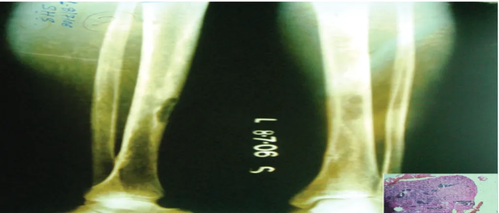
With detailed past history, patient was found to be known case of left tibial adamantinoma diagnosed six years back. He presented with pain and swelling in middle part of left leg in June 2006. There was no history of trauma. Swelling measured about 3x2 cms and plain X-ray of left leg showed lytic lesion in the anteromedial cortex of the middle third of tibial shaft surrounded by dense sclerosis [Figure 1]. He was operated and bone grafting was done. Histopathological examination of swelling revealed adamantinoma of left tibia (basaloid and spindled subtype) [Figure1:Inset]. In August 2007 he presented with swelling at same site. MRI showed a recurrent neoplasm [Figure 2]. PET scan showed
active disease at same site. He was then reoperated and titanium rod was placed in tibial shaft [Figure 3]. Histopathological examination revealed recurrent adamantinoma similar to primary tumor.
Now on examination of left inguinal region a freely mobile swelling measuring 4x3 cms, with cystic consistency was seen. Right inguinal region was normal. A linear scar mark about 8 cm was seen on medial aspect of middle of left leg with stretched and shiny skin. No palpable mass was felt at scar mark. There was no hepatosplenomegaly. Liver function tests were within normal limits. Ultra Sonography (USG) of swelling suggested a cystic lesion. Chest X-ray was within normal limits. Fine needle aspiration cytology (FNAC) showed occasional clusters of basaloid cells and no definite opinion was possible.
Medica Innovatica
December 2014, Volume 3 - Issue 2
100
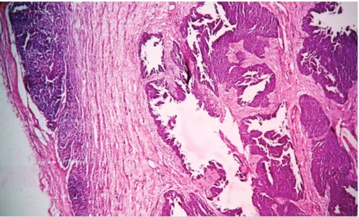
There were no squamous cells. Focal mucoid areas were also seen [Figure 4 and 5]. PAS stain showed basement membrane like material in intercellular areas. Reticulin stain showed reticulin fibres in intercellular portions. Immunohistochemistry showed tumor cells with bcl-2 granular positivity
and negative for CK, EMA and CD34. A diagnosis of metastatic adamantinoma (basaloid and spindled subtype) in left inguinal
Medica Innovatica
December 2014, Volume 3 - Issue 2
102
Figure 5. Microphotograph of lymph node showing tumor cells with oval to spindle shaped nuclei and scant cytoplasm [ H&E, X400 ]
Discussion
Adamantinoma is a rare neoplasm that tends to involve the tibia. The term adamantinoma comes from the histologic resemblance of the neoplasm to ameloblastoma of the jaw. Most of the patients are young adults, men and women are equally affected. The duration of symptoms can be remarkably long. Risk factors for recurrent or metastatic disease include male sex, pain, symptoms of less than five years duration and initial treatment by biopsy, curettage or excision[3]. The histogenesis of this tumor remains controversial[4]. Now that the presence of epithelial differentiation has been proved beyond doubt, the two most favoured possibilities are origin from intraosseous epithelial rests or epithelial metaplasia in a primary mesenchymal process. The epithelial cells have four different morphological patterns basaloid, tubular, squamoid and spindled types[5]. Basaloid and spindled pattern are most commonly encountered and have an aggressive behaviour[6]. Studies have shown that there is no clear relationship between primary subtype and rate of recurrence or metastasis. There is a tendency for shift to spindled cell pattern when
local recurrence or metastasis occurs[1]. Study done by Keeney et al showed that histological feature
associated with an increased recurrence rate is lack of squamous differentiation[3]. In the present case the primary tumor, recurrent tumor and metastatic tumor showed both basaloid and spindled subtypes and lack of squamous cells. Keeney et al[3] studied 85 cases of adamantinoma of long bones, out of which six had lymph node metastasis. Death was reported in one patient due to postoperative pulmonary embolus. One patient had lymph node resection and was alive without evidence of disease 10 years later. Khemiri et al[7] reported a case of adamantinoma of the tibia and fibula in a 50 year old female with pulmonary metastasis one year after complete resection of primary neoplasm. They concluded that even though wide resection of tumor usually gives good results of healing, long term follow up is mandatory. Bruce et al[8] reported a case of metastasis of adamantinoma of tibia to lungs sixteen years after knee amputation. They concluded that early aggressive treatment and long term follow up are mandatory. The currently preferred treatment options are wide enblock excision, limb reconstruction surgery or an amputation.
doing well without any additional lesions. To conclude adamantinoma is a locally aggressive tumor requires long term follow up for evidence of local recurrence or metastasis.
Metastasis of adamantinoma sixteen years after knee disarticulation. J Bone Joint Surg Am 1986; 68:772-76.
References
1. Hazelbag HM, Taminiau AH, Fleuren GJ, Hogendoorn PC. Adamantinoma of the long bones. A clinicopathological study of thirty two patients with emphasis on histological subtype, precursor lesion and biological behaviour. J Bone Joint Surg Am 1994; 76:1482-99.
2. Jain D, Jain VK, Vasishta RK, Ranjan P, K u m a r Y . A d a m a n t i n o m a : a clinicopathological review and update. Diagnostic Pathology 2008; 3:1-11.
3. Keeney GL, Unni KK, Beabout JW, Pritchard DJ. Adamantinoma of long bones. A clinicopathologic study of 85 cases. Cancer 1989; 64:730-7. 4. Beppu H, Yamaguchi H, Yoshimura N, Atarashi K, Tsukimoto K,
Nagashima Y. Adamantinoma of the rib metastasizing to the liver. Internal Medicine 1994; 33:441-45.
5. Schneider H, Enderle A. Differential diagnosis of a metastasizing adamantinoma of the tibia and fibula. Arch Orthop Trauma Surg 1979; 94:143-49.
6. Ramaswamy AS, Chatura KR, Chandrashekhar HR. Metastatic or metachronous adamantinoma: An enigma. International Journal of Applied and Basic Medical Research 2012; 2:132-35.
7. Khemiri C, Mrabet D, Mizouni H, Abbes I, Mnif E, Sellami S, Essaddem H. Adamantinoma of the tibia and fibula with pulmonary metastasis: an unusual presentation. B M J Case Reports 2011;doi:10.1136/ bcr.06.2011.4318
8. Cohn BT, Brahms MA, Froimson AI. Metastasis of adamantinoma sixteen years after knee disarticulation. J Bone Joint Surg Am 1986; 68:772-76.
Source of Support : Nil
![Figure 5. Microphotograph of lymph node showing tumor cells with oval to spindle shaped nucleiand scant cytoplasm [ H&E, X400 ]](https://thumb-us.123doks.com/thumbv2/123dok_us/8841296.1795090/4.612.54.564.64.319/figure-microphotograph-showing-tumor-spindle-shaped-nucleiand-cytoplasm.webp)
