Collagen: A Brief Analysis OMPJ
Collagen: A Brief Analysis
1Supriya Sharma, 2Sanjay Dwivedi, 3Shaleen Chandra, 4Akansha Srivastava, 5Pradkshana Vijay
OMPJ
REVIEW ARTICLE
10.5005/jp-journals-10037-1143
ABSTRACT
Collagen is the most abounding structural protein in a human body representing 30% of its dry weight and is significant to health because it designates the structure of skin, connective tissues, bones, tendons, and cartilage. Much advancement has been made in demonstrating the structure of collagen triple helices and the physicochemical premise for their stability. Collagen is the protein molecule which produces the major part of the extracellular matrix. Artificial collagen fibrils that exhibit some characteristics of natural collagen fibrils are now con-gregated using chemical synthesis and self-aggregation. The indigenous collagen fibrils lead further development of artificial collagenous materials for nanotechnology and biomedicine.
Keywords: Collagen, Structure of Collagen, Diagnostic Impor-tance, Collagen Disorders.
How to cite this article: Sharma S, Dwivedi S, Chandra S, Srivastava A, Vijay P. Collagen: A Brief Analysis. Oral Maxillofac Pathol J 2019;10(1):11-17.
Source of support: Nil
Conflict of interest: None
INTRODUCTION
Collagen is the unique, triple helical protein molecule which organizes the major part of the extracellular matrix.1
The word collagen was procured from the Greek word “kolla” which estates “glue producer”. Previously the col-lagen of tendons and bones was accustomed in the industry to manufacture glue. Also in organism collagen is a kind of glue.2 Collagen is a kind of biological macromolecule
which constructs a greatly organized, three-dimensional architecture and can transfer any component due to its network-like organizational nature. It is used as a biomate-rial because of its extensive applicability in diverse fields.
Its adaptable role is due to its immense properties such as biocompatibility, biodegradability and easy availability.1
They are centrally involved in the constructions of basement membranes along with diverse structures of the extracellular matrix, fibrillar and microfibrillar networks of the extracellular matrix. It establishes their fundamental fractional monetary unit and identifies crucial steps in the biosynthesis and supramolecular preparing of fibril-lar collagens.3 They are the most abundant structural
component of the connective tissue and are present in all multicellular organisms. In the light microscope, collagen fibers typically appear as the wavy structure of variable width and intermediate length.
They stain readily with eosin and other acidic dyes. When examined with a transmission electron micro-scope (TEM), collagen fibers appear as bundles of fine thread-like subunits. These subunits are collagen fibrils. Within individual fibers, the collagen fibrils are relatively uniform in diameter. In different locations and at different stages of development, however, the fibrils differ in size. In developing or immature tissues, the fibrils may be as small as 15 or 20 nm in diameter. In dense regular connective tissue of tendons or other tissues that are subject to considerable stress, they may measure up to 300 nm in diameter. Collagens are also known to form highly ordered aggregates. The perio-dicity in these macromolecular structures makes them suitable for investigation by means of X-ray diffraction.4-6
However, collagen attracts attention not only for com-mercial motives. Also from a clinical perspective, there is much mesmerized in collagens because many diverse diseases are concomitant to disarray in collagen. Genetic disorders of collagen metabolism customarily influence tissues in which the proper advancement and integrity of connective tissue are of preponderant importance. Thus, a better understanding of the spatial structure will give us more insight into collagen-related disorder diseases.1,7
Structure and Types of Collagen
It consists of 3 helically coiled linear chains, each of about 1,000 amino acids. Two of these chains (α1) are identical while the third (α2) has a different amino acid composi-tion. The amino acid composition of collagen is:
• 25% Glycine • 25% Proline • Hydroxyproline
1,5Senior Resident, 2Research Scientist, 3Professor and Head, 4Senior Dentist
1,3,5Department of Oral Pathology and Microbiology, Faculty of
Dental Sciences, King George’s Medical University, Lucknow, Uttar Pradesh, India
2Department of Plant Ecology and Climate Division CSIR-
National Botanical Research Institute, Lucknow, Uttar Pradesh, India
4JRMA Health Care, Navi Mumbai, India
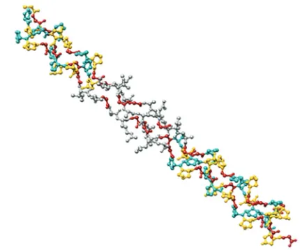
In α chain glycine is repeated every fourth residue. The triplets gly-pro-pro and gly-pro-hpro occur frequently. A protein consists of one or more polypeptide chains. Collagen consists of 3 polypeptide chains, each in the form of a left-handed helix. The explaining feature of collagen is a distinguished constructional motif in which three parallel polypeptide strands in a left-handed, polyproline type II helical conformation coil about each other with a one-residue stagger to organize a right-handed triple helix (Fig. 1).3,4,6,8
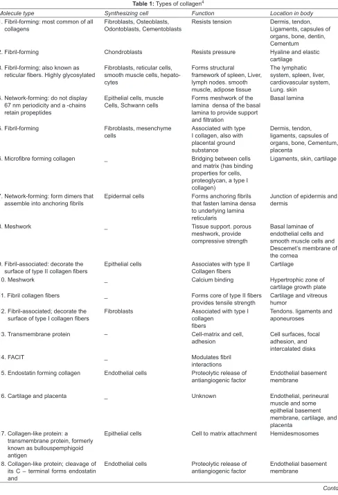
Until now, the molecule has been classified in 28 dif-ferent types that are grouped into eight families depend-ing on its structure, chain bonddepend-ing, and position in the human body (Table 1).4 Among the classifications, it can
be found the fibril-forming, microfibrillar, anchoring fibrils, hexagonal network-forming, basement membrane, fibril-associated collagens with interrupted triple helix (FACIT), transmembrane, and multiplexins.
Microscopic Appearance
Collagen fibers of connective tissue are generally less than 10 μm in diameter and are colorless, when unstained. They appear as long, wavy, pink fibers bundles after staining with Hematoxylin and Eosin. These fibers are constructed from parallel aggregates of thinner fibrils 10 to 300 nm in diameter and numerous micrometers in length.4
In electron micrographs of negatively stained prepa-rations, densely stained molecule fills the gap region, or holes between the ends of the adjacent collagen molecules and heavy metals display cross banding at regular inter-vals of 67 nm, a characteristic property of these fibers is seen. In hard tissues (bone, dentin, cementum), these holes are filled with mineral crystals. The banding pattern of the fibrils seen in thinly sectioned electron micrograph results from the differing amount of stain bound by a charged amino acid that is aligned in adjacent collagen molecules.9,10
Fig. 1: Collagen Triple-helix Structure3
Collagen Formation
The cells of the mesenchyme and their derivatives (fibroblasts, odontoblast, osteoblast, cementoblasts, and chondroblasts) are the active producers of collagen. Alterna-tive cell types forming collagen are epithelial, endothelial, Schwann and muscle cells. As an excretory protein, fibrous collagen is manufactured as a proprotein (procollagen). Messenger RNA controls the assembly of distinct amino acids into polypeptide chains on ribosome related with the rough endoplasmic reticulum (RER). The primary poly-peptide chains are around one and a half times lengthened than those in the consequent collagen molecule because they have N- and C-terminal distensions that are imperative for the aggregation of the triple helical molecule. As the chains are incorporated, they are transferred into the lumen of the rough endoplasmic reticulum, where considerable post-translational modification takes place.
The first conversion is innumerable hydroxylation of many of the lysine and proline by-products in the chain, which allows hydrogen bonding with the alongside chains as the triple helix is manufactured. The vitamin C reliant enzymes lysyl hydroxylase and prolyl hydroxylase are essential for this step. Through the effect of galactos-yltransferase in the rough endoplasmic reticulum, some of the hydroxylysine remnants are glycosylated by the inclusion of galactose.
The three polypeptide chains are assembled into the triple helix. Proper alignment of the chains then is achieved by disulfide bonding at the C-terminal extension and then the three chains twist around themselves to “zip up” the helix. The assembled helix then is transported to the Golgi complex, where glycosylation is completed by the addition of glucose to the O-linked galactose residues. Secretary granules containing the procollagen molecule are formed at the Trans face of the Golgi complex and are released subsequently by the exocytosis by the cell surface. The formation and secretion of the collagen mol-ecule take approximately 35 to 60 minutes.4,9,10
Fibroblast
Collagen: A Brief Analysis OMPJ
Table 1: Types of collagen4
Molecule type Synthesizing cell Function Location in body
1. Fibril-forming: most common of all
collagens Fibroblasts, Osteoblasts, Odontoblasts, Cemento blasts Resists tension Dermis, tendon, Ligaments, capsules of organs, bone, dentin, Cementum
2. Fibril-forming Chondroblasts Resists pressure Hyaline and elastic
cartilage 3. Fibril-forming; also known as
reticular fibers. Highly glycosylated Fibroblasts, reticular cells, smooth muscle cells, hepato-cytes
Forms structural
framework of spleen, Liver, lymph nodes. smooth muscle, adipose tissue
The lymphatic system, spleen, liver, cardiovascular system, Lung. skin
4. Network-forming: do not display 67 nm periodicity and a -chains retain propeptides
Epithelial cells, muscle
Cells, Schwann cells Forms meshwork of the lamina densa of the basal lamina to provide support and filtration
Basal lamina
5. Fibril-forming Fibroblasts, mesenchyme
cells Associated with type I collagen, also with placental ground substance
Dermis, tendon, ligaments, capsules of organs, bone, Cementum, placenta
6. Microfibre forming collagen _ Bridging between cells and matrix (has binding properties for cells, proteoglycan, a type I collagen)
Ligaments, skin, cartilage
7. Network-forming: form dimers that
assemble into anchoring fibrils Epidermal cells Forms anchoring fibrils that fasten lamina densa to underlying lamina reticularis
Junction of epidermis and dermis
8. Meshwork _ Tissue support. porous
meshwork, provide compressive strength
Basal laminae of endothelial cells and smooth muscle cells and Descemet’s membrane of the cornea
9. Fibril-associated: decorate the
surface of type II collagen fibers Epithelial cells Associates with type II Collagen fibers Cartilage
10. Meshwork _ Calcium binding Hypertrophic zone of
cartilage growth plate 11. Fibril collagen fibers _ Forms core of type II fibers
provides tensile strength Cartilage and vitreous humor 12. Fibril-associated; decorate the
surface of type I collagen fibers Fibroblasts Associated with type I collagen fibers
Tendons. ligaments and aponeuroses
13. Transmembrane protein – Cell-matrix and cell,
adhesion Cell surfaces, focal adhesion, and intercalated disks
14. FACIT _ Modulates fibril
interactions
15. Endostatin forming collagen Endothelial cells Proteolytic release of
antiangiogenic factor Endothelial basement membrane
16. Cartilage and placenta _ Unknown Endothelial, perineural
muscle and some epithelial basement membrane, cartilage, and placenta
17. Collagen-like protein: a
transmembrane protein, formerly known as bullouspemphigoid antigen
Epithelial cells Cell to matrix attachment Hemidesmosomes
18. Collagen-like protein; cleavage of its C – terminal forms endostatin and
Endothelial cells Proteolytic release of
antiangiogenic factor Endothelial basement membrane
19. FACIT _ Unknown Endothelial, perineural muscle and some epithelial basement membrane, cartilage, and placenta
20. FACIT – – Cornea (chick)
21. FACIT - - Stomach, kidney
22. FACIT - - Tissue junctions
23. Membrane-associated collagen
with interrupted triple helix - - Heart, retina
24. Fibrillar - - Bones, cornea
25. Membrane-associated collagen
with interrupted triple helix - - Brain, heart, testis
26. FACIT - - Testis, ovary
27. Fibrillar - - Cartilage
28. Microfiber forming collagen - - Dermis, sciatic nerve
fibroblast or fibrocyte is minor than the active fibroblast and is commonly spindle-shaped. It has a minor, darker, elongated nucleus; fewer processes and higher acido-philic cytoplasm with the much less rough endoplasmic reticulum. They have a branched cytoplasm surrounding an elliptical, speckled nucleus having one or two nucleoli. Active fibroblasts can be identified by their oval, pale- staining nucleus and higher amount of cytoplasm, Golgi apparatus, abundant rough endoplasmic reticulum, secretory vesicles and mitochondria (Fig. 2). They exhibit motility and contractility which are crucial during connec-tive tissue remodeling and generation and wound repair. In certain tissues, fibroblasts have significant contractile properties and are called as Myofibroblasts.4
Degradation of Collagen
The C-terminal extensions and at least part of the N-ter-minal extensions are removed by the action of procol-lagen peptidases. These condensed molecules range as a 5 unit, quarter-overwhelmed microfibrils, which then aggregate in a collateral function, giving advancement to a well-organized series of holes or gaps inside the fibrils. These gaps are the regions of the primary depos-its of mineral-related with the collagen fibrils in dentin, bones, and cementum. After the fibrils are assembled, the remaining portion of the N-terminal extension is removed by procollagen peptidase. The oxidization of some lysine and hydroxylysine residues by the extracellular enzyme lysyl oxidase, forming reactive aldehydes, results in inter-molecular cross-links that further stabilize the fibrils. 4,9,10
Factors involved in collagen degradation are:
• Highly strong to Proteolytic attack
• Collagenase 1, 2 and 3 degenerate types I, II, III, V collagen Collagenase 3 can degenerate type I, II, III, IV, IX,
Fig. 2: Shape of fibroblast: A: Nucleus; B: Nucleolus; C: Golgi appara-tus; D: Cytoplasm; E: Intermediate/transfer vesicles; F: Ribosomes; G: Mitochondria; H: Polyribosome; I: Rough endoplasmic reticulum; J: Col-lagen fibrils; K: Cell processes; L: Microtubules; M: Secretory granules.4
X, XI, fibronectin and another extracellular matrix component.11
Matrix metalloproteinases (MMP) is a great family of proteolytic enzymes that involves:
• Collagenases (MMP-1, 8, 13) • Metalloelastases (MMP-12) • Stromelysins (MMP-3, 10, 11) • Matrilysin (MMP-7)
• Gelatinases (MMP-2, 9) 4
Diagnostic Importance of Collagen in Various Conditions
Many histochemical stains have been accustomed to determine Collagen fibers which involve Weigert’s Resorcin Fuchsin, Modified Movat’s Stain, Goldner’s Trichrome method, Van Gieson, Masson’s Trichrome, etc, but the picrosirius red stain underneath the polarizing microscope is the most extensively used because of the inherent character of birefringence of collagen.11
Cysts
Collagen: A Brief Analysis OMPJ
et al. reported that under pathological conditions birefrin-gence represents various patterns in contrast with collagen in general tissue and demonstrated that type I collagen was massive, greatly birefringent red fibers while type III collagen exposed as fine weakly birefringent green fibers.12
Tumor
In a tumor, there is an enhanced density of collagen in the initial phase, which stimulates tumor initiation and metastasis.13
Odontogenic Fibroma
In odontogenic fibromas, mature type I collagen fibers are present. These fibers ran roughly parallel to the inac-tive epithelial strands and foci of mineralization were surrounded by concentric thick type I collagen bundles.12
Ameloblastoma
The collagen fibers in the basement membranes of amelo-blasts in ameloblastomas are found to be spatially orga-nized with thick type I collagen passing perpendicularly through the basement membrane zone and merging with the collagen in the capsule and fibrous septa between the epithelial follicles.12
Oral Submucous Fibrosis
One of the most customary precancerous conditions which are broadly pervasive in the Indian subcontinent is oral submucous fibrosis (OSMF). In a very early stage, fine fibrillar collagen distributed with pronounced edema and sturdy fibroblastic response demonstrating plump young fibroblasts including profuse cytoplasm will be observed. In early and moderately advanced stage collagen is seen as thickened separate bundles and moderately hyalinised respectively.14 Studies done in OSMF using picrosirius red
stain and polarizing microscopy revealed that there was a gradual decrease in the greenish-yellow color of the fibers and a shift to orange-red color with increase in severity of the disease which appeared that the tight packing of collagen fibers in OSMF progressively increased as the disease progressed from early to advanced stages.11
In Drug Delivery Systems and Tissue Engineering
Collagen is a predominant biomaterial in medical uti-lization due to its particular characteristics, like weak antigenicity and biodegradability. In the biomedical application, the chief reason for the usefulness of collagen is the fabrication of fibers with supplemental vitality and stability through its self-assembly and cross-linking and the in vivo metabolism of collagen is supervised by the
application of cross-linking agents, such as glutaralde-hyde, formaldeglutaralde-hyde, polyepoxy compounds, chromium tanning, acrylamide, and carbodiimides.15
A generalized view of collagen in the body
Collagens are a great family of triple helical proteins that are extensive throughout the body. They are crucial for a wide range of functions, involving cell migration, cancer, angiogenesis, tissue morphogenesis, tissue scaffolding, cell adhesion, and tissue repair. It is the main component of tissues such as fibrous tissue, bone, cartilage, valves of heart, cornea, and basement membrane etc.1,4,6,16
Collagen in Health
Collagen is seldom referred to as the body’s cement that keeps everything in place. It is crucial to health because it dictates the designs of skin, connective tissues, tendon, bone, and cartilage.16
Skin Health
Collagen plays an important role in skin health. Dermis layer of the skin is a connective tissue layer made up of dense and stout collagen fibers, fibroblast, and histio-cytes. Collagen type I constitute 70% of the collagen in the skin, with type III being 10% and a trace amount of collagen types IV, V, VI and VIII. Collagen conserves the toughness and elasticity of the skin. Collagen in the form of collagen hydrolysate keep skin hydrated. During the aging process, a lack of collagen becomes obvious as skin begins to sag and lines and wrinkles begin to form. In the development of scar tissue as a result of age or injury, there is variation in the abundance of types I and III col-lagen as well as their proportion to one another. Type III collagen synthesis reduces with age resulting in changes in skin tension, elasticity, and healing.4
Muscles
In muscle tissue, it serves as an utmost component of endomysium. In Smooth Muscle Cells (SMCs) collagen exist as a meshwork surrounding individual SMCs (type IV or basement membrane collagen) or as interstitial dense fibers that occupy a substantial volume of the tissue (type I and III fibrillar collagens).17,18,19
Wound Healing
Col-the elderly has been recorded and much of this age-related delayed wound healing is caused by diminished collagen synthesis and an advanced degradation. Increase in col-lagen and fibroblasts during healing proposed that a cor-relation might exist among a quantity of collagen, number of fibroblasts and tensile strength of a scar.4,16
Bone
Bone is a complex and dynamic tissue that renders structural support for the body, preservation of internal organs and acts as levers to which muscles are attached, allowing movement. In total, out of 22 to 25% of the organic component, the principal collagen type I is 94 to 98% along with other noncollagenous proteins and 2 to 5% are cells present in the bone tissue. The mixture of flexible collagen and hard mineral makes bone dense than cartilage without being brittle. A mixture of water and collagen mesh constructs a strong and slippery pad in the joint that shields the ends of the bones in the joint during muscle movement.
Cartilage, Tendon, Ligaments
In fibrous tissue, such as tendon and ligament, collagen in the form of elongated fibrils is predominantly present. It is a stretchy and flexible protein that is used by the body to support tissues and thus it plays an important role in the preservation of the cartilage, tendons, and ligaments. Normal tendon consists of soft and fibrous connective tissue that is consisted of densely packed col-lagen fibers bundles and surrounded by a tendon sheath also consisting of components of the extracellular matrix. Collagen comprises 75% of the dry tendon weight and functions principally to withstand and transfer large forces among muscle and bone. Collagen also constructs a great constituent of cartilages. Cartilage collagen fibrils composed of collagen II, the quantitatively few collagens IX and XI.
Dental Tissues
Dentin
The mature dentin is made up of nearly 20% organic material, 70% inorganic material and 10% water by weight. The organic phase is about 30% collagen (prin-cipally type I with few amounts of types III and V) with fractional inclusions of non-collagenous matrix proteins and lipids. Collagen type I serve as a scaffold that contains a great proportion (estimated at 56%) of the mineral in the pores and holes of fibrils.
The extracellular compartment of the matrix or pulp made up of collagen fibers and ground substance. The fibers are mainly types I and III collagen. The general collagen content of the pulp advances with age, the ratio among types I and III remain steady and the increased amount of extracellular collagen constructs into fiber bundles.
Cementum
Type I collagen (forms 90% of the organic matrix) is predominant collagen present in cementum. Collagens found in trace amount in cementum are types V, VI, and XIV. Different collagens related with cementum include type III, less cross-linked collagen found in high concentrations during advancement and reconstruction and repair of mineralized tissues and type XII that binds to type I collagen and also to non-collagenous matrix proteins.
Periodontal ligament
The periodontal ligament is made up of collagen fibers bundles connecting cementum and alveolar bone proper.
The chief collagens are types I, III and XII, with individual fibrils having an approximately minor average diameter than tendon collagen fibrils.
Principal fibers are the vast majority of collagen fibrils found in the periodontal ligament and are arranged in definite and distinct fiber bundles. The periodontal liga-ment has also the capability to adapt to functional changes. When the functional demand advances, the thickness of the periodontal ligament can advance by as much as 50 % and the fiber bundles also increase noticeably in thickness.
Basement membrane:
The epithelial basement membrane and neighboring area are termed the epithelial basement membrane zone. The lamina densa comprising of type IV collagen that is covered by heparin sulfate, a glycosaminoglycan and anchoring fibrils, that are made up of type VII collagen and widen from the lamina densa to the connective tissue.4
Collagen Disorders
These collagen disorders are classified as 20
Heritable/Genetic Collagen Disorders: • Ehlers-Danlos syndrome
Collagen: A Brief Analysis OMPJ
• Epidermolysis Bullosa • Marfan Syndrome
Collagen Vascular Disorders/Autoimmune Collagen Disorders:
• Systemic Lupus Erythematous • Systemic Sclerosis
• Oral submucous fibrosis • Sjogren’s syndrome • Scleroderma
• Rheumatoid Arthritis • Ankylosis Spondylitis
Miscellaneous:
• Human Atherosclerotic Plaques • Osteoporosis
• Scurvy
CONCLUSION
Collagen is the predominant structural material of the body and is the most bountiful mammalian protein accounting for about 20 to 30% of total body proteins. They are principal constituent of the extracellular matrix (ECM) that encourages the tissues. The ECM is defined as the diverse collection of proteins and sugars that sur-rounds cells in all solid tissues.
They have been classified by types that describe spe-cific sets of polypeptide chains that can form homo- and heterotrimeric assemblies. Collagen plays an impera-tive role in the development of tissues and organs and is involved in diverse functional expressions of cells. It is a good surface-active agent and has the capability to penetrate a lipid-free interface.4,5,6,21
REFERENCES
1. Muthukumar T, Sreekumar G, Sastry TP, Chamundeeswari M. Collagen as a potential Biomaterial in Biomedical Applica-tions. Rev. Adv. Mater. Sci 2018jun;53:29-39
2. Vander Rest M, Garrone R. Collagen family of proteins. FASEB 1991 Oct; 5(13):2818-2823.
3. Avila Rodr_ıguez MI, Rodr_ıguez Barroso LG, Sanchez ML. Collagen: A review of its sources and potential cosmetic applications. J Cosmet Dermatol 2018 Oct; 17:20-26.
4. Sandhu SV, Gupta S, Bansal H, Singla K. Collagen in Health and Disease. J Orofac Res 2012; 2(3):153-159.
5. Gelse T, Poschl E, Aigner T.Collagens-Structure, Function,and Biosynthesis. Advanced Drug Delivery Reviews 2003 Nov; 55(12):1531– 1546
6. Karsdal M. Biochemistry of Collagens Structure, Function, and Biomarkers. London, United Kingdom: Academic Press; 2016.
7. Bateman JF, Handford RPB, Lamandé SR. Genetic diseases of connective tissues: cellular and extracellular effects of ECM mutations. Nat Rev Genet 2009; -10:173-183.
8. Yazaki M, ItoY, Yamada M, Goulas S, Teramoto S, Nakaya M, Ohno S,Yamaguchi K. Oral Ingestion of Collagen Hydro-lysate Leads to the Transportation of Highly Concentrated Gly-Pro-Hyp and Its Hydrolyzed Form of Pro-Hyp into the Bloodstream and Skin. J. Agric. Food Chem2017Mar22;65(11): 2315-2322
9. Nanci A.Ten Cate’s Oral Histology: Development, Structure and Function. 6th ed. St Louis Missouri:Elsevier; 2003. 10. Nanci A.Ten Cate’s Oral Histology: Development, Structure
and Function. 7th ed. St Louis Missouri:Elsevier; 2008 11. Viswanathan S, Ramesh V, Balamurali PD. Assessment of
collagen fibers in the wall of odontogenic cysts: A possible role in the expansion? J. Nepal Dent. Assoc 2011;22:12-19. 12. Hangelbroek J, Raubenheimer EJ, Vorster R, Ngwenya SP.
Collagen In Odontogenic Tumours: A Histochemical- And Immunohistochemical Study of 19 Cases. Jitps 2012;26:14-18. 13. Provenzano PP, Inman DR, Eliceiri KW, Knittel JG, Yan L,
Rueden CT, White JG, Keely PJ. Collagen density promotes mammary tumor initiation and progression. BMC Medicine 2008;6:1-15.
14. Houli J, Jamil J. Digestive Articular Manifestations of Collagen Diseases. Study of 55 Patients. Ann RheumDis 1965; 24:52-55. 15. Nair R, Sevukarajan M, Mohammed T, Badivaddin C, Kumar
A. Collagen Based Drug Delivery Systems: A Review Jitps 2010;1:288-304.
16. Hongdong S, Bo Li. Beneficial Effects of Collagen Hydrolysate: A Review on Recent Developments. Biomed J Sci & Tech Res 2017 July;1(2):1-4.
17. Gartner LP, Hiatt JL. Extracellular matrix. In: Color Textbook of Histology (3rd ed). Saunders 2007:73-75.
18. Hand AR, Tencate AR. Cytoskeleton, junctions,and fibroblast. In: Nanci A, (Ed). Tencate’s oral histology: Development, structure,and function (6th ed). India: Elsevier 2006:54-60. 19. Kierszenbaum AL, Abraham L. Histology, and cell biology—
An introduction to pathology: St Louis (u.a): Mosby 2002: 101-103.
20. Yalovac A, Ulusu NN. Collagen and Collagen Disorders.Fabad J Pharm Sci 2007; 32:139-144.