© 2018, IRJET | Impact Factor value: 6.171 | ISO 9001:2008 Certified Journal | Page 162
A Survey on Detection and Classification of Brain Tumor from
MRI Brain Images using Image Processing Techniques
Shanti Parmar
1, Nirali Gondaliya
21Student, Dept. of Computer Engineering, AITS-Rajkot, Gujarat, India
2Assistant Professor, Dept. of Computer Engineering, AITS-Rajkot, Gujarat, India
---***---Abstract -Brain tumor is an abnormal growth of brain cells within the brain. Brain tumor detection and segmentation and is one of the most challenging and time consuming task in medical image processing. MRI (Magnetic Resonance Imaging) is a visualization medical technique, which provides plentiful information about the human soft tissue, which helps in the diagnosis of brain tumor. The detection of brain tumor is complicated procedures in medical field. The algorithm incorporates steps for pre‐processing, image segmentation, feature extraction and image classification. Various data mining algorithms like k-means, SVM, FCM, k-nearest neighbor, neural network used for this purpose. In this review paper, the review of different brain tumor methods using the MR images with pros and cons are given. It also gives comparative study of all the reviewed methods.
Key Words: Brain Tumor, Classification, Disease Identification, Magnetic Resonance Imaging (MRI), Segmentation, Tumor Detection.
1. INTRODUCTION
Brain is the center of human central nervous system. The brain is a complex organ as it contains 50-100 billions neurons forming a gigantic network. A brain tumor is a mass of unnecessary and abnormal cell growing in the brain or it can be defined as an intracranial lesion which occupies space within the skull and tends to cause a rise in intracranial pressure. Brain tumors are mainly classified into two i.e. Benign and Malignant. Benign tumors are non-cancerous and they seldom grows back where as malignant tumors are cancerous and they rapidly grows and invade to the surrounding healthy brain tissue.
In medical practices, the early detection and recognition of brain tumors accurately is very vital. In literature, there are many techniques has been proposed by different researchers for the accurate segmentation of brain tumor. Some discoveries such as X-rays, ultrasound, radioactivity, magnetic resonance imaging (MRI) or computed tomography and the development of tools that can generate medical images have facilitated the development of some of the most efficient exploration tools in medicine [11].
Magnetic resonance imaging (MRI) is high-quality medical imaging, particularly for brain imaging. MRI inside the human body is helpful to see the level of detail. Doctors have major technical and economic importance of reliable and fast
detection and classification of brain cancer, based on common practices. Most of the technicians are slow, less responsible, and that's hard to quantify possess a degree of subjectivity.
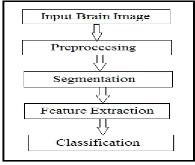
Detection of brain tumor from MRI images involves various Phases such as Preprocessing, Feature extraction, Segmentation and classification. Fig 1 shows different stages in brain tumor detection. Image Preprocessing techniques are applied to improve the quality of image [12].
Fig -1: Stages in Brain Tumor Detection
MRI Image segmentation is based on set of process of brain tumor detection; pixel intensity based features are extracted. Image Segmentation group pixels into regions and hence defines the object regions. Segmentation uses the features extracted from image. Classification is the last step in process of brain tumor image into normal or abnormal and classifies the abnormality type whether it is benign or malignant. This study evaluates various techniques which are used in tumor detection from brain MRI.
In this paper we are aiming to take review of different methods of brain tumor image segmentation. We are aiming to present the different MRI images segmentation methods and provide comparative study of all methods.
2. LITERATURE SURVEY
[image:1.595.334.532.331.498.2]© 2018, IRJET | Impact Factor value: 6.171 | ISO 9001:2008 Certified Journal | Page 163 processing techniques for brain MRI image segmentation is
classified as k-means, SVM, FCM, k-nearest neighbor, neural network, adaboost, genetic and other methods etc.
Parveen, Amritpal singh [2] purposed algorithm is a combination of SVM and fuzzy c-means, a hybrid technique for prediction of brain tumor. Here, the image is enhanced using contrast improvement, and mid-range stretch. Double thresholding and morphological operations are used for skull striping. Fuzzy c-means (FCM) clustering is used for the image segmentation. Grey level run length matrix (GLRLM) is used for extraction of feature. Then, Linear, Quadratic and Polynomial SVM technique is applied to classify the brain MRI images. Real data set of 120 patients MRI brain images have been used to detect 'tumor' and 'non-tumor' MRI images. The SVM classifier is trained using 96 brain MRI images, after that the remaining 24 brain MRI images was used for testing the trained SVM. SVM classifier with Linear, Quadratic and Polynomial kernel function give 91.66%, 83.33% and 87.50% accuracy respectively and 100% specificity.
Astina minz, Prof. Chandrakant Mahobiya [8] proposed an effective automatic classification method for brain MRI is projected using the Adaboost machine learning algorithm. The proposed system consists of three parts such as Preprocessing, Feature extraction and Classification. Preprocessing has removed noise in the raw data, it transform RGB image into grayscale, median filter and thresholding segmentation is applied. For feature extraction by using GLCM technique 22 features were extracted from an MRI. For classification boosting technique used (Adaboost). It gives 89.90% accuracy and result in normal brain or in Malignant or Benign type of tumor. In future work, we can work of quadratic and polynomial kernel function. The accuracy of the system will be increased by increasing training database images. Also the system can be implement for different types of classes like Glioma and Meningioma.
Garima Singh, Dr. M.A. Ansari [9] proposed, a novel technique which includes Normalization of Histogram and K-means Segmentation. First, input image is pre-processed in order to remove the unwanted signals or noise from it. To de-noise filters such as Median filter, Adaptive filter, Averaging filter, Un-sharp masking filter and Gaussian filter is used in the MRI images. The histogram of the pre-processed image is normalized and classification of MRI is done. Finally, the image is segmented using K-means algorithm in order to take out the tumor from the MRI. Efficient classification of the MRIs is done using NB Classifier and SVM so as to provide accurate prediction and classification. Naive Bayes and SVM Classifier give accuracy 87.23% and 91.49% respectively. SVM give better classification accuracy. For implementation MATLAB is used. The proposed method has some limitations that it could not find out the precise or accurate boundary of the tumor region.
In the future, improvement in the proposed algorithm can be done by working on the limitations, the quality of the output
images can be improved by using better morphological operations.
G Rajesh Chandra, Dr. Kolasani Ramchand, H Rao [4] proposed method in that MRI image of brain is de-noised using DWT by thresholding of wavelet co-efficient. Genetic algorithm is applied to detect the tumor pixels. A genetic algorithm is then used in order to determine the best combination of information extracted by the selected criterion. The present approach uses k-Means clustering methods into Genetic Algorithms for guiding this last Evolutionary Algorithm in his search for finding the optimal or sub-optimal data partition. This method achieved segmentation accuracy from 82 percent to 97 percent of detected tumor pixels based on ground truth. The limitation of this work is that wavelet transform require large storage and its computational cost is high.
Mukambika P. S., Uma Rani K. [1] Proposed Methodology in which Image is processed through: Preprocessing, Segmentation, Feature extraction Classification stages. In preprocessing, Morphology technique using double thresholding is applied to remove the skull out of the MRI brain images. The present work presents the comparison study of two techniques used for tumor detection of MRI images. One is based on the Level set method that uses the non parametric deformable models with active contour to segment the brain tumor from the MRI brain images. The other one is the K-means segmentation algorithm. After the segmentation decision making is performed in two stages: Feature extraction using Discrete Wavelet Transform and Gray Level Co-occurrence Matrix, and classification using the Support Vector Machine. Dataset of MRI brain tumor images includes T2 weighted 17 benign and 24 malignant tumor images of different patients. SVM with Level Set and K-Means segmentation classify image into normal brain, benign or Malignant tumor with 94.12% and 82.35% accuracy respectively. Level Set method gives better results than k-means segmentation.
K. Sudharani, Dr. T. C. Sarma, Dr. K. Satay Rasad [6] Proposed Methodology include methods like Histogram, Re-sampling, K-NN Algorithm, Distance Matrix. First, Histogram gives the total number of specified value of pixels distributed in a particular image. Re-sampling re-size image to 629X 839 for proper geometrical representation. Classification and identification of brain tumor by using k-NN which is based on training of k. In this work Manhattan metric has applied and calculated the distance of the classifier. The algorithm has been implemented using the Lab View. Algorithm has been tested on 48 images. The identification score for all images are about 95%.
© 2018, IRJET | Impact Factor value: 6.171 | ISO 9001:2008 Certified Journal | Page 164 images are converted in grey scale image. Median Filter is
applied to remove noise from mri image. Then Skull Masking is use to remove non-brain tissue from MRT brain image. Dilation and erosion are two elementary morphological operations used for skull masking. In feature extraction symmetrical, gray scale and texture features are extracted. When different machine learning techniques: Support Vector Machine (SVM), K- Nearest Neighbor (KNN) and Hybrid Classifier (SVM-KNN) is used to classify 50 images, it is observed from the results that the Hybrid classifier SVM-KNN demonstrated the highest classification accuracy rate of 98% among others.
Rasel Ahmmed, Anirban Sen Swakshar, Md. Foisal Hossain, Md. Abdur Rafiq[14] proposed method which include stages like image pre-processing, segmentation, feature extraction, SVM classification and tumor stage classification using Artificial Neural Network(ANN). In pre-processing three contrast enhancement techniques like adjusted, adaptive threshold and histogram imaging using both weiner2 and median2 filter is applied. Segmentation is done by TKFCM algorithm which is integration of the K-means and Fuzzy c-means with some modification. Feature extraction is done in two orders. In First order statistic features and in Second order region property based statistic features are derived. Then SVM classify brain MRI image into normal or tumor brain. Brain Tumor stage is classified by ANN classifier. The number of the used data for each MRI image of normal brain, malignant tumor, and benign tumor is obtained from 39 images where 3 normal, 9 benign, 17 malignant I, 6 malignant II, 3 malignant II, and 1 malignant IV stage tumor brain MRI images. The accuracy of proposed method is 97.44%.
3. COMPARATIVE STUDY OF DIFFERENT BRAIN TUMOR DETECTION AND CLASSIFICATION TECHNIQUES USING MRI IMAGES
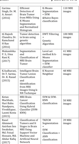
Comparative study of different brain tumor Detection and Classification techniques are summarized in compare table (Table I) with advantages and disadvantages. Most of the key features of methods are mentioned in Table I with respective limitations and benefits that make our work unique.
Table -1:Comparison of Brain tumor detection and classification techniques-I
Author Title Proposed
Technique Dataset
Parveen, Amritpal singh (2015)
Detection of brain tumor in MRI images, using
combination of FCM and SVM
FCM Segmentation + SVM classification 120 MRI images Astina Minz, Prof. Chndrakant Mahobiya (2017) MR Image classification using Adaboost for brain tumor type Adaboost & Neural Algorithms 50 MRI images Garima Singh, Dr. M. A. Ansari (2016)
Efficient Detection of Brain Tumor from MRIs Using K-Means Segmentation and Normalized Histogram K-Means Segmentation + SVM &Naïve Bayes classification 110 MRI images tG Rajesh Chandra, Dr. Ramchand H Rao (2016) Tumor detection in brain using genetic algorithm DWT Filtering + Genetic Algorithm 100 MRI images Mukambika P. S., Uma Rani K. (2017) Segmentation and Classification of MRI Brain Tumor Level set method & k-Means Segmentation + SVM classifier 41 MRI images K.Sudharani, Dr.T. Sarma, Dr. K. Rasad (2015) Intelligent Brain Tumor Lesion Classification and Identification from MRI Images Using k-NN Technique
K Nearest
Neighbor 48 MRI images
Ketan Machhale, Hari Babu Nandpuru, VivekKapur, Laxmi Kosta (2015) MRI Brain Cancer Classification Using Hybrid Classifier (SVM-KNN)
SVM & SVM-KNN
classification 50 MRI images
[image:3.595.302.566.61.544.2]Rasel Ahmmed, Anirban Sen Swakshar, Md. Foisal Hossain, Md. Abdur Rafiq (2017) Classification of Tumors and It Stages in Brain MRI Using Support Vector Machine and Artificial Neural Network TKFCM Segmentation + SVM Classification + ANN classification 39 MRI images
Table -2:Comparison of Brain tumor detection and classification techniques-II
Author Accuracy Benefits Limitations
Parveen, Amritpal singh (2015)
91.66% It combines clustering and classification algorithm Efficient method
Brain tumor type can’t be classified Difficult to choose SVM kernel function Astina Minz, Prof. Chndrakant Mahobiya (2017)
89.90% &
74.00% Minimize the error, Less time consuming
It can maximize the margin with respect to features that have already been selected. Garima
Singh, Dr. M. A. Ansari
91.49 &
87.23% Accurate results. Fast & efficient in
© 2018, IRJET | Impact Factor value: 6.171 | ISO 9001:2008 Certified Journal | Page 165
(2016) term of
computational time and cost
input data) of different size and Different density T.G. Rajesh
Chandra, Dr. Ramchand H Rao (2016)
90.00% Uses the ability of GA to solve optimization problems with large data set
Wavelet
transform require large storage and its computational cost is high.
Mukambika P. S., Uma Rani K. (2017)
94.12% & 82.35%
Increased Accuracy and Robust modeling
Potential of misidentificat-ion of what is supposed to be categorized
K.Sudharani , Dr.T. Sarma, Dr. K. Rasad (2015)
95.00 % Simple and flexible to implement, Handle multi-class cases
Large search problem to find nearest neighbor Storage of data
Ketan Machhale, Hari Babu Nandpuru, VivekKapur, Laxmi Kosta (2015)
98.00% Handle multi-class cases Increased Accuracy
When there is a change in dataset, fresh training dataset is required.
Rasel Ahmmed, Anirban Sen Swakshar, Md. Foisal Hossain, Md. Abdur Rafiq (2017)
97.44%. Increased Accuracy Classify brain tumor with brain tumor affected stages
Difficulty in selecting optimal features to distinguish different classes Time Consuming
4. CONCLUSIONS
In this paper we have accomplished a partial survey of various classification techniques for MRI brain image as well their advantages and disadvantages. A comparative study is made on various techniques. After evaluation of well-known technique it is clearly shown the various methods which can detect the tumor efficiently and provide accurate result. Though some algorithms producing accurate and reasonable results, at the same time they are having some limitations like it is not suitable for large data set and having longer computation time. One of the principal reasons might be the lack of standardized procedures. Computational time will also be considered to compare this technique efficiently. As the diagnosis tumor is a complicated and sensitive task, accuracy and reliability are always assigned much importance. For the future work we suggest to present more accurate, efficient as well as faster method for early detection and classification of brain tumors.
ACKNOWLEDGEMENT
I would like to thank the all faculty members of the Institute who helped us lot in calculating the facts and Figures related
to this review paper. I would also like to thank the researcher whom research on this field, provide important references for my paper. I am also thankful to all reviewers who gave their helpful review.
REFERENCES
[1] Mukambika P. S., Uma Rani K. “Segmentation and
Classification of MRI Brain Tumor” in International Research Journal of Engineering and Technology (IRJET) Volume: 04 Issue: 07 July -2017
[2] Parveen, Amritpal Singh “Detection of Brain Tumor in
MRI Images, using Combination of Fuzzy c-means and SVM” in 2nd International Conference on Signal Processing and Integrated Networks (SPIN) 2015
[3] Sathees B. Kumar, Dr. Anbu R. Selvi “Feature Extraction
Using Image Mining Techniques to Identify Brain Tumors” in IEEE Sponsored 2nd International Conference on Innovations in Information Embedded and Communication Systems ICIIECS 2015
[4] G Rajesh Chandra, Dr. Kolasani Ramchand H Rao “
TUMOR DETECTION IN BRAIN USING GENETIC ALGORITHM” in 7th International Conference on Communication, Computing and Virtualization 2016
[5] Sanjana Chawla, Rajiv Sharma “Application of Data
Mining in Bioinformatics ” in International Journal of Engineering Science and Computing, June 2016
[6] K. SUDHARANI, Dr. T. C. SARMA, Dr. K. SATYA RASAD
“Intelligent Brain Tumor Lesion Classification and Identification from MRI Images Using k-NN Technique” in International Conference on Control, lnstrumentation, Communication and Computational Technologies (lCCICCT) 2015
[7] Ashraf M. Said, Fatma S. Ibrahim “Comparative Study of
Segmentation Techniques for Detection of Tumors Based on MRI Brain Images” in International Journal of Bioscience, Biochemistry and Bioinformatics Issue September 2017
[8] Astina Minz, Prof. Chanddrakant Mahobiya “MR Image
classification using Adaboost for brain tumor type” in IEEE 7th International Advance Computing Conference (IACC) 2017
[9] Garima Singh, Dr. M. A. Ansari “Efficient Detection of
Brain Tumor from MRIs Using K-Means Segmentation and Normalized Histogram” in IEEE Issue 2016
[10] S.U.ASWATHY, Dr G.GLAN DEVA DHAS, Dr. S.S.KUMAR
© 2018, IRJET | Impact Factor value: 6.171 | ISO 9001:2008 Certified Journal | Page 166
[11] Ruchi D., Deshmukh et al “Study of Different Brain
Tumor MRI Image Segmentation Techniques” in International Journal of Computer Science Engineering and Technology( IJCSET) Vol 4, Issue 4,133-136 April 2014
[12] Mr.Deepak .C.Dhanwani, Prof. Mahip M.Bartere “Survey
on Various Techniques of Brain Tumor Detection from MRI Images” in International Journal of Computational Engineering Research Vol, 04 Issue, 1 Issn 2250-3005, January-2014
[13] Ketan Machhale, Hari Babu Nandpuru, Vivek Kapur,
Laxmi Kosta “MRI Brain Cancer Classification Using Hybrid Classifier (SVM-KNN) ” in international conference on instrumentation and control (ICIC) MAY 28-30, 2015.
[14] Rasel Ahmmed, Anirban Sen Swakshar, Md. Foisal