Comparative Evaluation of Sealing Ability of Various
Single-cone Systems: An in vitro Study
Tanika G Garg
ORIgInal aRtICle
Reader
Department of Conservative Dentistry and Endodontics, ITS Dental College, Greater Noida, Uttar Pradesh, India
Corresponding Author: Tanika G Garg, Reader, Department of Conservative Dentistry and Endodontics, ITS Dental College Greater Noida, Uttar Pradesh, India, e-mail: drtanikaendo@ gmail.com
ABSTRACT
Aim: Analyzing the sealing ability of contemporary single-cone obturation systems – gutta-percha/AH Plus, ActiV GP system,
and Resilon/Epiphany system, based on glucose filtration
method with spectrophotometer.
Materials and methods: Thirty-six extracted mandibular premolars were prepared using EndoWave rotary instruments. Samples were divided into three groups with 10 teeth each. Group I: Obturated with AH Plus sealer and gutta-percha cones (6%), group II: Obturated with ActiV GP sealer and ActiV GP cones (6%), group III: Obturated with Epiphany sealer and Resilon cones (6%), positive control (3 teeth): obturated without sealer with gutta-percha cones (6%), and negative control (3 teeth): Obturated as in group I and covered with sticky wax and nail varnish.
Results: Mean leakage of ActiV GP was significantly less
than gutta-percha/AH Plus and Resilon/Epiphany at 7th day (p < 0.05) and the mean leakages of gutta-percha/AH Plus, ActiV
GP system, and Resilon/Epiphany were not significant when
compared to each other at 14th, 21st, and 28th day (p > 0.05).
Conclusion: ActiV GP and Resilon/Epiphany produce a comparable seal to that of single gutta-percha cone with AH Plus sealer using a glucose penetration protocol.
Keywords: ActiV GP, EndoWave, Glucose penetration, Resilon/ Epiphany, Single-cone gutta-percha.
How to cite this article: Garg TG.Comparative Evaluation of Sealing Ability of Various Single-cone Systems: Anin vitroStudy. Int J Prev Clin Dent Res 2016;3(3):192-196.
Source of support: Nil
Conflict of interest: None
INTRODUCTION
One of the key goals of endodontic therapy is complete three-dimensional (3D) obturation of the root canal system. The ideal root canal obturating material should be well adapted to the canal walls and its irregularities and the entire length of the canal should be densely com-pacted with a homogenous mass of gutta-percha (GP).1
Lateral compaction of GP cones with a root canal sealer is considered to be a commonly accepted technique for obturation in endodontics. Despite close proximity to the dentinal walls, it has been shown that GP does not have a complete dentinal seal. Potential unfilled spaces may allow leakage along the sealer–dentin and sealer–root filling material interfaces or through voids within the sealer. Thus, the ability of a sealer to bond to the tooth structure and core material is of considerable importance.
Various types of sealers have been introduced to endodontics. Epoxy-resin sealers like AH Plus (Dentsply, Germany) have been used for many years and have been found to provide acceptable apical sealing. Studies have shown that it is biocompatible, has good tissue tolerance, and long-term dimensional stability.2,3
Interest in the application of adhesive technology to endodontics has led to the development of obturating systems with a specific focus on obtaining a monoblock. The systems that advertise this technology include Resilon/Epiphany, ActiV GP, and EndoREZ.
Resilon/Epiphany (Resilon Research LLC, Madison, CT), a new thermoplastic filled polymer, is composed of a parent polymer, polycaprolactone, which is bio-degradable aliphatic polyester. The material has the ability to bond with methacrylate-based resins since dimethacrylate monomers are blended into the polymer. The Epiphany system contains a self-etching primer and a dual-curable resin composite root canal sealer. By combin-ing self-etchcombin-ing adhesives and methacrylate-based resin sealers with Resilon, a monoblock bonding concept for the obturation of root canals is achieved.
ActiV GP Precision Obturation System (Brasseler USA, Savannah, GA) is a new glass-ionomer (GI)-based obturation system. The manufacturer claims the product to be superior to previous GI-based systems in terms of handling characteristics, working time, and radiopacity. Inadequate bonding between GI and GP is a drawback with GI-based sealers. To enhance the GI–GP bonding, ActiV GP has a 2 µm coating of GI particles on its surface.
Further, the advent of greater taper cones that closely match the geometry of nickel–titanium rotary systems in combination with resin-based sealer has rejuvenated interest in the single-cone obturation technique.4,5
Comparative Evaluation of Sealing Ability of Various Single-cone Systems: An in vitro Study IJPCDR
monoblock that prevents bacterial leakage and increases the fracture resistance of filled roots.
A wide variety of test methods have been used to assess the seal of endodontic materials, including dye penetration with methylene blue or India ink, dye extraction, fluid filtration, radioisotopes, electrochemical circuits, bacterial and endotoxin leakage, etc. Due to inadequacies associated with these types of testing methods as a result of nonexistence of a universally accepted model, glucose penetration studies might be meaningful and clinically relevant.
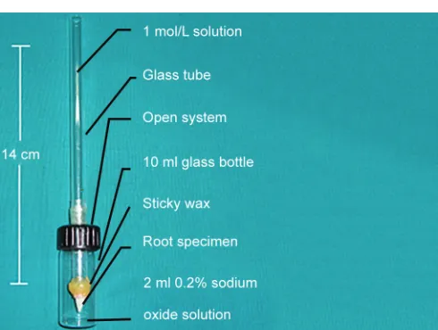
In 2005, Xu et al6 discussed a new model that allows
measurement of the glucose molecule. The model (Fig. 1) consists of a tube containing concentrated glucose solution, i.e., connected to the coronal aspect of the tooth, whilst the apical region is dipped in sodium azide solution. Glucose that accumulates in the apical chamber is measured with a spectrophotometer following an enzymatic reaction. Glucose has a low molecular weight of 180 Da, and may be used as an indication for toxins that might penetrate the canal. The glucose test was found to be more sensitive than the measurement of air-bubble movement.
Thus, the aim of the present study was to analyze the sealing ability of recently introduced single-cone obturation systems – GP/AH Plus, ActiV GP system, and Resilon/Epiphany system, based on the filtration rate of glucose along the root canal filling. The amount of solution leaked was quantified using enzymatic reaction with spectrophotometer.
MATERIALS AND METHODS
Thirty-six freshly extracted human permanent mandibu-lar premomandibu-lars were chosen for the study. The external surfaces of all the teeth were cleaned using ultrasonic instruments. Teeth were stored in 0.9% sodium chloride containing 0.02% sodium azide solution at 4°C for
pre-venting bacterial growth. Radiographs were taken to ensure that there was only a single canal in all the teeth.
For standardized samples preparation, the specimens were decoronated to create root segments that were approximately 14 mm long using diamond disk in a slow speed hand-piece under copious water irrigation.
The working length was established by passing #15-k file into the canal and was verified with the help of radiograph. The apex was sealed using sticky wax. The biomechanical preparation of the canals was performed with crown-down technique using EndoWave 0.06 taper NiTi rotary instruments. Canal preparation was started with the orifice shaper to prepare the coronal half of the canal, followed by the use of file #30/06 and then file #25/06 until it was 2 to 3 mm short of the estimated working length. Then with file #20/06 canal was prepared till full working length. Subsequently, #25/06 and #30/06 were used till full working length. This was followed by file #35/06 to get the desired apical preparation of #35. Preparation was done under constant irrigation with 1 mL of 5.25% NaOCl and recapitulation with # 15-k file between each instrument. Freshly prepared 17% ethylenediaminetetraacetic acid (EDTA) solution was used for smear layer removal, and EDTA was employed as final rinse followed by a rinse with distilled water.
All the samples were divided randomly into three groups of 10 teeth each based on the type of sealer and obturation material used. The remaining 6 teeth were equally divided into positive and negative control groups.
Group I (10 teeth): Obturated with AH Plus sealer and GP cones (6%).
Group II (10 teeth): Obturated with ActiV GP sealer and ActiV GP cones (6%).
Group III (10 teeth): Obturated with Epiphany sealer and Resilon cones (6%).
Positive control (3 teeth): Obturated without sealer with GP cones (6%).
Negative control (3 teeth): Obturated as in group A and dipped in molten sticky wax and further covered with nail varnish.
Radiographs were taken to ensure that there were no voids in the root canal filling. All specimens were stored in 100% relative humidity at 37°C in the incubator for 1 week to ensure complete set of the root canal sealer.
Statistical Analysis
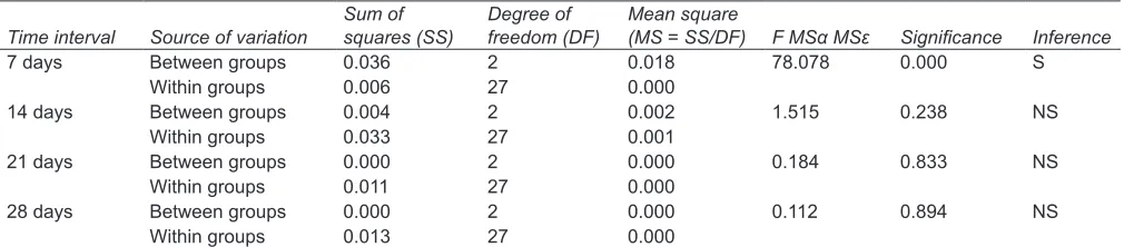
Descriptive statistics have been calculated for all the variables. One-way analysis of variance (ANOVA) with
post hoc analysis (Bonferroni) has been applied to see significance among the various groups at different time intervals (Table 1). The p-value of 0.05 has been consid-ered as statistically significant level.
RESULTS
All experimental obturating systems showed leakage at all time intervals: 7, 14, 21, and 28 days (Graph 1).
Leakage of GP/AH Plus sealer displays greater amount of leakage with increase in time interval from 7th day (0.166) to 14th day (0.272) to 21st day (0.418) till 28th day (0.465). Maximum leakage was found to be present at the end of 28th day.
Leakage of ActiV GP/ActiV GP sealer also displays greater amount of leakage with increase in time interval from 7th day (0.101) to 14th day (0.252) to 21st day (0.418) till 28th day (0.469). Maximum leakage was found to be present at the end of 28th day.
Leakage of Resilon/Epiphany sealer also displays greater amount of leakage with increase in time interval from 7th day (0.181) to 14th day (0.279) to 21st day (0.422) till 28th day (0.468). Maximum leakage was found to be present at the end of 28th day (Graph 2).
Positive control group displays greater amount of leakage with increase in time interval from 7th day (0.255) to 14th day (0.338) to 21st day (0.480) till 28th day (0.497). Maximum leakage was found to be present at the end of 28th day.
No leakage was observed in Negative control group at four time intervals for all three samples.
DISCUSSION
Three-dimensional sealing of the root canal with an impervious, biocompatible, and dimensionally stable filling material is one of the main goals of endodontic treatment. The use of GP in conjunction with a sealer– cement is currently one of the most commonly used methods for root canal obturation.
However, numerous in vitro studies have shown that the GP obturated teeth leak at high rate. The use of resin-based sealers in endodontics has been proposed as a means to create monoblocks within the root canal where the restoration and the tooth act as a single unit. Therefore, the search for the material that could adequately seal the root canal led to the development of obturating systems like Resilon/ Epiphany and ActiV GP.
The Resilon/Epiphany system consists of a resin-based sealer Epiphany and Resilon points. This polymeric root canal filling material bonds throughout the length of the root canal by creating micromechanical retention via formation of thin hybrid layer.
The ActiV GP obturation system was recently introduced and incorporates GI into the GP points to enhance the GI–GP bonding. It is used in conjunction with ActiV GP–GI sealer.
Graph 1: Mean leakage of all the three groups at
four time intervals Graph 2: Trend of leakage in all the three groups over a period of 28 days
Time interval Source of variation squares (SS) freedom (DF) (MS = SS/DF) F MSα MSε Significance Inference
7 days Between groups 0.036 2 0.018 78.078 0.000 S
Within groups 0.006 27 0.000
14 days Between groups 0.004 2 0.002 1.515 0.238 NS
Within groups 0.033 27 0.001
21 days Between groups 0.000 2 0.000 0.184 0.833 NS
Within groups 0.011 27 0.000
28 days Between groups 0.000 2 0.000 0.112 0.894 NS
Within groups 0.013 27 0.000
Comparative Evaluation of Sealing Ability of Various Single-cone Systems: An in vitro Study IJPCDR
With the application of adhesive dentistry to end-odontics and introduction of greater taper cones, there has been a renewed interest in the single-cone obturation technique.
Hence, we formulated a study to compare the sealing ability of contemporary single-cone obturation systems – Resilon/Epiphany, ActiV GP Precision System, and gutta-percha/AH Plus – with the help of spectrophotometry.
In the present study, cleaning and shaping procedure was followed for all the samples so as to obtain a uniform root canal preparation. The samples were divided into three groups and were obturated with the above three systems according to the manufacturer’s instructions.
Various studies have shown that none of the materials and techniques provides a leakproof seal. Hence, leakage tests are a relevant way to evaluate the apical seal. Methods used to evaluate leakage include dye penetration, electrochemical testing, radioisotope, bacterial leakage, fluid filtration, and glucose penetration.7
The fluid transport model has been shown to be more sensitive than dye penetration for the detection of through and through voids along root canals, and to be highly reproducible. However, the results of the fluid transport method may change by using different values of atmosphere pressure, capillary action, and the degree of canal dryness.8
Recently, the glucose penetration model, as a new possibility to evaluate the sealing ability of root canal fillings, has been introduced by Xu et al.6 We chose this
method for the evaluation of leakage as it has several advantages. Glucose is hydrophilic and chemically stable. It has a low molecular weight of 180 Da and may be used as an indication for toxins that might penetrate the canal.6 Glucose as a marker in leakage studies has
clinical relevance because it is an important nutrient for microorganisms.9 Sodium azide was used to inhibit
the growth of microorganisms that might influence the glucose readings.
In the glucose penetration model, the tooth is continu-ously subjected to the pressure of the glucose solution in the coronal chamber, for a period of 1 month. The fluid filtration model detects leakage usually after subjecting the filling to pressure for 3 hours. This enormous time difference might make the glucose test more sensitive, as it may result in the detection of small voids in the filling.9,10
As a limitation of the glucose penetration method for testing leakage, Shemesh et al11 reported that glucose
reacts with Portland cement, mineral trioxide aggregate and calcium hydroxide-based sealers. Therefore, these materials should not be evaluated for sealing ability with this method. Accordingly, we selected various obturation materials for our study.
The results of our study showed significantly greater (p < 0.05) leakage in the gutta-percha/AH Plus group (mean = 0.166) and Resilon/Epiphany group (mean = 0.181) when compared with the ActiV GP System (mean = 0.101) after the interval of 7 days. This is because the GI barrier after obturation with the ActiV GP system provides an immediate seal which is significantly more resistant to fluid movement than the seal provided by the other systems, thus making it an effective method of limiting coronal microleakage.12
At the end of the 4th week, there was no statistically significant difference (p > 0.05) in the leakage among all the three tested materials: Gutta-percha/AH Plus, ActiV GP system, and Resilon/Epiphany. This finding was in agreement with the results of Fransen et al,13 who tested
the sealing ability of the same materials via bacterial leakage method.
CONCLUSION
• The leakage shown by all the samples increased over the time and was maximum on the 28th day.
• Mean leakage of ActiV GP was significantly less than GP/AH Plus and Resilon/Epiphany at 7th day (p < 0.05).
• Mean leakages of GP/AH Plus, ActiV GP system, and Resilon/Epiphany were not significant when compared to each other at 14th, 21st, and 28th day (p > 0.05).
Within the limitation of this study, it can be concluded that the two contemporary single-cone obturation systems – ActiV GP and Resilon/Epiphany – produce a comparable seal to that of single GP cone with AH Plus sealer. Using a glucose penetration protocol, long-term evaluation of the sealing ability of root canal filling materials can be done with a logical approach. Further studies are required for these newer systems to improve their value as obturation materials.
REFERENCES
1. Epley SR, Fleischman J, Hartwell G, Cicalese C. Completeness of root canal obturations: epiphany techniques versus gutta-percha techniques. J Endod 2006 Jun;32(6):541-544.
2. De Moor RJ, Hommez GM. The long-term sealing ability of an epoxy resin root canal sealer used with five gutta-percha obturation techniques. Int Endod J 2002 Mar;35(3):275-282. 3. Drukteinis S, Peciuliene V, Maneliene R, Bendinskaite R.
In vitro study of microbial leakage in roots filled with EndoREZ sealer/EndoREZ Points and AH Plus sealer/conventional gutta-percha points. Stomatologija 2009;11(1):21-25.
4. Tay FR, Loushine RJ, Monticelli F, Weller RN, Breschi L, Ferrari M, Pashley DH. Effectiveness of resin-coated gutta-percha cones and a dual-cured hydrophilic methacrylate resin-based sealer in obturating root canals. J Endod 2005 Sep;31(9): 659-664.
6. Xu Q, Fan MW, Fan B, Cheung GSP, Hu HL. A new quan-titative method using glucose for analysis of endodontic leakage. Oral Surg Oral Med Oral Path Oral Radiol Endod 2005 Jan;99(1):107-111.
7. Verissimo DM, Vale MSD. Methodologies for assessment of apical and coronal leakage of endodontic filling materials: a critical review. J Oral Sci 2006 Sep;48(3):93-98.
8. Bodrumlu E, Semiz M. Antibacterial activity of a new end-odontic sealer against Enterococcus faecalis. J Can Dent Assoc 2006 Sep;72(7):637.
9. Sluis LWM, Shemesh H, Wu MK, Wesselink PR. An evalua- tion of the influence of passive ultrasonic irrigation on the seal of root canal fillings. Int Endod J 2007 May;40(5): 356-361.
structure and filled root canals. Int Endod J 2007 Nov;40(11): 866-872.
11. Shemesh H, Souza EM, Wu MK, Wesselink PR. Glucose reactivity with filling materials as a limitation for using the glucose leakage model. Int Endod J 2008 Oct;41(10):869-872. 12. Jack RM, Goodell GG. In vitro comparison of coronal microle-akage between Resilon alone and gutta-percha with a glass-ionomer intraorifice barrier using a fluid filtration model. J Endod 2008 Jun;34(6):718-720.