P R O T O C O L
Open Access
Protocol for a systematic review,
meta-analysis, and trial sequential analysis of
clinical outcomes following accelerated
versus conventional corneal collagen
cross-linking for corneal ectasia
Siddharth Nath
1,2*, Carl Shen
1, Alex Koziarz
3and Laura Banfield
4Abstract
Background:Collagen cross-linking (CXL) is an evolving procedure that enhances the biomechanical rigidity of the cornea and can slow or halt ectatic disease. CXL requires delivery of 5.4 J/cm2of ultraviolet A (UVA) radiation to a cornea saturated with riboflavin in order to induce cross-link formation. The conventional CXL procedure achieves this fluence by exposing the cornea to a 3 mW/cm2UVA lamp for 30 min; however, some surgeons have proposed accelerated protocols which achieve the same fluence in a shorter period of time by using a higher power light source. Whether accelerated protocols are as effective in arresting ectasia as the established conventional procedure remains unclear. Accordingly, this study will systematically review and synthesise the evidence on accelerated CXL and compare it to the conventional approach across an array of clinical outcomes.
Methods:We will search 16 electronic databases, including MEDLINE, Embase, and the Cochrane Library, from
inception to February 1, 2019. We will include all randomised controlled trials comparing accelerated and conventional CXL for any corneal ectatic disease. Two reviewers will independently screen search results to identify eligible articles, complete data collection, and conduct quality assessment. The quality of individual trials will be assessed using the Cochrane Collaboration’s Risk of Bias Assessment Tool. Our primary outcome will be the change in maximal keratometry (Kmax) at 12 months following treatment. Additional outcomes will
include: incidence of disease progression, incidence of serious adverse events, as well as change in Kmax at
longest follow-up, mean stromal demarcation line depth, mean uncorrected distance visual acuity, mean corrected distance visual acuity, meanKmax, mean endothelial cell density, mean central corneal thickness, mean spherical
equivalent, mean intraocular pressure, and mean corneal power, at 12 months following treatment. We will calculate relative risks and 95% confidence intervals (CIs) for dichotomous outcomes and weighted mean differences and corresponding 95% CIs for continuous outcomes. Prespecified subgroup analyses will be performed to investigate heterogeneity. We will rate the overall quality of evidence using the Grading of Recommendations Assessment, Development, and Evaluation (GRADE) approach.
(Continued on next page)
© The Author(s). 2019Open AccessThis article is distributed under the terms of the Creative Commons Attribution 4.0 International License (http://creativecommons.org/licenses/by/4.0/), which permits unrestricted use, distribution, and reproduction in any medium, provided you give appropriate credit to the original author(s) and the source, provide a link to the Creative Commons license, and indicate if changes were made. The Creative Commons Public Domain Dedication waiver (http://creativecommons.org/publicdomain/zero/1.0/) applies to the data made available in this article, unless otherwise stated. * Correspondence:[email protected]
1Division of Ophthalmology, Department of Surgery, McMaster University,
Hamilton, Ontario, Canada
2Department of Biochemistry and Biomedical Sciences, Michael G. DeGroote
(Continued from previous page)
Discussion:This review will provide a comprehensive evaluation of the evidence on accelerated and conventional CXL approaches and serve to inform clinical practice, medical device design, and future research. Evaluating variations of the CXL protocol aimed at reducing treatment duration is of critical importance and a prerequisite to expanding treatment availability to more patients.
Systematic review registration:PROSPERO CRD42018104151
Keywords:Cross-linking, Accelerated, Corneal ectasia, Keratoconus, Systematic review
Background
Corneal ectasias are progressive, degenerative ocular dis-eases which result from thinning, bulging, and structural changes in the peripheral, central, or paracentral cornea. These structural alterations distort vision, restrict lens fitting, and result in a substantial reduction in quality of life [1]. The most common corneal ectasia is keratoco-nus, which affects approximately 1 in 375 individuals [2]. Despite extensive research into the cellular, molecu-lar, and genetic mechanisms underlying ectatic pro-cesses, the pathophysiology of these diseases remains unclear, and there is a paucity of disease-modifying treatments [3]. Although many patients with ectasia may initially be managed with spectacles or rigid gas perme-able lenses, a subset can have continued disease progres-sion that requires a corneal graft [1].
Corneal collagen cross-linking (CXL) is a minimally invasive procedure proposed in the late twentieth cen-tury as a potential disease-modifying intervention for keratoconus [4]. The aim of CXL is to increase the bio-mechanical rigidity of the cornea by inducing cross-links both within and between collagen fibrils in the cor-neal stroma. This is achieved by using riboflavin as a photosensitiser, which, upon exposure to ultraviolet A (UVA) light, induces cross-link formation via the nat-ural lysyl oxidase pathway [4, 5]. These increased cross-links serve to stabilise the structure of the cor-nea and enhance its stiffness, thereby protecting against further degeneration. Animal studies investi-gating the effect of CXL on corneal structure have been largely supportive of this hypothesis, finding sig-nificantly increased biomechanical rigidity in treated corneas [6–8]. CXL was approved for treating kerato-conus and other corneal ectasias in the early 2000s across Europe and in 2016 in the USA [9, 10].
The conventional approved CXL technique, commonly referred to as the‘Dresden protocol’, begins with removal of the central 7 mm of corneal epithelium, followed by the administration of riboflavin both prior to and during exposure of the cornea to UVA light. A fluence of 5.4 J/ cm2was determined to be ideal for cross-link formation, and this protocol achieves it through exposure to a 3 mW/cm2 lamp for 30 min [9]. Multiple aspects of the
conventional protocol have been modified by surgeons in efforts to refine the procedure and achieve improved safety and efficacy.
In particular, clinicians have focused on minimising the UVA light exposure time, in order to decrease the dur-ation of the procedure. Harnessing the Bunsen-Roscoe law of reciprocity, which states that a biological effect is proportional only to the energy dose and not the method of energy delivery, some surgeons have introduced accel-erated CXL [11]. This modified CXL protocol achieves the same 5.4 J/cm2fluence of UVA radiation in a shorter dur-ation of time by implementing a stronger UVA light source. The decrease in exposure time is inversely propor-tional to the increase in UVA light power (e.g., 3 mW/cm2 for 30 min translates to 9 mW/cm2for 10 min). Shorten-ing the treatment duration may improve patient comfort and minimise adverse events linked to prolonged corneal exposure [12]. Moreover, with a shorter procedure, clini-cians can rapidly expand treatment availability to a greater number of patients and intervene to arrest progressive ectasia more swiftly.
Why it is important to do this review
Although accelerated CXL is rooted in principles of physics and offers considerable theoretical benefits, it re-mains unclear whether it induces similar biomechanical changes in the cornea as the conventional protocol. Whether the increased UVA light power is associated with additional adverse events is also uncertain. Rando-mised trials and observational studies comparing accel-erated and conventional CXL have shown promising results; however, the field has yet to reach a consensus [13, 14]. Thus, we will undertake a comprehensive sys-tematic review and meta-analysis in order to evaluate the safety and efficacy of accelerated CXL in comparison to the conventional protocol.
Methods
Objective
Registration
This study protocol has been developed in line with the Preferred Reporting Items for Systematic Reviews and Meta-Analysis Protocols (PRISMA-P) [15] statement and is registered with the International Prospective Register of Systematic Reviews (PROSPERO; registration number: CRD42018104151). We will conduct our sys-tematic review and meta-analysis in accordance with the Preferred Reporting Items for Systematic Reviews and Meta-Analyses (PRISMA) [16, 17] guidelines and the Cochrane Collaboration’s Handbook for Systematic Re-views of Interventions [18]. If there are insufficient ran-domised trials (fewer than three) available to conduct a systematic review and meta-analysis, we will include ob-servational studies and implement guidelines set forth by the Meta-analysis of Observational Studies in Epi-demiology (MOOSE) consensus statement [19]. This protocol will be updated as required by PRISMA-P cri-teria [15], and amended versions will be made available on PROSPERO.
Literature search
We will conduct a detailed search of the following elec-tronic databases: MEDLINE, Embase, Web of Science,
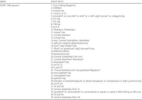
Cochrane CENTRAL, Cochrane DSR, CINAHL, Open-Grey, metaRegister of Controlled Trials, LILACS, Clinical-Trials.gov, WHO Clinical Trials Database, WangFangData, CQVIP, INSPEC, COMPENDEX, and CNKI, from incep-tion through February 1, 2019. We will use medical subject heading (MeSH) terms and keywords related to corneal ectasia and the cross-linking approach (accelerated or con-ventional) as well as clinical outcomes. Search strategies will be designed by a multidisciplinary research team composed of clinicians, researchers, and academic librarians with ex-pertise in conducting systematic reviews. The proposed search strategy for the MEDLINE database is provided in Table 1. Our electronic search will be supplemented by manually screening the references of eligible articles, reviewing the proceedings of relevant meetings, and con-tacting clinical experts in the field. We will conduct our search without any restrictions on publication type, lan-guage, or time.
Study selection
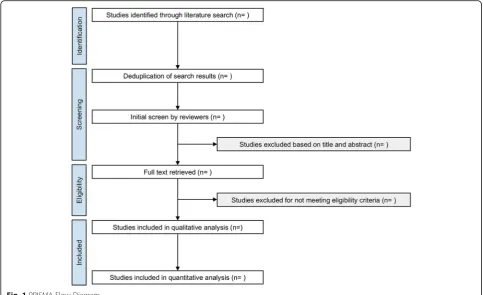
Search results will be evaluated independently by two re-viewers (SN, CS) against predefined eligibility criteria to identify relevant studies (Fig. 1). Results from all searched databases will be exported as .RIS, .XML, or
Table 1Search strategy for the MEDLINE electronic database using the Ovid interface
Database Search terms
MEDLINE 1946–present 1 Cross-Linking Reagents/
2 CORNEA/ 3 cornea*.mp. 4 1 and (2 or 3)
5 ((crosslink* or cross link* or xlink* or x link*) adj3 (cornea* or collagen)).mp. 6 CCL.mp.
7 CXL.mp. 8 C3R.mp. 9 or/4–8
10 Dilatation, Pathologic/ 11 ectasia*.mp. 12 corneal diseases/ 13 cornea*.mp.
14 exp. Corneal Dystrophies, Hereditary/ 15 pellucid marginal degeneration.mp. 16 (fuch* adj2 atroph*).mp.
17 ((fuch* or groenouw*) adj2 dystroph*).mp. 18 KERATOCONUS/
19 keratoconus.mp.
20 Corneal Endothelial Cell Loss/ 21 Corneal Wavefront Aberration/ 22 keratopath*.mp.
23 or/10–22 24 9 and 23
25“Transendothelial and Transepithelial Migration”/ 26 trans-epitheli*.mp.
27 transepitheli*.mp. 28 epitheli*.mp.
29 (dresden or phototherapeutic or photo-therapeutic or iontophoresis or tight junctions).mp. 30 or/25–29
31 24 and 30
32 remove duplicates from 31
33 (accelerat* or nonaccelerat* or conventional or regular or rapid or flash-linking or KXL).mp. 34 24 and 33
.CIW files containing the complete reference, and End-Note X9 (Clarivate Analytics, Philadelphia, USA) soft-ware will be used for reference management. Reviewers will screen titles and abstracts against inclusion criteria, and full articles will be retrieved for all references that meet these criteria, or where there is any ambiguity. In cases of ambiguity, the complete report of the specific study will be screened independently by both reviewers in order to reach a judgement on inclusion. Disagree-ments between reviewers will be resolved by deliberation and consensus, and, if needed, including an impartial third reviewer (AK), or contacting the trial authors.
Eligibility criteria
Our inclusion criteria will be:
Population:patients of any demographic undergoing CXL for treatment of corneal ectasia following refractive surgery, keratoconus, or pellucid marginal degeneration.
Intervention:accelerated CXL Control:conventional CXL
Outcomes:clinical outcomes such as change in maximal keratometry (Kmax) at 12 months after
treatment (primary outcome), incidence of serious adverse events, as well as incidence of disease progression, change inKmaxat longest follow-up,
mean stromal demarcation line depth, meanKmax,
mean uncorrected distance visual acuity (UDVA), mean corrected distance visual acuity (CDVA), mean endothelial cell density, mean central corneal thickness, mean spherical equivalent, mean intraocular pressure, and mean corneal power, at 12 months following treatment.
infection) will also be excluded. We will record reasons for excluding studies.
Data management and collection
Data from included studies will be collected independ-ently by two reviewers (SN, CS) and confirmed for ac-curacy by a third reviewer (AK). Prior to data extraction, the complete reports of all studies meeting our inclusion criteria will be collated, and reviewers will develop and pilot data extraction forms. For studies not published in English, the complete article will be translated into Eng-lish and a clinical expert fluent in the original language of the study will be consulted. Discrepancies in data ex-traction will be resolved collaboratively by discussion amongst the two primary reviewers (SN, CS), conferring with an independent third reviewer (AK), or contacting the original trial authors. Where data in included studies is incomplete or ambiguous, we will contact study au-thors for further information and clarification.
We will extract data from eligible studies using forms with fields for the following: study first author, year of publication, journal of publication, language, study de-sign, included centres, included countries, number of pa-tients, number of males, number of females, recruitment period, eligibility criteria, method of randomisation, indi-cation for CXL, number of patients in accelerated and conventional groups, age of patients in accelerated and conventional groups, procedure for follow-up, number of patients with disease progression 12 months following CXL in accelerated and conventional groups, Kmax
be-fore CXL in accelerated and conventional groups, Kmax
12 months after CXL in accelerated and conventional groups, Kmaxat longest follow-up after CXL in
acceler-ated and conventional groups, UDVA before CXL in ac-celerated and conventional groups, UDVA 12 months after CXL in accelerated and conventional groups, CDVA before CXL in accelerated and conventional groups, CDVA 12 months after CXL in accelerated and conventional groups, central corneal thickness before CXL in accelerated and conventional groups, central corneal thickness 12 months after CXL in accelerated and conventional groups, endothelial cell density before CXL in accelerated and conventional groups, endothelial cell density 12 months after CXL in accelerated and con-ventional groups, intraocular pressure before CXL in ac-celerated and conventional groups, intraocular pressure 12 months after CXL in accelerated and conventional groups, corneal power before CXL in accelerated and conventional groups, corneal power 12 months after CXL in accelerated and conventional groups, spherical equivalent before CXL in accelerated and conventional groups, spherical equivalent 12 months after CXL in ac-celerated and conventional groups, stromal demarcation line depth after CXL in accelerated and conventional
groups, method for epithelium removal, method for transepithelial riboflavin application, postoperative pain following accelerated and conventional CXL, time to best CDVA following accelerated and conventional CXL, time to best UDVA following accelerated and conven-tional CXL, power of UVA light in accelerated group, UVA light exposure time in accelerated group, incidence of significant complications (e.g., corneal melt, persistent epithelial defects, scarring, and persistent stromal haze) after accelerated and conventional CXL, and protocol preference as reported by authors.
Risk of bias in individual studies
All included trials will be assessed using the Cochrane Collaboration’s Risk of Bias Assessment Tool by two in-dependent authors (SN, CS) [20]. Studies will be assessed to determine the risk of selection, performance, detection, attrition, reporting, and other biases. Discrep-ancies in quality assessment will be resolved collegially by deliberation and consensus, and consultation with an impartial third reviewer (AK). If there is insufficient in-formation available to make a judgement on risk within an individual domain, we will rank that domain as‘ un-clear’and the original trial authors will be contacted for further information. Studies with one or more domains assessed to be‘high risk’will be categorised as having an overall high risk of bias.
Definition of outcomes
Our primary outcome will be the change in Kmax (in
dioptres, D) over 12 months. Additional outcomes shall be the following: incidence of disease progression (de-fined as an increase of the Kmax by ≥1.0 dioptres (D)
over 12 months) after treatment, as well as change in: mean UDVA (in logMAR) at 12 months following treatment, mean CDVA (in logMAR) at 12 months fol-lowing treatment, mean endothelial cell density (in cells/ mm2) at 12 months following treatment, mean Kmax(D)
at longest follow-up after treatment, mean stromal de-marcation line depth (inμm) after treatment, mean cen-tral corneal thickness (in μm) at 12 months following treatment, mean spherical equivalent (D) at 12 months following treatment, mean intraocular pressure (in mmHg) at 12 months following treatment, mean corneal power (D) at 12 months following treatment, and the mean Kmax (D) at 12 months after treatment. We will
also examine the incidence of serious adverse events (e.g., corneal melt, persistent epithelial defects, scarring, and persistent stromal haze).
Data synthesis
progression and incidence of serious adverse events, we will summarise our analyses by calculating the relative risk (RR) with corresponding 95% confidence interval (CI). For continuous outcomes, such as change inKmax,
mean Kmax, mean UDVA, mean CDVA, mean central
corneal thickness, mean stromal demarcation line depth, mean endothelial cell density, mean intraocular pressure, mean corneal power, and mean spherical equivalent, at 12 months following treatment, we will compute the weighted mean difference (MD) with 95% CI. Moreover, we will determine pooled estimates of all incidences across studies for the accelerated group, and then separ-ately for the conventional group. The DerSimonian and Laird random effects model will be used to conduct our meta-analysis, and weights will be calculated using the inverse variance method [21]. The threshold for type I error for statistical significance shall be α= 0.05. Between-study heterogeneity will be assessed using Cochran’s Q test and quantified by the I2statistic, with I2
values in excess of 25%, 50%, and 75%, graded as low, moderate, and high heterogeneity, respectively [22]. Pub-lication bias will be examined qualitatively by visual in-spection of funnel plot symmetry and quantified by Begg and Mazumdar’s [23] and Egger’s tests [24].
We will undertake prespecified subgroup analyses to investigate whether covariates exist and to examine het-erogeneity in our primary outcome. Analyses will be per-formed for subgroups stratified by patient age and sex, CXL technique (transepithelial versus epithelium-off ), severity of disease at time of treatment, and study risk of bias (low versus high). In addition, we will conduct a sensitivity analysis by performing trial sequential analysis (TSA) [25, 26]. TSA adjusts for the risk of type I error and constructs monitoring boundaries to determine whether an intervention is beneficial, harmful, or futile in comparison to control. We will perform TSA with an overall power of 80% and type I error risk of 5%.
The quality of available evidence will be summarised using the Grading of Recommendations Assessment, De-velopment, and Evaluation (GRADE) approach [27]. Evi-dence will be assessed for the domains of risk of bias, consistency, precision, reporting bias, directness, and other bias. The evidence for each outcome will be ranked as being of high, moderate, low, or very low quality in ac-cordance with criteria set by the GRADE working group.
All statistical analyses will be conducted using Com-prehensive Meta-analysis v.3.3.070 (Biostat, Englewood, New Jersey, USA), and TSA will be performed using Trial Sequential Analysis v0.9.5.10 Beta (Copenhagen Trial Unit, Copenhagen, Denmark).
Discussion
The results of this review are expected to considerably in-form clinical practice, future research, and the management
of patients with ectatic disease. By comprehensively exam-ining the evidence on accelerated and conventional CXL approaches, this study is expected to influence how clini-cians perform this evolving treatment and how UVA cross-linking devices are engineered and, potentially, im-prove patient outcomes. Moreover, through analysing the safety and efficacy of accelerated CXL, this study may incite changes leading to shorter operative times and increased treatment availability. Findings from this study will be sub-mitted to a peer-reviewed journal for publication and will be presented at conferences and seminars.
Abbreviations
CDVA:Corrected distance visual acuity; CI: Confidence interval; CXL: Corneal collagen cross-linking; GRADE: Grading of Recommendations Assessment, Development, and Evaluation;Kmax: Maximal keratometry; MD: Mean
difference; MeSH: Medical subject heading; PRISMA: Preferred Reporting Items for Systematic Review and Meta-Analyses; PRISMA-P: Preferred Reporting Items for Systematic Review and Meta-Analysis Protocols; PROSPERO: International Prospective Register of Systematic Reviews; RR: Relative risk; TSA: Trial sequential analysis; UDVA: Uncorrected distance visual acuity; UVA: Ultraviolet A
Acknowledgements
Not applicable.
Funding
This research received no specific grant from any funding agency in the public, private, or not-for-profit sectors.
Availability of data and materials
Data sharing is not applicable to this article as no datasets were generated or analysed during the current study.
Authors’contributions
SN and CS conceived and designed the study. SN and LB developed the search strategy and SN, LB, and CS piloted it in electronic databases. SN and CS developed the review protocol, eligibility criteria, risk of bias assessment strategy, and data management processes. SN, CS, and AK established the data synthesis methodology. All authors critically revised and commented on the intellectual content of the manuscript. All authors read and approved the final manuscript.
Ethics approval and consent to participate
Not applicable.
Consent for publication
Not applicable.
Competing interests
The authors declare that they have no competing interests.
Publisher’s Note
Springer Nature remains neutral with regard to jurisdictional claims in published maps and institutional affiliations.
Author details
1Division of Ophthalmology, Department of Surgery, McMaster University,
Hamilton, Ontario, Canada.2Department of Biochemistry and Biomedical
Sciences, Michael G. DeGroote School of Medicine, Faculty of Health Sciences, McMaster University, 1200 Main Street West, Room 4N53, Hamilton, Ontario L8N 3Z5, Canada.3Faculty of Medicine, University of Toronto,
Toronto, Ontario, Canada.4Health Sciences Library, McMaster University,
Received: 26 September 2018 Accepted: 25 March 2019
References
1. Maharana PK, Dubey A, Jhanji V, Sharma N, Das S, Vajpayee RB. Management of advanced corneal ectasias. Br J Ophthalmol. 2016;100:34–40.
2. Godefrooij DA, de Wit GA, Uiterwaal CS, Imhof SM, Wisse RP. Age-specific incidence and prevalence of keratoconus: a nationwide registration study. Am J Ophthalmol. 2017;175:169–72.
3. Romero-Jiménez M, Santodomingo-Rubido J, Wolffsohn JS. Keratoconus: a review. Cont Lens Anterior Eye. 2010;33:157–66.
4. Spoerl E, Huhle M, Seiler T. Induction of cross-links in corneal tissue. Exp Eye Res. 1998;66:97–103.
5. O’Brart DPS. Corneal collagen cross-linking: a review. J Optom. 2014;7:113–24. 6. Spoerl E, Huhle M, Seiler T. Erhöhung der Festigkeit der Hornhaut durch
Vernetzung. Ophthalmologe. 1997;94:902–6.
7. Bueno JM, Avila FJ, Martinez-Garcia MC. Quantitative Analysis of the Corneal Collagen Distribution afterIn VivoCross-Linking with Second Harmonic Microscopy. BioMed Research international. 2019;1–12.https://doi.org/10. 1155/2019/3860498.
8. Spoerl E, Schreiber J, Hellmund K, Seiler T, Knuschke P. Untersuchungen zur Verfestigung der Hornhaut am Kanichen. Ophthalmologe. 2000;97:203–6. 9. Wollensak G, Spoerl E, Seiler T. Riboflavin/ultraviolet-a-induced collagen
crosslinking for the treatment of keratoconus. Am J Ophthalmol. 2003;135:620–7. 10. Mohammadpour M, Masoumi A, Mirghorbani M, Shahraki K, Hashemi H.
Updates on corneal collagen cross-linking: indications, techniques and clinical outcomes. J Curr Ophthalmol. 2017;29:235–47.
11. Bunsen RW, Roscoe BA. Photochemical researches–part V. On the measurement of the chemical action of direct and diffuse sunlight. Proc R Soc Lond. 1862;12:306–12.
12. Woo JH, Iyer JV, Lim L, et al. Conventional versus accelerated collagen cross-linking for keratoconus: a comparison of visual, refractive, topographic and biomechanical outcomes. Open Ophthalmol J. 2017;11:262–72.
13. Sadoughi MM, Einollahi B, Baradaran-Rafii A, Roshandel D, Hasani H, Nazeri M. Accelerated versus conventional corneal collagen cross-linking in patients with keratoconus: an intrapatient comparative study. Int Ophthalmol. 2018;38:67–74.
14. Elbaz U, Shen C, Lichtinger A, et al. Accelerated (9-mW/cm2) corneal collagen
crosslinking for keratoconus–a 1-year follow-up. Cornea. 2015;33:769–73. 15. Shamseer L, Moher D, Clarke M, et al. Preferred reporting items for
systematic review and meta-analysis protocols (PRISMA-P) 2015: elaboration and explanation. BMJ. 2015;350:g7647.
16. Moher D, Liberati A, Tetzlaff J, et al. Preferred reporting items for systematic reviews and meta-analyses: the PRISMA statement. BMJ. 2009;339:b2535. 17. Liberati A, Altman DG, Tetzlaff J, et al. The PRISMA statement for reporting
systematic reviews and meta-analyses of studies that evaluate healthcare interventions: explanation and elaboration. BMJ. 2009;229:b2700. 18. Cochrane handbook for systematic reviews of interventions. 5.1.0 ed. The
Cochrane Collaboration, 2011.
19. Stroup DF, Berlin JA, Morton SC, et al. Meta-analysis of observational studies in epidemiology: a proposal for reporting. Meta-analysis of Observational Studies in Epidemiology (MOOSE) group. JAMA. 2000;283:2008–12. 20. Higgins JP, Altman DG, Gøtzsche PC, et al. The Cochrane Collaboration’s
tool for assessing risk of bias in randomised trials. BMJ. 2011;343:d5928. 21. DerSimonian R, Laird N. Meta-analysis in clinical trials. Control Clin Trials.
1986;7:177–88.
22. Higgins JP, Thompson SG, Deeks JJ, et al. Measuring inconsistency in meta-analyses. BMJ. 2003;327:557–60.
23. Begg CB, Mazumdar M. Operating characteristics of a rank correlation test for publication bias. Biometrics. 1994;50:1088–101.
24. Egger M, Davey Smith G, Schneider M, et al. Bias in meta-analysis detected by a simple, graphical test. BMJ. 1997;315:629–34.
25. Brok J, Thorlund K, Gluud C, Wetterslev J. Trial sequential analysis reveals insufficient information size and potentially false positive results in many meta-analyses. J Clin Epidemiol. 2008;61:763–9.
26. Wetterslev J, Thorlund K, Brok J, Gluud C. Trial sequential analysis may establish when firm evidence is reached in cumulative meta-analysis. J Clin Epidemiol. 2008;61:64–75.