R E V I E W
Open Access
Intersection of pathological tau and
microglia at the synapse
Thomas Vogels
1,2, Adriana-Natalia Murgoci
2and Tomá
š
Hromádka
1,2*Abstract
Tauopathies are a heterogenous class of diseases characterized by cellular accumulation of aggregated tau and
include diseases such as Alzheimer
’
s disease (AD), progressive supranuclear palsy and chronic traumatic
encephalopathy. Tau pathology is strongly linked to neurodegeneration and clinical symptoms in tauopathy
patients. Furthermore, synapse loss is an early pathological event in tauopathies and is the strongest correlate of
cognitive decline. Tau pathology is additionally associated with chronic neuroinflammatory processes, such as
reactive microglia, astrocytes, and increased levels of pro-inflammatory molecules (e.g. complement proteins,
cytokines). Recent studies show that as the principal immune cells of the brain, microglia play a particularly
important role in the initiation and progression of tau pathology and associated neurodegeneration. Furthermore,
AD risk genes such as Triggering receptor expressed on myeloid cells 2 (TREM2) and Apolipoprotein E (APOE) are
enriched in the innate immune system and modulate the neuroinflammatory response of microglia to tau
pathology. Microglia can play an active role in synaptic dysfunction by abnormally phagocytosing synaptic
compartments of neurons with tau pathology. Furthermore, microglia are involved in synaptic spreading of tau
–
a
process which is thought to underlie the progressive nature of tau pathology propagation through the brain.
Spreading of pathological tau is also the predominant target for tau-based immunotherapy. Active tau vaccines,
therapeutic tau antibodies and other approaches targeting the immune system are actively explored as treatment
options for AD and other tauopathies. This review describes the role of microglia in the pathobiology of
tauopathies and the mechanism of action of potential therapeutics targeting the immune system in tauopathies.
Keywords:
Tau pathology, Tau immunotherapy, Microglia, Astrocytes, Synaptic dysfunction, Complement,
Neurodegeneration, Neuroinflammation, APOE4, TREM2
Introduction
The role of microglia in tauopathies
Pathological tau protein is observed a wide range of
neu-rodegenerative disorders (NDD) and is the key defining
feature of a heterogeneous class of diseases called
tauo-pathies. Alzheimer’s disease (AD) is the most common
tauopathy - affecting approximately 45 million people
worldwide
–
and is additionally characterized by
extra-cellular plaques composed of amyloid beta (A
β
) [265].
Less common tauopathies include Picks’
disease (PiD),
corticobasal degeneration (CBD), progressive
supra-nuclear palsy (PSP), argyrophilic grain disease (AGD),
and chronic traumatic encephalopathy (CTE). In AD
and other tauopathies, tau pathology closely correlates
with neurodegeneration and functional decline [11,
115,
147,
211,
232]. Additionally, tauopathies are
character-ized by early synaptic dysfunction. Tau-induced damage
in synaptic compartments ultimately leads to major
syn-apse loss, which is the closest correlate of cognitive
de-cline
[76,
148,
263,
264].
Furthermore,
synaptic
connections are the principal sites at which pathological
tau can spread from diseased to healthy neurons
–
a
process which is thought to underlie the progressive
na-ture of tau pathology throughout the brain [218].
Tauo-pathies are also characterized by reactive gliosis and an
increase in inflammatory molecules such complement
proteins and pro-inflammatory cytokines
–
collectively
referred to as neuroinflammation [65,
79,
106,
144,
205,
233,
244,
267,
271,
272,
274]. The purpose of the
neu-roinflammatory state is to remove the cause (e.g.
© The Author(s). 2019Open AccessThis article is distributed under the terms of the Creative Commons Attribution 4.0 International License (http://creativecommons.org/licenses/by/4.0/), which permits unrestricted use, distribution, and reproduction in any medium, provided you give appropriate credit to the original author(s) and the source, provide a link to the Creative Commons license, and indicate if changes were made. The Creative Commons Public Domain Dedication waiver (http://creativecommons.org/publicdomain/zero/1.0/) applies to the data made available in this article, unless otherwise stated. * Correspondence:[email protected]
1Axon Neuroscience R&D Services SE, Bratislava, Slovak Republic 2Institute of Neuroimmunology, Slovak Academy of Sciences, Bratislava,
pathogens, protein aggregates, damaged cells) and return
the tissue to homeostasis. However, it is not clear if
neu-roinflammation in tauopathies is mostly protective or
damaging and how this depends on disease stage.
Multiple cell types can have immune functions in the
brain, for example microglia, astrocytes, perivascular
macrophages, meningeal macrophages, choroid plexus
macrophages, and infiltrating peripheral myeloid cell
types [304]. However, microglia are of particular interest
as they are the principal macrophages of the CNS and
exciting recent research has shown novel roles for these
immune cells in both health and disease. Additionally,
genome-wide association studies (GWAS) have
identi-fied several late onset AD (LOAD) risk variants that are
found in proteins that are predominantly expressed in
the innate immune system and microglia (e.g. APOE,
TREM2, ABCA7, CD33, CR1) [207]. This strongly
impli-cates microglia as central players in the development of
LOAD [124]. Given the central role of tau pathology in
AD and other tauopathies, there is now increasing
inter-est in how microglia are involved in the pathobiology of
tau protein. It is currently unclear if altered microglial
function is a cause, consequence, or contributor to tau
pathology. Secreted factors from microglia may lead to
initiation of tau aggregation in neurons [116]. Microglia
may be also involved in tau-induced synapse loss and
tau spreading, and play an important role in the
mech-anism of action of tau immunotherapy and other
thera-peutics aimed at treating tauopathies [12,
99,
135,
192].
This review provides an overview of how interaction of
tau pathology and microglia leads to synaptic
dysfunc-tion in tauopathies. Furthermore, we provide an
over-view of the published preclinical in vivo studies of tau
immunotherapy and immune-related pathways for the
treatment of tauopathies.
Tau pathology
Tau is an abundant protein that is predominantly
expressed in the axonal compartment of neurons, but
also at lower levels in oligodendrocytes and astrocytes
[16]. The main function of tau is to regulate the
assem-bly, nucleation and bundling of microtubules and to
modulate axonal transport [122]. In addition, recent
re-search suggests that tau may also have a multitude of
other physiological functions [282]. Tau protein is
encoded by the microtubule-associated protein tau
(MAPT
) gene on chromosome 17q21.31, and this gene
can be mutated, inverted, duplicated, and abnormally
methylated. All these modifications have been associated
with increased risk of developing tauopathy and the
gen-etic evidence therefore clearly links tau to
neurodegener-ation [18,
138,
139,
176].
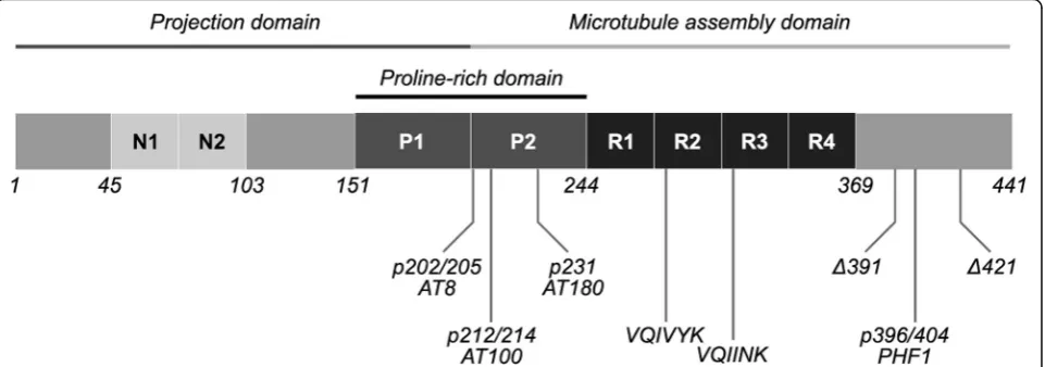
The human brain contains 6 isoforms generated by
al-ternative splicing of exons 2, 3 and 10 of the
MAPT
gene
[314]. Tau can have either 0, 1 or 2 N-terminal inserts
and either 3 or 4 pseudo-repeats (R), resulting in
iso-forms ranging from 352 to 441 amino acids (aa) (36.7–
45.9 kDa) (Fig.
1) [112]. Tau protein can be subdivided
into several domains: a structurally disordered
N-terminal, the proline rich mid-domain and a highly
con-served C-terminal which includes microtubule binding
repeats (MTBR). Tau is also subject to a wide range of
post-translational modifications (PTMs) (e.g.
phosphor-ylation, acetphosphor-ylation, truncation), which alter its structure,
function, and subcellular localization [171,
325]
.
The six
isoforms in combination with the multitude of potential
PTMs make the biology of tau extraordinarily complex.
Belonging to the class of natively unfolded or
intrinsic-ally disordered proteins, tau proteins lack clearly defined
secondary and tertiary structures.
The MTBR of tau contains two hexapeptides that can
form intermolecular beta sheet rich structures: aa275–
280 (VCIINK) in R2 and aa306–311 (VQIVYK) in R3
[307,
308]. Pathological conformations of tau can
inter-act with physiological tau, leading to aggregation and
ul-timately formation of highly structured insoluble fibrils
which deposit into the cell as neurofibrillary tangles
(NFTs). This process is referred to as templated
misfold-ing, seeded nucleation, or simply seeding [97]. As tau is
a highly soluble protein and the initial aggregation phase
is thermodynamically unfavorable, it is currently unclear
how tau shifts from its dynamic physiological structure
to a misfolded monomer that is prone to aggregation
[212,
270]. Specific patterns of PTMs may change the
conformation of the protein, causing tau to become
seed-competent [62,
77]. Moreover, dynamic
phosphor-ylation of the residues in the MTBR or flanking regions
regulates the affinity of tau for tubulin and
hyperpho-sphorylation may thereby increase the pool of free tau
available for aggregation [160]. The phosphorylation of
tau is regulated by both kinases (e.g. cdk5, GSK-3
β
,
p38-MAPK) and phosphatases (e.g. PP2A) [142].
Phosphoryl-ation at a number of sites on tau has been linked to tau
pathology (e.g. Ser202/Thr205, Thr212/Ser214, Thr231,
Ser396/Ser404, Fig.
1) [314]. Abnormal cleavage can
po-tentially play an important role in tauopathies, as several
truncated fragments have an increased propensity for
ag-gregation and their overexpression leads to
neurofibril-lary pathology in rodents [87,
329]. As will be discussed
later in this review, factors secreted from microglia can
lead to abnormal patterns of PTMs and may therefore
play a role in the initiation of tau aggregation.
the extracellular space and taken up by healthy neurons
[98,
121,
162]. This process may be of critical
import-ance as it is thought to underlie the progression of tau
pathology throughout the brain. Interestingly, it has
already been observed in the classical Braak staging
scheme that the progression of tau pathology seems to
occur along neuronal connections [43]. It has been
dem-onstrated using a variety in vitro and in vivo approaches
that tau pathology predominantly spreads along synaptic
connections [48,
73,
318]. Recent studies have made
sig-nificant progress in showing that this also occurs in the
brain of Alzheimer’s patients: seed-competent tau is
present in axons of white matter tracts and
synapto-somes, and tau seeding occurs in synaptically connected
areas before the occurrence of hyperphosphorylated tau
in these regions [78,
100,
158,
159]. It is currently
un-clear what the major mechanism of synaptic tau
secre-tion is, but the evidence so far suggests: (1) release from
synaptic vesicles [242] (2) secretion in extracellular
vesi-cles such as exosomes [241,
256,
313] and ectosomes
[80], (3) direct translocation across the membrane [157,
208] or (4) tunneling nanotubes [1,
295]. Similarly,
sev-eral tau uptake mechanisms have been identified which
are not mutually exclusive: (1) bulk endocytosis [98,
121,
259,
317] macropinocytosis by heparin sulfate
proteogly-cans [84,
132,
162,
248,
288,
328] or (3)
clathrin-mediated endocytosis [49,
82]. After tau seeds enter the
neuron they can seed physiological monomers, thereby
propagating the disease process [85].
Neuronal stress or neuronal damage induced by
intra-cellular tau pathology can also impact nearby immune
cells, such as microglia [174]. Furthermore, microglia
can be affected by extracellular tau secreted by neurons
with tau pathology and tau filaments leaking from
dying cells [257]. Microglia may also be directly
in-volved in tau-induced synapse loss and synaptic
spread-ing of tau pathology [12,
75]. Understanding how
microglia contribute to synaptic dysfunction is therefore
of critical importance and will be discussed in more detail
below.
Microglia and their role at the synapse
to be involved in activity-dependent formation and
re-moval of synapses [316].
During neurodevelopment, microglial contact induces
synapse formation in the cortex [213]. Furthermore,
de-velopmental pruning by microglial phagocytosis is
crit-ical for normal brain development [234,
262]. Knockout
of the chemokine (C-X3-C motif ) ligand 1 (Cx3cl1)
re-ceptor leads to reduced microglial synaptic pruning,
al-tered synaptic function, neural connectivity, and social
behavior [39,
136,
234,
326]. It is unclear how loss of
CX3C chemokine receptor 1 (Cx3cr1) leads to pruning
deficits, but it is possible that the chemokine Cx3cl1 acts
as a soluble
“find-me”
signal for microglia. In addition,
P2Y12 purinergic receptors may also act as receptors
that respond to
“find me”
signals from synapses. P2Y12
receptors are required for process outgrowth to
dam-aged tissue [125,
193] and also modulate synaptic
plasti-city in visual cortex [280]. A more direct pathway is a
surprising new role for the complement system.
Com-plement initiation factor C1q tags synapses for removal
in an activity dependent manner. This subsequently
leads to deposition of complement component 3 (C3)
and microglial phagocytosis via complement receptor 3
(C3R) [262]. This pathway seems to be reactivated under
neurodegenerative conditions and this will be discussed
in later sections of this review. A comprehensive
under-standing of the signals that lead to localization of C1q at
synapses is still missing, but it is known that microglia
are the dominant source of C1q [92]. Additionally,
astro-cytic TGF-
β
signaling can induce C1q expression in
de-veloping retinal neurons and blocking this pathways
blocks synapse removal [31]. Astrocytes also secrete
interleukin-33, which acts on microglial interleukin 1
receptor-like 1 to promote synapse phagocytosis [302].
Microglial synapse phagocytosis via triggering receptor
expressed on myeloid cells 2 (TREM2)
–
which is
encoded by a LOAD risk gene - also plays a role in
nor-mal development of neural circuits [88]. Developmental
synaptic pruning by microglia is a tightly regulated
process as microglia also respond to
“don‘t eat me”
sig-nals such as cluster of differentiation 47 (CD47) to
pre-vent excess pruning [180].
Microglia also play an important role in maintaining
synaptic structure and function later in life. Microglia
are for example required for maintenance of synaptic
structure and synaptic transmission in the adult retina
[312]. Microglia-synapse contacts were also shown to
enhance synaptic activity and promote neuronal network
synchronization [4]. Furthermore, activated microglia
can protect the adult brain by migrating towards
inhibi-tory synapses and displacing them from cortical neurons
[55]. Interestingly, microglia play a role in the adult
brain by learning dependent synapse formation via
se-cretion of brain-derived neurotrophic factor (BDNF)
[235]. Microglial cytokines interleukin (IL)-1 beta (1B),
IL-2, IL-6, IL-8, IL18, interferon (IFN)-alpha,
INF-gamma and tumor necrosis factor alpha (TNF-a) are all
involved in synaptic plasticity, learning, and memory
[225]. Low levels of even pro-inflammatory cytokines
might therefore be necessary for normal synaptic
tion. Microglia thus have important physiological
func-tions at the synapse in both the developing and adult
brain.
Microglia in the aging brain
When trying to understand the effects of pathological
protein aggregates such as tau pathology on the brain, it
is important to note that in humans these effects are
often superimposed on the normal effects of aging. In
rats, for example, viral delivery of tau protein to young
and aged animals led to more microgliosis, neuronal
loss, and behavioral deficits in the aged group [166]. It
is therefore also important to understand the normal
alterations of microglia in the aging brain. For
ex-ample, a somatic mutation in microglia precursor cells
leads to late-onset neurodegeneration [201], which
suggests that genetic phenotypes of microglia can
manifest themselves in the context of the aging. It is
therefore possible that the effects of late onset AD risk
mutations in proteins expressed in microglia only
become apparent at advanced age. Indeed,
haploinsuffi-ciency of AD risk gene
TREM2
only leads to impaired
response of microglia to injury in old mice [261].
Furthermore, in old age, microglia operate in an aged
environment. For example, age-related myelin
fragmen-tation overloads the microglial lysosomal system and
contributes to microglial senescence and immune
dys-function in aging [255].
together, microglial senescence may impair their ability
to keep the aging brain clean.
Bidirectional effects of tau pathology and
microglial neuroinflammation
The effects of tau pathology on microglia
In AD, microglia were previously predominantly studied
in the context of plaque pathology and
plaque-associated microglia were indeed already observed by
Alois Alzheimer [7]. However, reactive microglia,
react-ive astrocytes, and inflammation-associated molecules
are also observed around neurofibrillary tangles (NFTs)
and ghost NFTs in AD brains [65,
79,
119,
233,
244,
269,
271,
274]. Furthermore, the same is also observed in
pri-mary tauopathies such as PiD, CBD, PSP, Guam
Parkin-son, Anti-IgLON5 disease [21,
56,
105,
106,
127,
128,
144,
237,
267], and tau transgenic animals [13,
140,
260,
289,
322,
331,
332]
.
As will be described in more detail
later, tau pathology is also robustly associated with
acti-vation of classical complement cascade and the release
of pro-inflammatory cytokines such as IL1B, IL6 and
TNFa [182]. A variety of factors can potentially mediate
tau-induced neuroinflammation (Fig.
2a).
The most obvious one is that tau aggregates directly
activate microglia. Tau oligomers co-localize with
micro-glia, astrocytes, and pro-inflammatory cytokines in the
brains of tauopathy patients and transgenic mice [222].
When applied in vitro, tau monomers, oligomers, and
fi-brils directly cause alterations in microglial morphology
and secretion of pro-inflammatory cytokines [216,
238].
Microglia have the capacity to phagocytose tau
aggre-gates in vitro and in vivo [12,
44,
73,
74,
99,
135,
192]
and recent research shows that the process is partly
dependent on Cx3cr1 receptors [37,
38]. Although the
phagocytic capacity of microglia for tau aggregates
seems to be relatively modest [135,
196], tau aggregates
are consistently found in reactive microglia in patient
brains [135,
229]. It is unclear if tau aggregates cause
microglial activation after phagocytosis or if they are
rec-ognized by microglial surface receptors leading to
pro-inflammatory cytokine release. Overexpression of
full-length tau in microglia causes their activation, but it is
unclear how this finding relates to the uptake of tau by
microglia in the brain [310].
Interestingly, application of AD-derived soluble tau to
cultured microglia causes their degeneration [251,
257]
and dystrophic microglia in the aged marmoset often
contain hyperphosphorylated tau [249]. Indeed, during
aging and AD, altered cytoskeleton, morphology, and
senescence of microglia have stronger correlation with
tau pathology than microglial activation [17,
72,
249,
291,
292,
297]. It is therefore possible that microglia first
have the ability to phagocytose extracellular tau, but they
are ultimately not able to keep up with degrading
insoluble material around them. This leads them to
be-come dystrophic and lose their normal homeostatic
functions [135,
290]. Microglia also show regional
vari-ation in clearance of dying neurons and dysfunctional
synapses, which may contribute to regional vulnerability
to tauopathy [15]. Neuronal tau pathology leads to
accu-mulation of senescent microglia and astrocytes and
re-moval of these senescent cells from a mouse model of
neuronal tauopathy led to decreased tau pathology and
improved cognition [47]. The presence of dysfunctional
glial cells can thus directly contribute to neuronal tau
pathology.
Neurons that are trying to cope with tau pathology
ex-press factors such as Cx3Cl1 acting on microglial
recep-tor Cx3cr1 [174]. This signaling mechanism limits
overactivation of microglia, and these types of pathways
are therefore referred to as immune checkpoints [129].
In aged mice or animal models with A
β
plaque
depos-ition, receptors for immune checkpoints are
downregu-lated in microglia [161]. Once tau-induced degeneration
of the neuron progresses, intracellular components,
myelin debris, and intracellular tau aggregates may
acti-vate microglia. Live neurons with tau filaments expose
phosphatidylserines, which act as an
“eat-me”
signal to
microglia. Microglia then secrete the opsonin
milk-fat-globule EGF-factor-8 and nitric oxide, leading to live
phagocytosis of the neuron [44]. It is currently unclear,
however, if this is process is harmful or helpful.
Micro-glial phagocytosis of stressed-but-viable neurons may
lead to cognitive decline via disintegration of neuronal
networks [46]. On the other hand, preventing
phagocyt-osis of live neurons or neuronal compartments with tau
filaments may cause inflammation and leakage of
aggre-gated tau which can spread to healthy neurons [293].
vasculature downstream of tau pathology, but could in
turn also get affected by vascular abnormalities and
alter-ations in the BBB. Interestingly, astrocytes also contain
tau inclusions in primary tauopathies such as PSP, CBD,
PiD, as well as in the aging brain [131]. A mouse model of
astrocytic tau pathology contains tau inclusions in the
astrocytic endfeet associated with vasculature. This is
ac-companied by accumulation of IgG and albumin around
the blood vessels, indicative of mild BBB disruption that
may in turn lead to microglial activation [95].
Microglia contribute to tau pathology
Whether inflammation is a cause, a contributor, or a
con-sequence of tau pathology is one of the central questions
relating to the role of microglia in tauopathies [330].
Sev-eral studies have used genetic approaches in mice to
examine the relationship between microglia,
inflamma-tion, and tau pathology. As mentioned previously, Cx3cl1
acts on microglial receptor Cx3cr1 to limit
microglia-induced neuroinflammation. Cx3cl1 overexpression in a
tauopathy mouse model decreases tau
hyperphosphoryla-tion of tau, neurodegenerahyperphosphoryla-tion, and cognitive deficits
–
likely by suppressing microglial activation via Cx3cr1 [89,
221]. The opposite effect was observed in CX3CR1
recep-tor knockouts [22,
29,
178,
199]. Curiously, knockout of
tau also rescued inflammation-mediated
neurodegenera-tion in mice lacking the Cx3cr1 receptor [198]. This
indi-cates
that
endogenous
tau
may
protect
against
inflammation and its downstream effects via a
yet-unknown mechanism. The deletion of small GTPase
RhoA
specifically from microglia led to microglial activation,
astrogliosis,
increased
transcription
levels
of
pro-inflammatory cytokines, neurodegeneration, and
accumu-lation of hyperphosphorylated tau in wild-type mice [281].
This suggests that microglia-induced inflammation could
not only aggravate existing tau pathology, but potentially
also initiate accumulation of hyperphosphorylated tau. In
addition to the effects of specific genes, transgenic animals
bred on a background that is more prone to
neuroinflam-mation have increased neurofibrillary pathology, despite
similar expression levels of truncated tau [289]. These
studies show that inflammation can directly lead to
initi-ation or aggraviniti-ation of tau pathology and its associated
consequences.
Administration of anti- or proinflammatory stimuli or
compounds has been used to demonstrate that microglia
and inflammation are linked to tau pathology.
Treat-ment of mouse models of tauopathy with
anti-inflammatory drugs led to a decrease in tau pathology
[104,
322]. Furthermore, depletion of microglia with
drugs that block colony-stimulating factor-1, which is
critical for microglial survival, led to a decrease in
accu-mulation of hyperphosphorylated tau in a mouse model
of tauopathy [12], but not in the 3xTG mouse model
that develops both tau pathology and A
β
plaques [68].
Additionally, reduction of microglia using the same
ap-proach in an aged aggressive tauopathy model did not lead
to changes in tau pathology or neurodegeneration [26].
Approaches to reduce inflammation in mouse models in
tauopathy will be described in more detail in later sections
of this review and are summarized in Table
1.
Induction or exacerbation of inflammation is also likely
linked to tau pathology. Both administration of LPS and
virus-induced inflammation led to increased
hyperpho-sphorylated and insoluble tau in 3xTg mice and this effect
could be rescued by blocking the kinase GSK-3B [165,
294]. LPS also accelerates accumulation of
hyperpho-sphorylated tau in the aggressive rTg4510 tauopathy
model, but this was not associated with more
Gallyas-positve NFTs [177]. Importantly, LPS was even shown to
induce accumulation of phosphorylated tau in wild-type
mice [102,
250]. In addition, administration of viral mimic
polyriboinosinic-polyribocytidilic acid also led to
periph-eral inflammation, release of pro-inflammatory cytokines,
missorting of tau to the somatodendritic compartment,
and accumulation of hyperphosphorylated tau in
wild-type mice [173]. Thus, neuroinflammation could not only
exacerbate ongoing tau pathology but potentially also lead
to the earliest pathological events of tau pathology. How
microglial inflammation might worsen or even possibly
initiate tau pathology is an important question, which will
be the topic of the next sections.
The role of the complement pathway in tau pathology
One consistently upregulated pathway in tauopathies is
complement [305]. The complement system is part of
the innate immune system and enhances the ability of
antibodies and phagocytes to clear pathogens and
dam-aged cells. Complement consists of three potential
initi-ating pathways that all converge on the formation of a
C3 convertase which cleaves C3 into C3a and C3b [305],
which then cleaves C5 into C5a and C5b. C3a and C5a
are anaphylatoxins that play an important role in
attract-ing immune cells and increasattract-ing inflammation [305].
C3b on the other hand binds to pathogens or damaged
cells and interacts with C3R on phagocytes such as
microglia to enhance phagocytosis [305]. C5b plays an
import role in the membrane-attack-complex (MAC).
The MAC disrupts the integrity of the cell membrane
and leads to death and lysis of the cell [305].
neuroinflammation, synapse loss, neurodegeneration,
and cognitive deficits [190]. More downstream
compo-nents of the complement pathway are most likely also
involved in tauopathy. C5a receptors (e.g. C5aR) were
shown to be closely associated with NFTs in human
brains [93] and C5aR antagonists decrease tau pathology
in 3xTG-AD [90]. Proteins of the MAC are also located
on neurons with NFTs [146,
206,
287,
315,
324] and an
increase in MAC formation was shown to lead to
in-creased tau pathology and neuron loss [45]. Curiously,
however, knocking out C1q - the initiating factor of the
classical complement pathway
–
had no effect on
neuro-inflammation and tau pathology in the 3xTG-AD mouse
model [91]. Collectively, these results show that the
mul-tiple parts of the complement pathway regulate tau
ac-cumulation and its downstream consequences.
The role of microglial secreted factors in tau pathology
The mechanism of inflammation-induced tau pathology
seems to be at least partly mediated through the direct
effect of pro-inflammatory cytokines. The best
character-ized cytokine involved in this regard is IL1B, which is
cleaved into its active form by caspase 1
–
downstream
of NLRP3 inflammasome activation [276]. Indeed, the
inflammasome is robustly upregulated in response to
ag-gregated tau [284]. IL1B increased the accumulation of
hyperphosphorylated tau and was associated with
reduc-tions in synaptic marker synaptophysin in vitro [169,
185]. This effect was replicated in vivo and a number of
studies have now shown using a variety of genetic and
pharmacological approaches that this effect was
medi-ated via the inflammasome and ultimately leads to
hyperphosphorylation of tau by the kinases cdk5/p25,
GSK-3
β
and p38-MAPK [29,
57,
107,
164,
197,
199,
273]. The cytokine IL-18 is also a product of the NLRP3
inflammasome and was shown to induce kinases that led
to tau hyperphosphorylation [230]. The strongest
evi-dence for inflammation-induced initiation of tau
path-ology currently exists for TNFa. This cytokine is almost
exclusively expressed in microglia and can cause
forma-tion of tau aggregates in neuronal neurites in vitro via
the formation of reactive oxygen species [116].
Further-more, overexpression of TNFa in 3xTG-AD mice led to
increased tau pathology [150]. Knockout of TNF-R2 or
Table 1
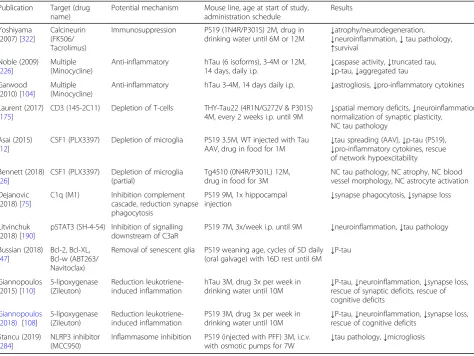
Pharmacological approaches to target microglial inflammation in mouse models of tauopathy
Publication Target (drug name)
Potential mechanism Mouse line, age at start of study, administration schedule
Results
Yoshiyama (2007) [322]
Calcineurin (FK506/ Tacrolimus)
Immunosuppression PS19 (1N4R/P301S) 2M, drug in drinking water until 6M or 12M
↓atrophy/neurodegeneration,
↓neuroinflammation,↓tau pathology,
↑survival Noble (2009)
[226]
Multiple (Minocycline)
Anti-inflammatory hTau (6 isoforms), 3-4M or 12M, 14 days, daily i.p.
↓caspase activity,↓truncated tau,
↓p-tau,↓aggregated tau Garwood
(2010) [104]
Multiple (Minocycline)
Anti-inflammatory hTau 3-4M, 14 days daily i.p. ↓astrogliosis,↓pro-inflammatory cytokines
Laurent (2017) [175]
CD3 (145-2C11) Depletion of T-cells THY-Tau22 (4R1N/G272V & P301S) 4M, every 2 weeks i.p. until 9M
↓spatial memory deficits,↓neuroinflammation, normalization of synaptic plasticity,
NC tau pathology
Asai (2015) [12]
CSF1 (PLX3397) Depletion of microglia PS19 3.5M, WT injected with Tau AAV, drug in food for 1M
↓tau spreading (AAV),↓p-tau (PS19),
↓pro-inflammatory cytokines, rescue of network hypoexcitability
Bennett (2018) [26]
CSF1 (PLX3397) Depletion of microglia (partial)
Tg4510 (0N4R/P301L) 12M, drug in food for 3M
NC tau pathology, NC atrophy, NC blood vessel morphology, NC astrocyte activation
Dejanovic (2018) [75]
C1q (M1) Inhibition complement
cascade, reduction synapse phagocytosis
PS19 9M, 1x hippocampal
injection ↓
synapse phagocytosis,↓synapse loss
Litvinchuk (2018) [190]
pSTAT3 (SH-4-54) Inhibition of signalling downstream of C3aR
PS19 7M, 3x/week i.p. until 9M ↓neuroinflammation,↓tau pathology
Bussian (2018) [47]
Bcl-2, Bcl-XL, Bcl-w (ABT263/ Navitoclax)
Removal of senescent glia PS19 weaning age, cycles of 5D daily (oral galvage) with 16D rest until 6M
↓P-tau
Giannopoulos (2015) [110]
5-lipoxygenase (Zileuton)
Reduction leukotriene-induced inflammation
hTau 3M, drug 3x per week in drinking water until 10M
↓P-tau,↓neuroinflammation,↓synapse loss, rescue of synaptic deficits, rescue of cognitive deficits
Giannopoulos (2018) [108]
5-lipoxygenase (Zileuton)
Reduction leukotriene-induced inflammation
PS19 3M, drug 3x per week in drinking water until 10M
↓P-tau,↓neuroinflammation,↓synapse loss, rescue of cognitive deficits
Stancu (2019) [284]
NLRP3 inhibitor (MCC950)
Inflammasome inhibition PS19 (injected with PFF) 3M, i.c.v.
with osmotic pumps for 7W ↓
both TNF-R1 and TNF-R2 in the same mouse model led
to increased plaque and tau pathology [214,
215]. It is
therefore possible that both TNFa receptors have
com-plex and opposing effects on the development of tau
pathology, but more studies in mouse models of pure
tauopathies are needed. As mentioned previously, the
cytokine IL6 is also consistently upregulated in
tauopa-thy mouse models. IL6 leads to phosphorylation of tau
at AD-associated residues via deregulation of the cdk5/
p35 pathway [245]. In addition to the effects of cytokines
on kinases and phosphates, it was recently shown that
metalloproteinase MMP-9 causes tau aggregation via
deacetylase HDAC6 [299]. Furthermore, the leukotrine
5-Lipoxygenase is upregulated in tauopathies, worsens
tau pathology, neuroinflammation, and increases synapse
loss [58,
108–111,
184,
300,
301]. More studies are
needed to identify if and how microglia can initiate tau
aggregation, rather than mere aggravation of existing tau
pathology.
The role of microglia in synaptic spreading of tau
Microglia can phagocytose extracellular tau, and
aggre-gated or hyperphosphorylated tau is observed in
micro-glia of mice and humans with tau pathology [37,
38,
44,
73,
99,
192,
196,
216,
229,
238]. Furthermore, microglia
can phagocytose synapses or entire neurons that contain
aggregated tau [44,
75]. Microglia, however, may also
play a critical role in spreading of tau protein [12].
When mice were injected with an adeno-associated virus
(AAV) that led to overexpression of human mutated tau
in the entorhinal cortex, spreading of human tau from
the entorhinal cortex to the dentate gyrus was observed
at 1 month post injection. Since neurons in the
entorhi-nal cortex connect to neurons in the dentate gyrus via
the perforant pathway, this spreading was likely
medi-ated through synaptic connections. However, depletion
of microglia led to a reduction of human tau detected in
the dentate gyrus. Knock out of TREM2 adapter protein
DAP12 in a similar model also led to inhibition of
syn-aptic tau spreading [14]. Therefore, it will be important
to characterize microglial pathways that are involved in
opsonization, degradation, and secretion of pathological
tau. An interesting recent in vitro study examined the
ability of primary microglia derived from various human
tauopathy cases or the rTg4510 mouse model to degrade
pathological tau [135]. The authors cultured the
micro-glia for multiple days and then applied to conditioned
medium a sensitive Förster resonance energy transfer
biosensor assay to measure tau seeding activity. Indeed,
microglia from human tauopathy cases as well as the
rTg4510 mouse secreted seed-competent tau. Microglia
also phagocytosed seed-competent tau, however, rather
than fully degrading it, they secreted tau back into the
extracellular space. Although a portion of tau spreading
might be mediated via neuron-to-neuron transfer or via
glial cells such as astrocytes [200,
218,
323], available
evidence suggests that microglia might play an
import-ant role in tau spreading as well.
Effects of AD risk genes on microglia and tau pathology
Many LOAD risk genes are predominantly expressed in
the innate immune system and enriched in microglia
[124]. The research on the links between tau pathology
and AD risk genes is still at an early stage, with new
as-sociations such as BIN1 reported very recently [96].
Studies that have studied the risk factors in the context
of neuroinflammation and tau pathology have so far
fo-cused on the strongest risk factors:
APOE
(apolipopro-tein E)
ε4
and
TREM2
.
APOEε4
is a common variant of
the
APOE
gene and the strongest risk factor for LOAD.
TREM2
risk mutations are substantially less common
than the
APOEε4
allele, but their risk effect for LOAD is
almost of the same magnitude [276]. Interestingly, two
recent
studies
independently
identified
a
unique
TREM2
-dependent transcriptional network in
disease-associated microglia (DAM) that is disease-associated with a
wide range of disease and neurodegenerative conditions
[161,
172]. Indeed, similar transcriptional networks were
described in mouse models of tauopathy [156,
190,
202,
309]. The DAM identity is distinct from the classically
described pro-inflammatory microglial phenotype that
can be induced by stimuli such as LPS or interferon
gamma. Like classic pro-inflammatory microglia, DAM
upregulate pro-inflammatory genes (e.g.
IL1B
,
CCL2
)
and downregulate homeostatic genes (e.g.
P2ry12
,
Tmem119
). However, in contrast to the LPS-induced
microglia, DAM upregulate other genes like
APOE
and
TREM2
. In addition to being part of the DAM genetic
network,
TREM2
and
APOE
have also been shown to
physically interact with each other and this pathway was
important for the phagocytosis of A
β
[276]. Interestingly,
APOE was also shown to directly bind to C1q, thereby
acting as an immune checkpoint inhibitor of
inflamma-tion in response to amyloid plaques [321]. However, the
effects of both genes on progression of plaque pathology
are complex and dependent on disease stage [276]. The
research on the effects of
TREM2
and
APOE
on tau
pathology is at an early stage, but the findings so far will
be discussed below.
TREM2
is
a
transmembrane
receptor
of
the
immunoglobulin-superfamily that in the brain is
pre-dominantly
expressed
on
microglia.
Activation
of
down TREM2 using a lentivirus led to increased levels
of pro-inflammatory cytokines, kinases,
hyperphosphory-lated tau, increased neurodegeneration, and behavioral
deficits [153]. Overexpression of murine TREM2 instead
of a knockdown led to exactly the opposite phenotype and
additional upregulation of homeostatic genes in microglia
[154]. Accordingly, knockdown and overexpression of
TREM2 in a neuron-microglia co-culture showed that
TREM2 prevents the effects of microglial activation and
pro-inflammatory signaling on tau phosphorylation [155].
TREM2
gene knock-out in the mild hTau model that
ex-presses all six human isoforms led to exacerbation of tau
pathology [23]. However, knockout of
TREM2
in the more
aggressive PS19 mouse model at later stages showed a
marked reduction in neurodegeneration and
DAM-associated genes [183]. Surprisingly, a recent study using
the same conditions showed that TREM2
haploinsuffi-ciency led to more severe tau-induced neurodegeneration
compared to the full knockout [261]. Knockout of TREM2
adaptor protein DAP12 in PS19 mice at early disease
stages led to increased hyperphosphorylated tau [14],
which was also associated with alterations in
electro-physiological readouts and cognitive deficits. The data
available on TREM2 and downstream effectors (e.g.
DAP12 and SYK) thus are contradictory and more studies
in different tauopathy models and varying stages of
tau-induced neurodegeneration are warranted.
APOE is a lipid carrier that is predominantly expressed
in astrocytes and to a lesser degree in microglia. The
hu-man brain contains three different alleles:
ε
2,
ε
3 and
ε
4.
One copy of
ε
4 increases AD risk by about 3 times,
whereas
ε
4/
ε
4 increases risk 12 times [276]. Surprisingly,
however, APOE
ε
4-negative prodromal AD patients had
greater tau pathology load, cortical atrophy and faster
cognitive decline compared to APOE
ε
4 carriers [203,
204]. In AD, APOE
ε
4 only associates with tau pathology
in the presence of amyloid pathology [86]. However, in
frontotemporal dementia with MAPT mutations that
lead to familial tauopathy, APOE
ε
4 lowers the age of
on-set independent of amyloid plaques [168]. In contrast,
another study found that APOE
ε
2 was associated with
increased tau pathology burden in PSP [327]. So far, only
two studies have experimentally examined the role of
different APOE alleles on tau-induced
neuroinflamma-tion and neurodegeneraneuroinflamma-tion in tau transgenic animals.
When PS19 mice were crossed with knock-in mice for
the different APOE alleles, the APOE
ε
4 group had the
most widespread phospho-tau staining in the
hippocam-pus despite similar levels of insoluble tau. The staining
was characterized by a dotted and grainy appearance.
This staining pattern was most strongly associated with
lower hippocampal volume and was completely absent
in the APOE knockout mice. Notably, the APOE
ε
4
group had no dense tangle-like neurons in the
phospho-tau staining, but no staining for NFTs was performed in
this study. The APOE
ε
4 group also had more severe
microgliosis, astrocyte activation and neurodegeneration
compared to the APOE
ε
2 and APOE
ε
3 groups [277].
Fur-thermore, in the same study, APOE knockout mice were
less affected on all these measures compared to all the
other APOE groups. Intriguingly, a recent study showed
dramatically different results when inducing tau pathology
using AAVs in knock-in mice for the different APOE
al-leles [327]. The APOE
ε
2 group had substantially increased
tau pathology and showed increased astrocyte reactivity.
However, there was no microgliosis or neurodegeneration
in any of the APOE groups compared to the control group
that just overexpressed GFP. The use of different mouse
models potentially representing different stages of tau
pathology could explain the apparent discrepancy between
these studies. More work, however, needs to be done to
determine how different APOE alleles affect tau-induced
neuroinflammation and neurodegeneration. For example,
microglia expressing APOE
ε
4 display increased
phagocyt-osis of apoptotic neurons [219]. Since APOE is expressed
in both astrocytes and microglia, cell-type specific
knock-in or knockout models would contribute greatly towards
determining the role of different cell types in tauopathy. It
would also be particularly informative to further
investi-gate different APOE alleles in various primary tauopathies
and tauopathy mouse models at different disease stages.
Finally, it is important to keep in mind that APOE has
prominent non-immune system related functions and the
different APOE alleles therefore likely also influence
tau-mediated neurodegeneration via other pathways [20].
Intersection of tau pathology and microglia at the
synapse
Effects of microglia on tau-induced synaptic dysfunction
Intracellular tau pathology can damage the synapses
from within via a multitude of pathways [148].
Aggrava-tion of intracellular tau pathology by microglia can
therefore indirectly lead to more tau-induced synapse
loss. Microglia, however, can also play a direct role in
neurodegeneration-induced synaptic dysfunction (Fig.
2b). One particularly compelling example is reactivation
of complement-mediated synaptic pruning, which was
first described in neurodevelopment [286]. This pathway
starts with synaptic tagging of C1q and downstream
syn-aptic deposition of C3, which leads to opsonization of
the synapse via the C3R on microglia [262]. Reactivation
of this pathway has been previously demonstrated in
multiple mouse models of neurodegenerative disease,
in-cluding glaucoma [286], FTD [191], and AD [134].
There is also a dramatic upregulation of C1q in normal
aging (~ 300-fold in certain brain regions) and
age-related cognitive decline was prevented in C1q and C3
upregulated in tauopathy patients as it was shown to
co-localize with neuronal and astrocytic tau pathology in
PiD [279]. Furthermore, C1q is detected alongside
hyperphosphorylated tau in AD-derived synaptosomes
[75] and decorates both the A
β
plaques and
NFT-bearing neurons in AD brain sections [2,
40,
206,
267,
272]. Indeed, complement-mediated pruning of
excita-tory synapses is strongly re-activated in the PS19 mouse
model of tauopathy and this was reversed after
intrace-rebral injection of an anti-C1q antibody [75].
It is unclear how tau pathology leads to C1q-mediated
tagging of synapses but a possible pathway could include
local apoptotic mechanisms, leading to the exposure of
phosphatidylserines on the synapse to which C1q can
bind [44,
123]. Furthermore, activation of the
metabo-tropic glutamate receptor 1 was shown to lead to
local C1q mRNA synthesis at the synapse in a mouse
model of AD. This led to phagocytosis of the synapse
by microglia [33]. Additionally, sialic acids in the cell
membrane prevent C1q binding and microglia
phago-cytosis through C3R [189]. It is therefore possible
that intracellular tau pathology decreases sialic acid
coating on the extracellular side of the synaptic cell
membrane. It has been shown recently that TREM2
adaptor protein DAP12 plays an important role in
tau-induced induction of C1q [14]. Although the
same study could not find similar effects by knocking
out TREM2, it would be interesting to study if
TREM2 itself could induce synapse opsonization by
microglia as has been observed in neurodevelopment
[88]. Finally, fibrinogen leakage from blood vessels
can also directly lead to microglial phagocytosis of
spines via CR3 in mouse models of AD [209].
Tau-induced vascular or BBB damage may therefore lead
to increased microglial synapse phagocytosis. More
studies, however, are needed to uncover and
under-stand the mechanistic link(s) between tau pathology,
C1q-mediated tagging of synapses and microglial
phagocytosis of synaptic compartments.
Tau pathology-induced alterations in microglial
se-creted factors may also adversely affect synaptic
func-tion. Microglia in the adult brain are important for
learning-induced synapse formation via secretion of
neurotrophic factor BDNF [235]. Microglia are known
to downregulate many homeostatic genes in response to
neurodegeneration, and it is possible that neurotrophic
support from microglia to synapses is disrupted in
tauo-pathy [129]. Similarly, tau pathology also induces a
pro-inflammatory phenotype in microglia, leading to chronic
elevation of pro-inflammatory cytokines. Describing the
individual synaptic effects of these cytokines is beyond
the scope of this review (see [225]). However, IL1B, IL6
and TNFa have, for example, been shown to modulate
various synaptic deficits in mouse models of AD, viral
infection, addiction, Creutzfeldt Jakob disease, obesity,
and aging [28,
64,
81,
103,
181,
266,
311]. Factors
se-creted from microglia may also have an indirect effect on
synapses. For example, activated microglia secrete
extra-cellular vesicles with miRNAs that downregulate synaptic
proteins and ultimately lead to loss of excitatory synapses
[243]. Chronically increased levels of pro-inflammatory
cytokines and dysregulation of other secreted factors
from microglia throughout the decades of developing tau
pathology may therefore adversely affect synaptic
func-tion in tauopathy patients. The exact contribufunc-tions of
these pathways to tau pathology are still unknown.
The role of astrocytes in tau-induced synaptic dysfunction
Microglia also have bidirectional signaling cascades with
as-trocytes. Astrocytes are a highly heterogenous population
that make up approximately 20% of brains cells and are
de-rived from the same progenitors as neurons [6]. Astrocytes
have a wide range of functions, including providing nutrient
support to neurons, forming part of the BBB, and
modulat-ing the flow of CSF in the brain as part of the glymphatic
system [6,
247]. Astrocytes have highly ramified processes
and it is estimated that a single cortical astrocyte can
con-tact up to 100,000 synapses in mice and up to 2,000,000
synapses in humans [5]. Indeed, astrocytes play a critical
role in neuronal connections by regulating glutamate
homeostasis, secreting gliotransmitters (e.g. ATP), secreting
factors that promote assembly and plasticity of synapses
(e.g. thrombospondins), and synaptic phagocytosis (e.g. via
MERTK and MEGF10) [6]. Under a variety of disease and
neurodegenerative conditions, microglial cytokines (IL1a,
TNFa and C1q) can induce a unique transcriptional profile
in astrocytes that is characterized by dramatic upregulation
of complement protein C3. This was associated with a
neurotoxic phenotype termed
“A1 astrocytes”,
character-ized by secretion of neurotoxic factors, loss of neurotrophic
functions, and impairments in several homeostatic synaptic
functions [188]. A1 astrocytes can be induced in normal
aging mice and are associated with more severe
neurode-generation in a mouse model of tauopathy [36,
61,
277].
Interestingly, microglia also secrete factors (e.g. TGF
α
,
VEGF-B) that limit the pathogenic activities of astrocytes
[253]. Furthermore, C3 upregulation in astrocytes is not
only the result of microglial inflammation, but the
down-stream cleavage product C3a can in turn dramatically
in-crease the synaptic toxicity of microglia in mouse models
of amyloidosis and tauopathy by binding to microglial
C3aRs [186,
187,
190]. Cross-signaling between microglia
and astrocytes therefore plays a key role in modulating
syn-aptic dysfunction and neurodegeneration (Fig.
2b).
synaptic dysfunction [239]. It is expected that
neurofibril-lary pathology-induced loss of astrocytic glutamate
homeostasis causes neuronal network dysfunction and
po-tential excitotoxicity. However, an interesting study shows
that healthy subjects with NFTs had more activated
astro-cytes with increased glutamate transporter 1 expression
compared to AD cases with dementia [167]. This raises
the possibility that at least some astrocytic phenotypes
ob-served in tauopathies may be beneficial rather than
dam-aging. Astrocytes can also prune synapses in the healthy
brain [59,
60] or under disease conditions, such as
ische-mia [217] and sleep deprivation [19]. Furthermore,
astro-cytes were shown to phagocytose apoptotic cells via the
C1q-MEGF10 pathway [143]. This raises the possibility
that not only microglia, but also astrocytes can use the
classical complement pathway to phagocytose synapses on
living neurons. Astrocytes were also shown to clear
trophic neurites in a mouse model of AD [114]. Since
dys-trophic neurites in AD patients often contain aggregated
tau, it is possible that astrocytes phagocytose pathological
tau species. Furthermore, the close proximity of astrocytes
to the pre- and postsynaptic compartments also raises the
possibility that astrocytes can pick up secreted
extracellu-lar tau or digest damaged synapses with
hyperphosphory-lated tau [73]. Intriguingly, recent studies show that
astrocytes may also be involved in tau spreading along
neuronal connections or from astrocyte-to-astrocyte [200,
220]. Finally, a transgenic mouse model of astrocytic tau
pathology displayed reduced expression and function of
glutamate transporter-1, and motor impairments already
before disease stages with overt hyperphosphorylated tau
accumulation [67]. This indicates that astrocytic tau
path-ology may lead to alterations in synaptic glutamate
homeostasis, neuronal network dysfunction, and
associ-ated functional impairments .
The role of microglia in therapeutic approaches
targeting the immune system in tauopathies
Given the increasing recognition of microglia as central
players in the pathogenesis of tauopathies, it is perhaps
not surprising that there is increasing interest in
target-ing inflammatory pathways for these diseases (Table
1).
Anti-inflammatory compounds such as FK506 and
min-ocycline were shown to reduce tau pathology and
down-stream neurodegeneration, but their mechanism of
action in relation to tau pathology is unclear [104,
226,
322]. Depletion of immune cells such as microglia or
T-cells may also be efficacious when initiated at early
stages of tau pathology [12,
175]. However, it is
import-ant to keep in mind that the immune system in the
per-iphery and the brain plays an important physiological
role. Non-specific suppression of the immune system
could leave the patient vulnerable to increased risk of
in-fections and accumulation of cellular debris in the
context of neurodegeneration. Targeted pharmacological
removal of dysfunctional cells from the brain may in
it-self be an efficacious therapeutic approach for
tauopa-thies [47]. Furthermore, restoring homeostatic microglial
phagocytosis with a CD22-blocking antibody rescued
cognitive deficits in aged mice, indicating that normal
microglial function is critical for maintaining a normal
cognitive state [240]. There is therefore increasing focus
on targeting specific inflammatory targets such as
com-plement [75,
190], leukotrienes [108,
110], or the NLRP3
inflammasome [284]. Antibodies against C1q can
dir-ectly reduce tau pathology-induced synapse phagocytosis
by microglia [75]. It remains to be determined to what
extent microglial synapse phagocytosis plays a central
role in the pathogenesis of tauopathies, and if there are
other mechanisms through which microglia affect
tau-induced synaptic dysfunction that could be targeted with
therapeutic agents.
Most research has focused on the development of
anti-bodies that neutralize extracellular tau and inhibit synaptic
spreading of pathological tau [227,
320]. Tau antibodies
could neutralize extracellular tau oligomers before they
have the chance to damage glial cells and the vasculature.
As mentioned previously, both microglia and astrocytes
are involved in propagation of tau pathology [12,
200].
Antibody-mediated neutralization of tau seeds before they
reach these types of cells, may therefore diminish
down-stream exosomal tau spreading. Additionally, extracellular
tau aggregates may lead to reactive gliosis, which can
po-tentially be inhibited by anti-tau antibodies. Extracellular
tau aggregates neutralized by tau antibodies need to be
re-moved from the brain. Clearance of extracellular tau
or other macromolecules is mediated by the
glympha-tic system and impairment of this system worsens tau
pathology [[141,
236]. In addition to global clearance,
tau-antibody complexes in immunized mice can also
be cleared locally by means of opsonization via
microglial Fc receptors and degradation in the
lyso-somes [8,
10,
74,
99,
179,
319]. Effectorless antibodies
incapable of actively engaging microglia retained their
therapeutic effect [179], indicating that tau antibody
complexes can also be cleared via additional pathways
in immunized mice. Indeed, peripheral or
AAV-mediated delivery of tau scFv (without an Fc domain)
seems to be an effective therapeutic approach in
tauo-pathy mice [145,
224,
283,
306]. More research is
therefore needed to describe the impact of actively
engaging
microglia
with
tau
immunotherapeutic
approaches.
Concluding remarks & future directions
Microglia are fascinating cells and the large number of
excellent recent studies demonstrates increasing
recog-nition of microglia as critical players in the pathobiology
of tau protein. It is currently not fully resolved if
inflam-mation is a cause, contributor or consequence of tau
Table 2
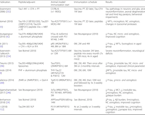
Preclinical studies using active immunotherapy
in vivo
Publication Peptide/adjuvant Animal model/
immunization start
Immunization schedule Results
Rosenmann (2006) [252]
Tau1-441 + CFA + PT C57BL/6
(+/- MOG)
Vaccine, PT 2D later, Tau-CFA 1W later
Tau pathology in neurons and glia, severe
neuroinflammation, axonal degeneration, tail and hind leg paralysis, behavioral impairments
Boimel (2010)
[35]
Tau195-213[P202/205], Tau207-220[P212/214], Tau224-238[P231] peptide mix + MBT + PT
Tau-K257T/P301S (+/-MOG) 4M
Vaccine, PT 2D later, peptides
1W later ↓
NFTs,microgliosis, NCastrogliosis, changes in lysosomal proteases
Boutajangout (2010) [42]
Tau379–408[pS396/S404] + aluminium phosphate
hTau (6 isoforms) crossed with PS1 M146L 3-4M
See Boutajangout (2010) ↓P-tau, NCmicro- and astrogliosis, improved cognition
Bi (2011) [30]
Tau395–406[pS396/S404] + CFA + KLH or IFA
pR5 (4R2N/P301L) 4M, 8M or 18M
0W, 2W and 4W ↓P-tau,↓NFTs,↑astrogliosisin aged group
Rozenstein-Tsalkovich (2013) [254]
See Boimel (2010) Tau-K257T/P301S 6M
or 12M
Vaccine, booster 2W later, peptide mix every month (7x in 12M mice, 4x in aged mice)
Severeneuroinflammation, neurological deficits
Theunis (2013) [296]
Tau393-408[pS396/pS404] liposomes
Tau.P301L (2N4R/P301L) 6M
0W, 2W, 4W. Then once after 3M or 2-monthly intervals
↓P-tau,↓insoluble tau, NCmicro- and astrogliosis,improved clinical parameters
Ando (2014) [9]
PHF + aluminium phosphate THY-Tau22 (4R1N/G272V & P301S) 12M
0W, 2W, 6W, 10W ↓NFTs,↓insoluble tau, NCmicro- and astrogliosis
Selenica (2014) [268]
2N4R or 2N4R/P301L + Quil-A Tg4510 (4R0N/P301L) 5M
0W, 2W, 4W, then 10W rest and followed by 3x 3-weekly boosters
↓P-tau,↓micro- and astrogliosis
Rajamohamedsait (2017) [246]
See Boutajangout (2010) 3xTg (4R02/P301L, PS1 M146V, APPSWE) 3M
See Boutajangout (2010) ↓P-tau,↓MC1,↓insoluble tau,
↓microgliosis, NCastrogliosis,
↓plaque burden Benhamron
(2018) [24]
See Boimel (2010) APPSwe/PSEN1dE9-tg
14M
See (Boimel, 2010) ↓P-tau,↓Aβburden,↑microgliosis, NCastrogliosis, improved cognition
Ji (2018) [152]
Tau294-305 VLP PS19 (4R1N/P301S)
3M
4x at 2-weekly or 3-weekly intervals
↓P-tau,↓insoluble tau,↓microgliosis,
pathology. Pro-inflammatory mediators secreted from
microglia (e.g. cytokines, complement) can initiate tau
pathology and play a critical role in tau-induced
neuro-degeneration. The strongest evidence for
inflammation-induced initiation of tau pathology currently exists for
TNFa, as this cytokine was shown to lead to formation
of tau aggregates in neurites [116]. More preclinical
work, however, is needed to fully characterize the
im-mune pathways involved in tau pathology and efforts
should be made to validate them in both AD and
pri-mary tauopathy patients. Many studies report the effects
of neuroinflammatory processes on tau phosphorylation
only. Future studies should also focus on the effects
of neuroinflammation on oligomerization or
accumu-lation of insoluble tau aggregates. Furthermore, risk
genes for AD or other tauopathies will have to be
in-vestigated in multiple mouse models of tauopathy,
without plaque pathology as a confounding factor.
Since the immune response to tau pathology changes
as the disease progresses, future studies should also
examine the evolution of neuroinflammatory pathways
at multiple stages of the disease. Furthermore, more
studies are needed on what events cause the initial
neuroinflammation in response to tau pathology and
via what pathways.
Microglia are also directly involved in tauopathies as
they have been shown to pathologically phagocytose
synapses of neurons with tau pathology. Currently, the
pathways underlying microglia-mediated synapse loss
are not fully characterized and a multitude of potential
pathways have been identified in neurodevelopment.
Complement-mediated synapse loss via microglial
syn-apse phagocytosis under neurodegenerative conditions is
now described in multiple disease models. However, it is
not known what causes the binding of C1q to synapses
and if this happens indiscriminately or only targets
vul-nerable synapses. In neurodevelopment, there are signals
(e.g. CD47) that protect synapses from microglial
prun-ing. We need to understand better the function of these
signals in the normal brain and determine if they are
downregulated in response to neurofibrillary pathology.
Additionally, since astrocytes play a critical role in both
synaptic function and neuroinflammation, more studies
are needed on bidirectional microglia-astrocyte signaling
in tauopathies.
As principal macrophages of the brain, microglia
phagocytose tau and may play a role in spreading tau
pathology throughout the brain. Determining the exact
contribution of microglia to the disease pathogenesis
re-mains an important topic for future investigations. More
studies are needed on the mechanisms of tau
internaliza-tion in microglia and if this is associated with activainternaliza-tion
of pro-inflammatory pathways. Furthermore, better
iden-tification
of
intracellular
pathways
that
lead
to
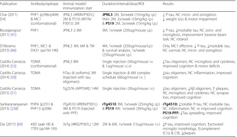
Table 3
Preclinical studies using passive immunotherapy
in vivo
Publication Antibody/epitope Animal model/ immunization start
Duration/interval/dose/ROI Results
Chai (2011) [54]
PHF1 (p396/p404) & MC1
(conformational)
JPNL3 (4R0N/P301L) 2M & PS19 (4R1N/ P301S) 2M
JPNL32M, 3x/week (15mg/kg i.p.) then 2M, 2x/week (10mg/kg i.p.) &PS192M, 2x/week (15mg/kg i.p.)
↓P-tau, NCmicro- and astrogliosis,
↓weight loss & motor impairment
Boutajangout (2011) [41]
PHF1 JPNL3 2-3M 3M, 1x/week (250ug/mouse i.p.) ↓P-tau,↓insoluble tau, NCastro- and
microgliosis, improvement traverse beam task, NC rotarod
D'Abramo (2013) [66]
PHF1, MC1 & DA31 (aa150-190)
JPNL3 3M, 6M & 7M 4M, 1x/week (250ug/mouse/i.p.) & survival analysis, 1x/week (250ug/mouse i.p.)
Only MC1 effective.↓P-tau,↓insoluble tau, NC survival, NCmicro- and astrogliosis
Castillo-Carranza (2014) [53]
TOMA (conformational)
JPNL3 8M Single injection (30ug/mouse i.v.
& 1ug/mouse i.c.v)
↓Tau oligomers, NCmicrogliosisand cytokines, improved cognition & motor deficits
Castillo-Carranza (2014) [50]
TOMA hTau (6 isoforms) 3M
(injected with tau oligomers)
Single injection & 6M complex
schedule (60ug/mouse i.v. ) ↓
tau oligomers, NC inflammation, improved cognition
Castillo-Carranza (2015) [51]
TOMA Tg2576 (APPSWE) 14M Single injection (30ug/mouse i.v.) ↓tau oligomers,↓Aβoligomers,↑plaques, NCmicrogliosisand cytokines, NC synapse loss, improved cognition
Sankaranarayanan (2015) [258]
PHF6 (p231) & PHF13 (p396)
rTg4510 (4R0N/P301L) 3M & PS19 (injected with PFF)
rTg45103M, 1x/week (25mg/kg i.p.) &PS194W, 1x/week (30mg/kg i.p.)
rTg4510:↓Soluble P-tau, NC insoluble tau, NCinflammation, NC or improved cognition.
PS19-PFF:↓Tau spreading, improved cognition
Dai (2017) [69] 43D (aa6-18) & 77E9 (aa184-195)