of Difficult Samples
Jane J. Pappas, André Toulouse, and W. E. C. Bradley
Abstract
The bisulfite genomic sequencing protocol is a widely used method for analyzing DNA methylation. It relies on the deamination of unmethylated cytosine residues to uracil; however, its high rates of DNA degradation and incomplete cytosine to uracil conversion often lead to failed experiments, uninformative results, and false positives. Here, we report the addition of a single-step multiple restriction enzyme digestion (MRED) designed to differentially digest polymerase chain reaction products amplified from unconverted DNA while leaving those of converted DNA intact. We show that for our model system,RARB2P2 promoter, use of MRED increased informative sequencings ninefold, and MRED did not alter the clonal representation in one fully methylated cell line, H-596, treated or not with 5-azadeoxycytidine, a methylation inhibitor. We believe that this method may easily be adapted for analyzing other genes and provide guidelines for selecting the most appropriate MRED restriction enzymes.
Key words: bisulfite genomic sequencing, multiple restriction enzyme digestion, methylation.
1. Introduction
The bisulfite genomic sequencing (BGS) protocol (1, 2) is a method of choice for analyzing DNA methylation at the nucleotide level. Sodium bisulfite is used to convert unmethylated cytosine residues to uracil residues in single-stranded DNA. In particular, bi-sulfite conversion consists of three sequential chemical reactions: sulfonation of cytosine to cytosine-6-sulfonate, deamination to uracil-6-sulfonate, and desulfonation to uracil. However, since 5-methylcytosine residues are nonreactive, they remain intact. The
Shulin Li (ed.),Biological Procedures Online, Volume 11, Number 1
© to the author(s) 2009
bisulfite-converted DNA is then amplified with specific primers designed for converted DNA, and purified polymerase chain reac-tion (PCR) products, which are usually subcloned, are sequenced. Bisulfite conversion is so powerful that it has been paired with numerous techniques other than traditional sequencing, including: methylation-specific PCR (3), combined bisulfite restriction en-zyme analysis (4), methylation-sensitive single nucleotide primer extension (5), methylation-sensitive single-strand conformation analysis (6), MethyLight (7), oligonucleotide microarray methods (8), denaturing high-performance liquid chromatography with bi-sulfite genomic sequencing (9), pyrosequencing methylation anal-ysis (10), and methylation-sensitive high-resolution melting-curve analysis (11), among others (see(12) for a review). In addition, many methylation analysis kits are also commercially available.
Unfortunately, high rates of DNA degradation and incom-plete conversion reactions often lead to decreased efficiency of the assay. Many attempts have been made to minimize template degradation and/or maximize cytosine conversion (13–19), but overall, the bisulfite conversion protocol has remained un-changed, and no other high resolution or positive display methyl-ation analysis protocol exists. As a result, the BGS protocol, as well as any technique paired with the bisulfite conversion reaction (and, hence, founded on the assumption that conversion is com-plete) often generate few or no informative results.
In our studies of theRARB2 P2 promoter (20), we found that incomplete conversion was an insurmountable challenge even after modifying the protocol in numerous ways. We, therefore, aimed to circumvent these issues altogether by depleting the PCR populations of products amplified from partially converted or unconverted DNA using a multiple restriction enzyme diges-tion (MRED) approach. We found that informative sequencings were increased ninefold using it. We believe that this method may easily be adapted for analyzing the detailed methylation sta-tus of other genes presenting incomplete cytosine to uracil con-version, and we provide guidelines for selecting the most appropriate restriction enzymes (REs).
2. Materials
and Methods
2.1. Cell Culture and Genomic DNA Extraction
obtained from the American Type Culture Collection (Rockville, MD). The CALU-1 daughter cell lines, C-19 and C-59, are
RARB2-transfectants that were established in our laboratory (21). MM-1 was also established in our laboratory (6). NCI-H23, NCI-H82, NCI-H125, NCI-H157, NCI-H520, and NCI-H596 were supplied by Dr. Adi Gazdar (NCI, NIH, Bethesda, MD). NBE-E6E7(22) was provided by Dr. Jean Viallet
(Gemin X Biotechnologies Inc., Montreal, Québec). SW 1222 was given to us by Dr. Clifford Stanners (McGill University, Montreal, Québec). Qu-DB was provided by Dr. Barbara Campling (Queen’s University, Kingston, Ontario). T47D, MDA-MB-231 (MB-231), ZR-75B, and HS-578T were kindly provided by Dr. Morag Park (McGill University, Montreal, Québec).
2.1.2. Cell Culture CALU-1, CACO-2, SW-1222, and LS-180 were grown in
α-MEM medium (Invitrogen, Carlsbad, CA) supplemented with 10% heat-inactivated fetal calf serum (FCS; Wisent Bioproducts, Saint-Jean-Baptiste de Rouville, Québec). NBE-E6E7 was grown
in keratinocyte-serum free medium (Invitrogen), supplemented with 50 µg/ml bovine pituitary extract, and 5 ng/ml recombi-nant human epidermal growth factor (Invitrogen). All other cells were grown in RPMI-1640 medium (Invitrogen) supplemented either with 5% (SK-MES, NCI-H23, NCI-H125, NCI-H520, Qu-DB, and HS-578T) or 10% FCS (NCI-H82, NCI-H157, MM-1, T47D, MDA-MB-231, ZR-75B, 201, COLO-205, and HCT-15). Where indicated, cells were treated with 1 µM 5-azadeoxycytidine.
2.1.3. Genomic DNA Extraction
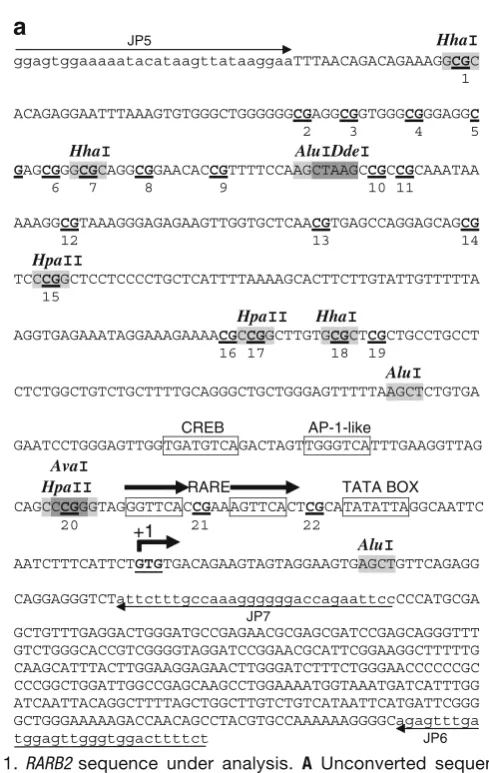
Genomic DNA was extracted using the standard phenol-chloro-form technique followed by proteinase K treatment to ensure complete protein removal (23). DNA was then digested with thePstI RE (New England BioLabs, Ipswich, MA) according to the supplier′s directives to shorten the fragment (2.95 kb) con-taining the targetRARB2 P2 promoter sequence investigated (541 bp; Fig. 1), thereby reducing the possibility for regional double-strand formation (24). PstI was the only RE available for the sequence under analysis.
2.2. Bisulfite Conversion
at 37°C, and solutions were carefully mixed by inversion with minimal aeration. Two hundred fifty microliters of freshly pre-pared 3.6 M sodium bisulfite (Sigma–Aldrich), pH 5.0, was added to the tubes while at 37°C, and solutions were again carefully mixed by inversion with minimal aeration. Reaction volumes were overlaid with mineral oil and incubated at 55°C for 16 h in the dark. Aqueous phases were transferred to new tubes and desalted
JP5 HhaI
ggagtggaaaaatacataagttataaggaaTTTAACAGACAGAAAGGCGC 1
ACAGAGGAATTTAAAGTGTGGGCTGGGGGGCGAGGCGGTGGGCGGGAGGC 2 3 4 5
HhaI AluIDdeI GAGCGGGCGCAGGCGGAACACCGTTTTCCAAGCTAAGCCGCCGCAAATAA 6 7 8 9 10 11
AAAGGCGTAAAGGGAGAGAAGTTGGTGCTCAACGTGAGCCAGGAGCAGCG 12 13 14 HpaII
TCCCGGCTCCTCCCCTGCTCATTTTAAAAGCACTTCTTGTATTGTTTTTA 15
HpaII HhaI
AGGTGAGAAATAGGAAAGAAAACGCCGGCTTGTGCGCTCGCTGCCTGCCT 16 17 18 19
AluI
CTCTGGCTGTCTGCTTTTGCAGGGCTGCTGGGAGTTTTTAAGCTCTGTGA
CREB AP-1-like
GAATCCTGGGAGTTGGTGATGTCAGACTAGTTGGGTCATTTGAAGGTTAG
AvaI
HpaII RARE TATA BOX CAGCCCGGGTAGGGTTCACCGAAAGTTCACTCGCATATATTAGGCAATTC 20 21 22
AluI
AATCTTTCATTCTGTGTGACAGAAGTAGTAGGAAGTGAGCTGTTCAGAGG
CAGGAGGGTCTattctttgccaaaggggggaccagaattccCCCATGCGA JP7
GCTGTTTGAGGACTGGGATGCCGAGAACGCGAGCGATCCGAGCAGGGTTT GTCTGGGCACCGTCGGGGTAGGATCCGGAACGCATTCGGAAGGCTTTTTG CAAGCATTTACTTGGAAGGAGAACTTGGGATCTTTCTGGGAACCCCCCGC CCCGGCTGGATTGGCCGAGCAAGCCTGGAAAATGGTAAATGATCATTTGG ATCAATTACAGGCTTTTAGCTGGCTTGTCTGTCATAATTCATGATTCGGG GCTGGGAAAAAGACCAACAGCCTACGTGCCAAAAAAGGGGCagagtttga tggagttgggtggacttttct JP6
+1
with Wizard Magic Miniprep DNA purification resins (Promega, Madison, WI). DNA was eluted with 120 µl distilled water and residual alcohol was removed using speedvac centrifugation. Ten microliters 3N NaOH was added to the remaining 100 µl and allowed to incubate for 15 min at 37°C. DNA was precipitat-ed with 33 µl 10 M sodium acetate pH 7.8 and 300 µl chillprecipitat-ed eth-anol using glycogen as a carrier in an ice-water bath for 10 min and then centrifuged at 13,000×gfor 60 min. The precipitate was resuspended in 100μl TE pH 8.0.
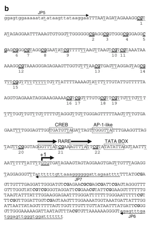
2.2.2. PCR Amplification A 541-bp sequence containing 22 CpG dinucleotides (Fig.1A) was identified within the human RARB P2 promoter (20). PCR amplification consisted of two rounds of amplification: round 1 primers consisted of the upper primer JP5 (5′
-GGAGTGGAAAAATATATAAGTTATAAGGAA-3′) and the
lower primer JP6 (5′-AAAAAAATCCACCCAACTCCAT
JP5
ggagtggaaaaatatataagttataaggaaTTTAATAGATAGAAAGGCGT
1
ATAGAGGAATTTAAAGTGTGGGTTGGGGGGCGAGGCGGTGGGCGGGAGGC 2 3 4 5
GAGCGGGCGTAGGCGGAATATCGTTTTTTAAGTTAAGTCGTCGTAAATAA
6 7 8 9 10 11
AAAGGCGTAAAGGGAGAGAAGTTGGTGTTTAACGTGAGTTAGGAGTAGCG 12 13 14
TTTCGGTTTTTTTTTTGTTTATTTTAAAAGTATTTTTTGTATTGTTTTTA 15
AGGTGAGAAATAGGAAAGAAAACGTCGGTTTGTGCGTTCGTTGTTTGTTT 16 17 18 19
TTTTGGTTGTTTGTTTTTGTAGGGTTGTTGGGAGTTTTTAAGTTTTGTGA
CREB AP-1-like
GAATTTTGGGAGTTGGTGATGTTAGATTAGTTGGGTTATTTGAAGGTTAG
RARE TATA BOX
TAGTTCGGGTAGGGTTTATCGAAAGTTTATTCGTATATATTAGGTAATTT 20 21 22
AATTTTTTATTTTGTGTGATAGAAGTAGTAGGAAGTGAGTTGTTTAGAGG
TAGGAGGGTTTattttttgttaaaggggggattagaattttTTTATGCGA JP7
GTTGTTTGAGGATTGGGATGTCGAGAACGCGAGCGATTCGAGTAGGGTTT GTTTGGGTATCGTCGGGGTAGGATTCGGAACGTATTCGGAAGGTTTTTTG
TAAGTATTTATTTGGAAGGAGAATTTGGGATTTTTTTGGGAATTTTTCGT TTCGGTTGGATTGGTCGAGTAAGTTTGGAAAATGGTAAATGATTATTTGG ATTAATTATAGGTTTTTAGTTGGTTTGTTTGTTATAATTTATGATTCGGG GTTGGGAAAAAGATTAATAGTTTACGTGTTAAAAAAGGGGTagagtttga tggagttgggtggattttttt JP6
+1
CAAACTCT-3′); round 2 semi-nested primers consisted of J P 5 a n d t h e l o w e r p r i m e r J P 7 ( 5′- A A A A T T C T A
ATCCCCCCTTTAACAAAAAAT-3′). Cycling conditions
were: 94°C/4 min × 1 cycle; 94°C/1 min, 61°C/2 min, 72°C/2 min × 5 cycles; 94°C/1 min, 61°C/1.5 min, 72°C/ 1.5–2 min × 25 cycles; 72°C/5 min × 1 cycle. Primers were designed following the guidelines found in (25). In particular, they were designed not to contain CpG dinucleotides so that PCR amplifications were not biased according to methylation status. The minimum number of non-CpG-cytosines available for measuring the rate of cytosine to uracil conversion, for quality control assessment, is 74. This does not include the one non-CpG-cytosine within the region complementary to JP5 and the seven non-CpG-cytosines within the region com-plementary to JP7 (see section 2.3.2, Special Considerations).
2.3. MRED
2.3.1. Restriction Enzyme Selection
The original (Fig.1A) and converted (Fig.1B) sequences were en-tered into NEBcutter V.2 athttp://tools.neb.com/NEBcutter2/ index.php(New England BioLabs), and RE maps and lists were made. Potential MRED isoschizomers were screened based on the following criteria: (1) RE sites should selectively cut unconverted DNA while leaving converted DNA intact; (2) RE sites may or may not contain CpG-cytosines but should contain at least one CpG-cytosine; (3) if RE sites do not contain at least one non-CpG-cytosine, then RE sites should not contain either of the follow-ing: (a) a CpG-cytosine, (b) a 3′-C if immediately followed by a G within the downstream sequence, or (c) a 5′-G if immediately pre-ceded by a C within the upstream sequence (for a summary of these criteria,seeTable1).
2.3.2. Special Considerations
(1) Since primers can anneal to DNA sequences with less than perfect complementarity and since this may potentially involve primer adenines annealing to unconverted non-CpG-cytosines, we omitted all MRED enzymes with sites within primer sequences.
Table 1
Guidelines for choosing (MRED) restriction enzymes
1 The RE site must contain at least one non-CpG-cytosine
2 The RE site must be abolished following cytosine to uracil conversion
3 The most 5′and 3′nucleotides of the sequence must not form CpG dinucleotides with upstream or downstream sequences respectively
4 The RE site preferably does not have a site within the primer sequences
We reasoned that not omiting them might incorporate some incompletely converted molecules. (2) It may be noteworthy to clarify that methylation-sensitive RE may indeed be used since the DNA being cleaved is synthesized in vitro (via PCR) and, hence, not methylated. (3) The five RE we chose (AluI,AvaI,
DdeI, HhaI, andHpaII) have a total of 11 sites within the target sequence, and each RE contains one non-CpG-cytosine, except
AvaI, which contains two. Since potential causes for lack of sin-gle-strandedness (incomplete denaturation, reannealing of com-plementary strands, or formation of secondary structures between complementary segments within a same strand) can occur anywhere within the entire sequence, we selected a group of enzymes having sites more or less evenly distributed across the en-tire sequence. RE sites are shown inFig.1A, and their character-istics are listed inTable2. (4) REs with star activity should be avoided. None of the enzymes chosen here have star activity, and conditions that are known to potentially cause star activity in certain REs (including high levels of glycerol or Mn2+, low or high pH, low or high ionic strength, or presence of DMSO or 2-mercaptoethanol) were also avoided.
2.3.3. Multiple Restriction Enzyme Digestion
Eighty microliters (~2 µg) of PCR products were digested with 10–20 units each, AluI, AvaI, DdeI, HhaI, and HpaII (New England BioLabs) as per the supplier’s directives for 2.5 to 4 h.
2.4. Gel Extraction and Subcloning
MRED digestions were ethanol precipitated, resuspended in TE buff-er pH8.0, and electrophoresed on 3% agarose gels. Undigested prod-ucts (the 541-bp band) were precisely excised using a new scalpel blade and extracted using the Sephaglas™BandPrep Kit (GE Health-care, Uppsala, Sweden). Gel extracted products were subcloned into
Table 2
Characteristics of the MRED restriction enzymes used for
RARB2
RE Site No. of non-CpG-C within RE siteA No. of sites within sequenceB A*B
AluI AGCT 1 3 3
AvaI CCCGGG 2 1 2
DdeI CTNAG 1 1 1
HhaI GCGC 1 3 3
HpaII CCGG 1 3 3
Total 12
pBluescript (Stratagene, La Jolla, CA) or pCR2.1 (Invitrogen, Carls-bad, CA) vectors using T4 DNA ligase (NEB) and transformed into competent DH5αEscherichia colicells (Invitrogen).
2.5. Sequencing Plasmid DNA was purified with Qiagen Maxi or Midi kits (Qiagen, Valencia, CA) and sequenced using universal T3 and/or T7 pri-mers. Sequencings were performed in-house or at BioS&T, Inc., Montreal, Canada.
2.5.1. Special Consideration
Each sample was derived from an independent bisulfite-treated DNA sample (i.e., only one bacterial colony was sequenced per bisulfite reaction to ensure that sequencings were not derived from the same PCR DNA template).
3. Results
and Discussion
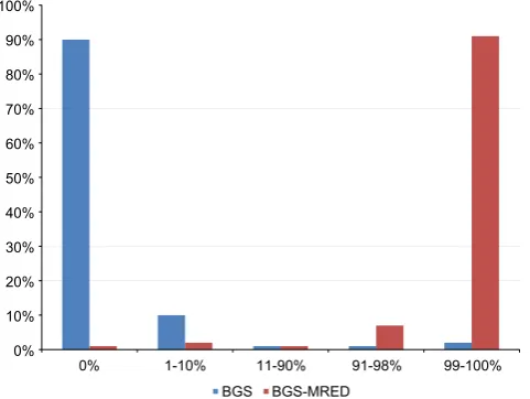
In order to compare the efficiencies of the original and the mod-ified protocols, we investigated the rates of conversion of the non-CpG-cytosine residues within the RARB2 P2 promoter region under analysis. These sites are not normally methylated and are, therefore, expected to be fully converted. There are 74 non-CpG-cytosine residues within this region (excluding those found within regions complementary to primers JP5 and JP7): we ran-domly set the threshold for the status of informativity to 73/74 (99%) conversions to uracil and used this threshold to distinguish fully converted sequencings from partially converted ones. In par-ticular, for a sample to be labeled as fully converted, it must have reached ≥99% conversion of these non-CpG-cytosines. Upon comparison, we found that there was a dramatic increase in the number of informative sequencings using our modified protocol: while only 10% of samples sequenced using the original protocol (n=200) achieved 99% conversion of non-CpG-cytosines, 91% of samples sequenced using the modified protocol (n=176) achieved 99% conversion (Fig.2). It is interesting to note that the majority of the remaining sequencings using the modified protocol were nearly fully converted (91–98%). In contrast, nearly all sequenc-ings using the original protocol were nearly fully unconverted (0% and 1–10%).
The use of MRED (using AluI, AvaI, DdeI, HhaI, and
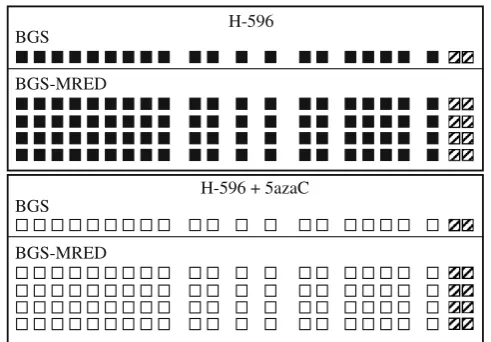
contained within the RE sites (1, 7, 15, 17, 18, 20 inFig.1), we applied MRED to DNA samples previously analyzed using BGS alone. DNA samples previously extracted from H-596 lung adenosquamous carcinoma cells treated or not with 5-azadeoxy-cytidine, a methylation inhibitor. Using BGS, they were found to be fully methylated from untreated cells or fully unmethylated following treatment with 5-azadeoxycytidine (Fig.3). When they were resequenced using the modified protocol, the results were identical: 8/8 sequencings displayed complete methylation (Fig.
3, top) or complete demethylation (Fig. 3, bottom) at all 20 in-formative sites. This clearly demonstrates that the introduction of the MRED step does not introduce a bias at any of the 20 infor-mative sites.
Using the BGS protocol (1) in over 400 sequencings, even with some modifications, we found that incomplete cytosine to uracil conversion and DNA degradation formed insurmountable challenges. In a first effort, numerous attempts to reduce the rate of target DNA degradation were made, including: (1) increasing the amount of genomic DNA from 1 to 10 µg; (2) decreasing the duration of the bisulfite conversion reaction (15); (3) incorporat-ing up to 5 µg salmon sperm DNA; or (4) usincorporat-ing agarose beads
0% 10% 20% 30% 40% 50% 60% 70% 80% 90% 100%
0% 1-10% 11-90% 91-98% 99-100% BGS BGS-MRED
(16). None of these modifications improved the rate of DNA deg-radation. In addition, the absence of PCR products could not be associated with any one factor (it was variable and unpredictable). In a second effort, numerous attempts to increase the rate of cytosine to uracil conversion were made, including: (1) digesting the genomic DNA with an endonuclease such asPstI to create smaller fragments containing the target sequence thereby reduc-ing the possibility for regional double strand formation (24); (2) denaturing the genomic DNA in an alkaline solution prior to BGS treatment, thereby beginning the BGS protocol with fully denatured DNA; (3) incubating the bisulfite reactions at 95°C (24) every 3 h, thereby aiming to maintain complete DNA dena-turation; (4) reducing the DNA quantity to as little as 100 ng (13); (5) increasing the sodium bisulfite concentration (6 M) (15), (6) using high-speed BGS (9 M sodium bisulfite for 20 min at 90°C or 40 min at 70°C) (19), (7) using a lower incu-bation temperature, such as 50°C (15), to increase the extent of cytosine conversion and/or to reduce the annealing of single-stranded DNA sequences during treatment; (8) monitoring the pH of the solutions to prevent incomplete desulfonation of pyrim-idine residues, which may inhibit DNA polymerases, leading to unsuccessful PCR amplifications (12); (9) changing PCR exten-sion time; (10) annealing temperature; (11) MgCl2
concentra-tion; (12) adding dimethyl sulfoxide to inhibit secondary structure formation (26). In all cases, PCR amplifications were
H-596 + 5azaC H-596 BGS
BGS-MRED
BGS
BGS-MRED
once again unpredictable, and when they were productive, none of these modifications increased the rate of occurrence of fully converted samples following BGS above 0–10%. Different primers were also designed, including fully nested as opposed to semi-nested primers as was the case here, to no avail.
Such resistance to deamination is a frequent characteristic of
RARB2(unpublished observation, Dr. Michael Trus, Juravinski Cancer Center, Hamilton, Ontario) and is not unique toRARB2
(e.g., (27–29)). Although high GC content has previously been suggested to cause incomplete conversion (30), the 541-bp re-gion we targeted has a GC content of 51%, 4% lower than that characterizing most CpG islands or promoters (31).
RARB2DNA methylation has been shown to be correlated with RARB2gene inactivation (32–38), and treatment with a methyltransferase inhibitor, 5-azadeoxycytidine, has been shown to be correlated with demethylation of exonic sequences and reacti-vation of gene expression (32,34,37,39). However, few studies have analyzed the detailed methylation pattern of the promoter region (33,40–42), and to our knowledge, studies have not analyzed isolat-ed alleles by sequencing only one subclone per bisulfite conversion reaction (seeSection2.5.1). The vast majority of studies have used methylation-specific PCR, pooling potentially mixed populations of alleles together, as previously described in ref. (38), and not allow-ing the direct assessment of cytosine to uracil conversion.
To our knowledge, this is the first report of a RE-based method to improve the BGS protocol. This modified protocol is not related to techniques in which RE digestion is used to re-veal and/or quantify DNA methylation-dependent sequence dif-ferences in PCR-amplified bisulfite-treated DNA (43) or with techniques in which methylation-dependent retention of preex-isting sites, such as BstUI (CGCG; following bisulfite-induced sequence conversion), are exploited to quantify DNA methyla-tion at specific loci, such as in the combined bisulfite restricmethyla-tion analysis (4). These techniques focus on specific CpG sites and are based on the assumption that conversion is complete. In contrast, the present protocol was designed to retain the fine resolution analysis capability of the original BGS protocol. It does so by digesting incompletely converted DNA molecules in the resulting mixed PCR population. Conversion efficiency is not assumed to be 100% but rather is measured directly for every sample.
and the growing interest in protocols providing internal quality control parameters. The guidelines for selecting REs are straight-forward and may be used for the methylation analysis of any gene. This method requires the addition of only one step, MRED, to the original protocol, adding only 4 h to the 3-day BGS process. While RE selection may be time consuming for some sequences, the same combination of RE may be used for all subsequent sequencings.
Acknowledgements
The authors thank Ms. Johane Morin (now at Montreal Diabetes Research Center, CR-CHUM—Technopôle Angus, Montreal, Canada) and Dr. Zhuo Li (Bio S&T) for their help with sequenc-ings. The authors also thank Dr. Mark Featherstone (School of Biological Sciences, Nanyang Technological University, Singa-pore) for his critical assessments of and invaluable input to the work in all its phases and Dr. Zeina Saikali (Juravinski Cancer Center, Hamilton, Ontario) for revising the manuscript.
Competing Interests Statement: The authors declare no competing interests.
Funding: This work was supported by grants from the Cancer Research Society. JJP was supported by the McGill University Center for Trans-lational Research in Cancer Fellowship Award, Israel Cancer Research
Foundation; the Bourse Fonds Robert Bourassa—Bourse de
l'Assem-blée Nationale du Québec; the Fondation Marc Bourgie; the Défi Cor-poratif Canderel; and the Institut du cancer de Montreal.
References
1. Clark SJ, Harrison J, Paul CL, Frommer M (1994) High sensitivity mapping of methyl-ated cytosines. Nucleic Acids Res 22:2990– 2997
2. Frommer M, McDonald LE, Millar DS, Collis CM, Watt F, Grigg GW, Molloy PL, Paul CL (1992) A genomic sequencing protocol that yields a positive display of 5-methylcytosine residues in individual DNA strands. Proc Natl Acad Sci U S A 89:1827–1831
3. Herman JG, Graff JR, Myohanen S, Nelkin BD, Baylin SB (1996) Methylation-specific PCR: a novel PCR assay for methylation status of CpG islands. Proc Natl Acad Sci U S A 93:9821–9826
4. Xiong Z, Laird PW (1997) COBRA: a sen-sitive and quantitative DNA methylation as-say. Nucleic Acids Res 25:2532–2534 5. Gonzalgo ML, Jones PA (1997) Rapid
quantitation of methylation differences at
specific sites using methylation-sensitive single nucleotide primer extension (Ms-SNuPE). Nucleic Acids Res 25:2529–2531 6. Bianco T, Hussey D, Dobrovic A (1999) Methylation-sensitive, single-strand confor-mation analysis (MS-SSCA): a rapid meth-od to screen for and analyze methylation. Hum Mutat 14:289–293
7. Eads CA, Danenberg KD, Kawakami K, Saltz LB, Blake C, Shibata D, Danenberg PV, Laird PW (2000) MethyLight: a high-throughput assay to measure DNA methylation. Nucleic Acids Res 28:E32 8. Gitan RS, Shi H, Chen CM, Yan PS,
Huang TH (2002) Methylation-specific ol-igonucleotide microarray: a new potential for high-throughput methylation analysis. Genome Res 12:158–164
J, Kerjean A (2003) DHPLC-based method for DNA methylation analysis of differential methylated regions from imprinted genes. Biotechniques 34:356–362
10. Dupont JM, Tost J, Jammes H, Gut IG (2004) De novo quantitative bisulfite se-quencing using the pyrosese-quencing tech-nology. Anal Biochem 333:119–127 11. Wojdacz TK, Dobrovic A (2007)
Methyla-tion-sensitive high resolution melting (MS-HRM): a new approach for sensitive and high-throughput assessment of methyla-tion. Nucleic Acids Res 35:e41
12. Fraga MF, Esteller M (2002) DNA methyl-ation: a profile of methods and applications. Biotechniques 33:632–649
13. Feil R, Charlton J, Bird AP, Walter J, Reik W (1994) Methylation analysis on individu-al chromosomes: improved protocol for bi-sulfite genomic sequencing. Nucleic Acids Res 22:695–696
14. Tasheva ES, Roufa DJ (1994) Densely methylated DNA islands in mammalian chromosomal replication origins. Mol Cell Biol 14:5636–5644
15. Raizis AM, Schmitt F, Jost JP (1995) A bi-sulfite method of 5-methylcytosine map-ping that minimizes template degradation. Anal Biochem 226:161–166
16. Olek A, Oswald J, Walter J (1996) A mod-ified and improved method for bisulfite based cytosine methylation analysis. Nucleic Acids Res 24:5064–5066
17. Paulin R, Grigg GW, Davey MW, Piper AA (1998) Urea improves efficiency of bisul-fite-mediated sequencing of 5′ -methylcyto-sine in genomic DNA. Nucleic Acids Res 26:5009–5010
18. Grunau C, Clark SJ, Rosenthal A (2001) Bisulfite genomic sequencing: systematic investigation of critical experimental param-eters. Nucleic Acids Res 29:E65
19. Shiraishi M, Hayatsu H (2004) High-speed conversion of cytosine to uracil in bisulfite genomic sequencing analysis of DNA methylation. DNA Res 11:409–415 20. Shen S, Kruyt FA, den Hertog J, van der Saag
PT, Kruijer W (1991) Mouse and human ret-inoic acid receptor beta 2 promoters: se-quence comparison and localization of retinoic acid responsiveness. DNA Seq 2:111–119
21. Houle B, Rochette-Egly C, Bradley WE (1993) Tumor-suppressive effect of the reti-noic acid receptor beta in human epidermoid lung cancer cells. Proc Natl Acad Sci U S A 90:985–989
22. Viallet J, Liu C, Emond J, Tsao MS (1994) Characterization of human bronchial epi-thelial cells immortalized by the E6 and E7 genes of human papillomavirus type 16. Exp Cell Res 212:36–41
23. Warnecke PM, Stirzaker C, Song J, Grunau C, Melki JR, Clark SJ (2002) Identification and resolution of artifacts in bisulfite se-quencing. Methods 27:101–107
24. Rein T, Zorbas H, DePamphilis ML (1997) Active mammalian replication origins are as-sociated with a high-density cluster of mCpG dinucleotides. Mol Cell Biol 17:416–426 25. Lowe T, Sharefkin J, Yang SQ, Dieffenbach
CW (1990) A computer program for selec-tion of oligonucleotide primers for poly-merase chain reactions. Nucleic Acids Res 18:1757–1761
26. Warnecke PM, Stirzaker C, Melki JR, Millar DS, Paul CL, Clark SJ (1997) Detection and measurement of PCR bias in quantitative methylation analysis of bisulfite-treated DNA. Nucleic Acids Res 25:4422–4426 27. Bearzatto A, Szadkowski M, Macpherson
P, Jiricny J, Karran P (2000) Epigenetic regulation of the MGMT and hMSH6 DNA repair genes in cells resistant to meth-ylating agents. Cancer Res 60:3262–3270 28. Boily G, Saikali Z, Sinnett D (2004)
Methyl-ation analysis of the glypican 3 gene in embry-onal tumours. Br J Cancer 90:1606–1611 29. Yin H, Blanchard KL (2000) DNA
methyl-ation represses the expression of the human erythropoietin gene by two different mech-anisms. Blood 95:111–119
30. Shen L, Guo Y, Chen X, Ahmed S, Issa JP (2007) Optimizing annealing temperature overcomes bias in bisulfite PCR methyla-tion analysis. Biotechniques 42:48–52 31. Gardiner-Garden M, Frommer M (1987)
CpG islands in vertebrate genomes. J Mol Biol 196:261–282
32. Bovenzi V, Le NL, Cote S, Sinnett D, Momparler LF, Momparler RL (1999) DNA methylation of retinoic acid receptor beta in breast cancer and possible therapeu-tic role of 5-aza-2′-deoxycytidine. Antican-cer Drugs 10:471–476
33. Bovenzi V, Momparler RL (2000) Quanti-tation of inhibition of DNA methylation of the retinoic acid receptor beta gene by 5-Aza-2′-deoxycytidine in tumor cells using a single-nucleotide primer extension assay. Anal Biochem 281:55–61
affecting the chromatin state of the retinoic acid receptor beta2 promoter in breast can-cer cells. Oncogene 19:1556–1563 35. Virmani AK, Rathi A, Zochbauer-Muller S,
Sacchi N, Fukuyama Y, Bryant D, Maitra A, Heda S, Fong KM, Thunnissen F et al (2000) Promoter methylation and silencing of the retinoic acid receptor-beta gene in lung carcinomas. J Natl Cancer Inst 92:1303–1307
36. Bovenzi V, Momparler RL (2001) Anti-neoplastic action of 5-aza-2′-deoxycytidine and histone deacetylase inhibitor and their effect on the expression of retinoic acid re-ceptor beta and estrogen rere-ceptor alpha genes in breast carcinoma cells. Cancer Chemother Pharmacol 48:71–76
37. Sirchia SM, Ren M, Pili R, Sironi E, Some-nzi G, Ghidoni R, Toma S, Nicolo G, Sac-chi N (2002) Endogenous reactivation of the RARbeta2 tumor suppressor gene epi-genetically silenced in breast cancer. Cancer Res 62:2455–2461
38. Cote S, Momparler RL (1997) Activation of the retinoic acid receptor beta gene by 5-aza-2′-deoxycytidine in human DLD-1 colon carcinoma cells. Anticancer Drugs 8:56–61
39. Cote S, Sinnett D, Momparler RL (1998) Demethylation by 5-aza-2′-deoxycytidine of specific 5-methylcytosine sites in the pro-moter region of the retinoic acid receptor beta gene in human colon carcinoma cells. Anticancer Drugs 9:743–750
40. Arapshian A, Kuppumbatti YS, Mira-y-Lopez R (2000) Methylation of conserved CpG sites neighboring the beta retinoic acid response element may mediate retinoic acid receptor beta gene silencing in MCF-7 breast cancer cells. Oncogene 19:4066–4070
41. Widschwendter M, Berger J, Hermann M, Muller HM, Amberger A, Zeschnigk M, Widschwendter A, Abendstein B, Zeimet AG, Daxenbichler G et al (2000) Methyla-tion and silencing of the retinoic acid recep-tor-beta2 gene in breast cancer. J Natl Cancer Inst 92:826–832
42. Pappas JJ, Toulouse A, Hebert J, Fetni F, Bradley WEC (2008) Allelic methylation bias of the RARB2 tumor suppressor gene promoter in cancer. Genes Chromosomes Cancer 47:978–993