www.ijper.org
Comparative Wound Healing Activity of two Polyherbal
Formulations GC-01 and GC-02 on Experimentally
induced Wounds in Rodents
Purnima Ashok*, Nishi Gupta, Niranjana Murthy1, Renuka Shukla and G Rohita Sri
Department of Pharmacology, KLE University’s College of Pharmacy, Bangalore-560010
1PentacareAyurPharma Industries, No. 1662, Srirampuram, Bangalore-003
ABSTRACT
The present study was undertaken to support the use of herbs in folklore medicine for wound healing activity. In the present investigation, a comparative study and screening was done on two polyherbal formulations GC-01 and GC-02 fortified with different herb extracts in ghee for wound healing activity in experimentally induced wounds in rodents. Wound healing activity was evaluated by using three wound models viz. incision, excision and burn. For the study, standard drug used was Framycetinsulphate cream for incision and excision wound model, while Silver sulfadiazine cream was used for burn wound model. Fresh ointments of 5%, 10% and 15% of polyherbal formulations in simple ointment base were prepared and evaluated by applying topically for a period of 10–21 days depending on the type of study. The parameters assessed were tensile strength in incision wound model whereas in excision and burn wound models reduction in mean wound area, percentage wound contraction, epithelization period, granulation tissue antioxidant status [estimation of superoxide dismutase (SOD) and reduced glutathione (GSH)], free radical (lipid peroxidation), connective tissue formation and maturation (hydroxy proline). The results have shown increased wound breaking strength and levels of hydroxy proline, superoxide dismutase andglutathione in the granulation tissue. Decreased mean wound area and lipid peroxidation was also observed. This could be the result of synergistic/potentiative action of individual medicinal herbs present in 01 and GC-02in gheeand diverse array of active principles present in them.
Keywords: Polyherbal formulation, incision, excision and burn wound model, antioxidant status.
DOI: 10.5530/ijper.48.2.11
Address for correspondence:
Dr. Purnima Ashok,
Professor and Head, Department of Pharmacol-ogy, K.L.E. University’s College of Pharmacy, Bangalore-560010, India. Mobile: +919341920793 E-mail: purnimaal1@yahoo. com
INTRODUCTION
Wounds are common and inescapable events of life which are a result of physical, chemical injury or microbial infections.1
Loss or breaking of cellular and anatomic or disturbances in functional continuity of living tissues are the reasons of wound.2 It
results in a variety of cellular and molecular sequelae, opening or breaking of the skin and
is also defined as a breach in the normal tis -sue continuum.3 On the other hand, wound
healing is a process involving a connective
tissue response and an acute inflammatory
phase followed by synthesis of collagens and other extra cellular macromolecules which later remodelsto form a scar.
Use of herbal extracts instead of crude herbs started with the aim to control quality and precise dosage for better results.
The plant extracts being more efficacious
are free from undesirable side effects.
Medicinal plants are used by folklore medicine in India for treatment of cuts, wounds and burns4 are abundantand due
to their specific healing property and
non-toxic actions, scientists are keen to evalu-ate drugs from them.5
wound healing potential in incision, excision and burn wound models along with the estimation of anti-oxidant status and biochemical param-eters. The present study gives substantial sup-port to the use of these herbs in folklore medicine for wound healing.
MATERIALS AND METHODS
Preparation of ointments of Polyherbal formulations: The polyherbal formulations, GC-01 and GC-02 were procured from Pentacare Ayur Pharma, Bangalore as gift samples. For the study, all the ointments were freshly prepared before topical application. Different concentrations ofPHF(5%, 10% and 15%) were incor-porated into simple ointment base I.P. and mixed in a mortar and pestle to obtain uniform consistency.
Animals: Adult, healthy albino Wistar rats of either sex
in the weight range of 180–250g were procured from
registered breeder during the experiment. Clearance from Institutional Animal Ethics Committee (IAEC) of KLE University’s College of Pharmacy, Bangalore was sought (IAEC/04/PA/2011–12) prior to experiment.
Standard drug: Framycetin sulphate cream (1%,
Sofra-mycin from Aventis Pharma
Limited) was selected as standard drug for incision and excision wound model whereas Silver sulfadiazine (1%, Burn heal from Curetech Skincare) was used in case of burn wound model.
Chemicals-Nitro blue tetrazolium chloride,
Phenazine methosulphate Trichloroacetic acid, Butylated hydroxyl toluene and p-dimethyl amino benzaldehyde were procured from M/s. SD Fine-Chem Limited, Mumbai. N-Butyl alcohol was from M/s. Nice Chemicals Pvt. Ltd., Kochi. Hydroxypro-line Standard, Di-thionitro benzoic acid and reduced glutathione were procured from M/s Sisco Research Lab,
Table 1. Composition of formulation of GC-01
Botanical name Indian name Family Quantity
Butea monosperma Lam. Palashapushpa Fabaceae 3 gm
Symplocos racemosa Roxb. Lodhra Symplocaceae 5 gm
Mimosa pudica Linn. Lajjalu, Mimosaceae 4 gm
Curcuma longa Linn. Haridra, Zingiberaceae 3 gm
Commiphora mukul Guggulu, Burseraceae 500 mg
Piper longum Linn. Pipalli Piperaceae 500 mg
Azadarchta indica Linn. Nimba, Meliaceae 2 gm
Pongamia glabra Karanja, Leguminosae 2 gm
Cocos nucifera Linn. Narikela Arecaceae 2 ml
Ghrita Ghee 8 ml
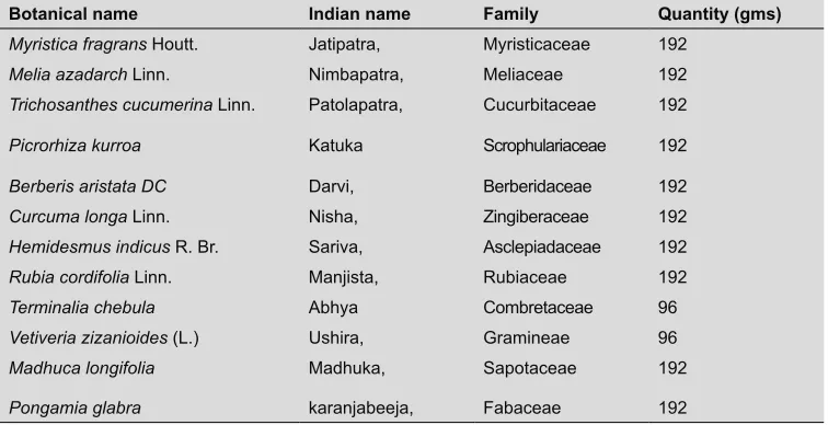
Table 2. Composition of Formulation of GC-02
Botanical name Indian name Family Quantity (gms)
Myristica fragrans Houtt. Jatipatra, Myristicaceae 192
Melia azadarch Linn. Nimbapatra, Meliaceae 192
Trichosanthes cucumerina Linn. Patolapatra, Cucurbitaceae 192
Picrorhiza kurroa Katuka Scrophulariaceae 192
Berberis aristata DC Darvi, Berberidaceae 192
Curcuma longa Linn. Nisha, Zingiberaceae 192
Hemidesmus indicus R. Br. Sariva, Asclepiadaceae 192
Rubia cordifolia Linn. Manjista, Rubiaceae 192
Terminalia chebula Abhya Combretaceae 96
Vetiveria zizanioides (L.) Ushira, Gramineae 96
Madhuca longifolia Madhuka, Sapotaceae 192
Pongamia glabra karanjabeeja, Fabaceae 192
Mumbai. Thiobarbituric acid was obtained from M/s. Spectrochem Pvt. Ltd., Mumbai.
Treatment protocol: The wound healing activity
was undertaken in incision, excision and burn wound models. In each model, eight groups of six animals each were used. In all the models group 1 was con-trol (no treatment), group 2 standard (Framycetin/ silver sulfadiazine), groups 3,4 and 5 were treated with 5%, 10% and 15% of GC-01 whereas groups 6, 7 and8 were treated with 5%, 10% and 15% of GC-02 respectively.
For wound healing study in incision wound model, the standard formulation and all test drug ointments (5%, 10% and 15% w/w) were applied topically on wound area once daily from 0 to 10th postoperative days where as in excision and burn wound models the standard for-mulation and all test drug ointments (5%, 10% and 15% w/w) were applied topically on wound area once daily from 0 to 21 postoperative days or till complete healing, whichever occurred earlier. The effect of GC-01 and GC-02 on various parameters (i) wound tensile strength (ii) physical parameters like mean wound area, percent-age wound contraction and epithelization period (iii) biochemical parameters like Malondialdehyde (MDA) and Hydroxy proline were estimated and compared with the standard and also with each other.
Experimental procedure: Adult, healthy albino Wistar
rats of either sex in the weight range of 180-250g were used for the study.
Incision wound model: The rats were anaesthetized
by administration of ketamine hydrochloride (50 mg/kg b.w., i.p.). On depilated back of the animal, two para-vertebral straight incision of 6 cm length each were made through the entire thickness of the skin, on either side of the vertebral column with the help of sharp sterile scalpel. Care was taken to see that the incisions were atleast 1 cm lateral to the vertebral column. After complete haemostasis the wounds were closed by means of interrupted suture placed at equidistance points about 1 cm apart using 2-zero surgical silk thread and a curved needle (No. 11). Wounds were then mopped with cotton swabs soaked in saline and were caged individually. Ani-mals ingroup 1 received no treatment and served as normal control. Standard (FSC) and all the test drug ointments (5%, 10% and 15% w/w of GC-01 and GC-02) were applied topically to the wound once a day for a period of 10 days. Sutures were removed on 8th day and tensile strength was measured on 10th day by continuous water flow technique of Lee.6 Three such readings were recorded for a given
incision wound and the procedure was repeated on the other wound of the same animal, thus
obtaining six readings for each animal. The mean breaking strength in each animal i.e. average of six readings was used to calculate the group mean.
Excision wound model: A round circular seal of 300
mm2 diameter was impressed on the dorsal thoracic
central region 5 cm away from the ears of anaesthetized rats. Full thickness skin from demarked area was excised to get a wound of approximate 300 mm2. After
achiev-ing the full haemostasis wound was blotted with cotton swab soaked in warm saline and animals were placed in their individual cages. The standard formulation (Framycetin), and all test drug ointments were applied as per the protocol.
Burn wound model: Partial thickness burn wound
was inflicted on overnight starved animals under light
ether anesthesia, by pouring hot molten wax at 800C into a metal cylinder of 300 mm2 on depilated back of
the animal. The molten wax in the cylinder was allowed to solidify which took about 10–12 minutes. Then the metal cylinder with wax adhered to the skin was removed which left a distinctly demarked partial thickness circular burn wound of 300 mm2 area. Treatment was
done as per the protocol.
Assessment of parameters: For assessing
parame-ters in excision and burn wound models, two wounds were induced simultaneously on depilated back of animal. One wound was used for measuring wound contraction and epithelization period whereas the sec-ond one was used for various biochemical estimations and anti-oxidant parameters. These estimations were done on 7th post wound day in the granulomatous tissue. Wound contraction7 was measured by tracing
wound on 1 mm2 graph paper on alternate days. The
calculated surface area was then employed to obtain the percentage wound contraction by taking the initial average size of wound 300 mm2 as 100% by using
fol-lowing equation:
Epithelialization period8:was recorded as the number
of days required for the clot (eschar) to fall off from the burn wound surface without leaving a raw wound behind.
Biochemical and antioxidant estimations9: On 7th
post wounding day of 2nd wound on animal, wounded
granulomatous tissue were excised from all the groups of both excision and burn wound modelsin such a way that wounded area was excised without contaminating it with normal skin and then homogenised. This tissue homogenate was used for the estimation of hydroxy-proline,10 superoxide dismutase11 (SOD), reduced
gluta-thione12,13 (GSH) and lipid peroxide14 (MDA).
Statistical analysis: Results were expressed as
mean values ± SEM (Standard Error of Mean) and were calculated for each group. Statistical differences between means were determined by Graph Pad Prism (5.0) using one-way ANOVA followed by Tukey’s post hoc test. Values p<0.05 were considered as statistically significant.
Results
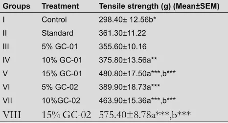
Effect on incision wound model: There was a
significant increase in tensile strength of the wound
treatedwith polyherbal formulations as compared to that of the control and standard groups. The results are shown in (Table 1). Higher doses of the formulation were found to be more effective. Topical application of 15% GC-01
and 10%, 15% GC-02 produced significant (p<0.0001)
increase in breaking tensile strength when compared with standard Framycetinsulphate treated group.
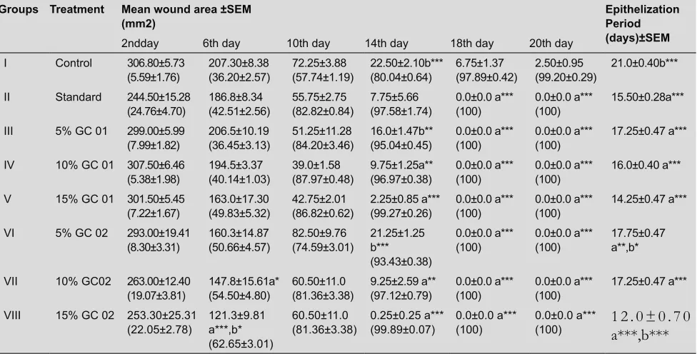
Effect on excision wound model: Both the
formulations showed faster healing when compared with control group and wound contraction was at a faster rate with the formulations as compared with control
group (Table 2). There was a significant decrease in the
number of daysrequired for complete healing in the ani-mals treated with5%, 10%, 15% of GC-01 (p<0.0001) and5% (p<0.001), 10% and 15% of GC-02 (p<0.0001) when compared to the control group animals. Higher concentrations of both the formulations have shown lesser epithelialization period or faster wound contrac-tion comparedto standard group. The number of days required for complete healing by 5%, 15% of GC-02 has
shown significant result (p<0.01, p<0.0001) respectively
when compared to that of the standard (FSC) group. Percentage wound contraction was found to behighest in10% and 15% of both the formulations compared to lower concentrations and standard group.
Hydroxy proline levels is a measure of collagen index and higher the concentration ofhydroxy prolinefaster
the rate of wound healing. Excision wound model
has shown a significant increase in hydroxyproline
content in 10% (p<0.01)and 15% (p<0.0001) of GC-01 and all concentrations of GC-02 (p<0.0001) formula-tion treated animals when compared to control group (Fig. 1). The animals received no treatment (group 1) had lowest content of hydroxy proline.
Measurement of malondialdehyde (MDA) is an index of lipid peroxidation. MDA levels were
significantly reduced in all the test groups (p<0.0001)
when compared with the control group and in 5% GC-02 (p<0.01) and 10% of GC-02 (p<0.001). MDA
levels were significantly reduced (p<0.0001) in stan -dard groups alsowhen compared with all the test groups as shown in (Fig. 2).
Estimation of antioxidant like SOD and glutathione (GSH )is relevant as it hastens the process of wound
healing. All the treatment groups have shown signifi -cant increase (p<0.0001) in SOD levels when compared
with control group. Significant increase was observed
in superoxide levels in 15% (p<0.0001) of both the formulations when compared with standard group as shown in (Fig. 3).
GSH levels were significantly increased in 5%, 10% and
15% of GC-02(p<0.0001) and in 10% GC-01(p<0.01) treated groups when compared with control and standard groups as shown in (Fig. 2).
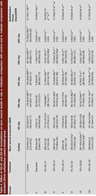
Effect on burn wound model: Both the formulations
showed faster healing of wound ( rate of wound con-traction) when compared with control group as shown
in (Table 3 and Fig. 4). There was a significant decrease
in the number of days required for complete healing of wound in the animals treated with 10% and 15% ofGC-01 (p<0.0ofGC-01),5%, 10%, 15% of GC-02 (p<0.00ofGC-01) and when compared to control group.Higher concen-trations of both the formulations have shown better results. 15% GC-02 was found to be more potent and equivalent to the standard drug.
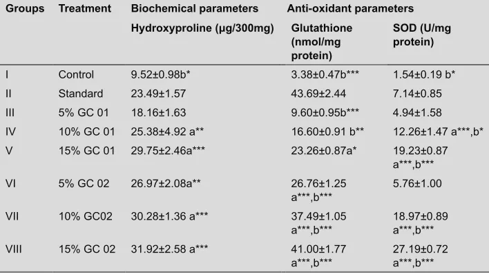
There was a significant increase in hydroxy proline
content in groups of 15% GC-01,10%, 15% of GC-02, formulation(p<0.0001)and 10% GC-01, 5% of GC-02 (p<0.01) when compared tocontrol group.
There was no significant change observed in MDA lev -els in GC-01 and GC-02 treated animals compared to control as well as standard group.
On comparing different concentrations of both the
formulations, there was significant increase in SOD
levels of 5% GC- 01 which was comparable to15% GC-01, 10%,15% of GC-02 and 10% GC-01 was com-pared with 15% of GC-02 (p<0.0001).
GSH levels were significantly increased in the treatment groups when compared with control group. Significant
Table 1. Effect of GC 01 and GC 02 on tensile strength in incision wound model on rats a- indicates comparison with control and b- indicate comparison with standard (FramycetinSulphate) treated group. Groups Treatment Tensile strength (g) (Mean±SEM)
I Control 298.40± 12.56b*
II Standard 361.30±11.22
III 5% GC-01 355.60±10.16
IV 10% GC-01 375.80±13.56a**
V 15% GC-01 480.80±17.50a***,b***
VI 5% GC-02 389.90±18.73a***
VII 10%GC-02 463.90±15.36a***,b***
Table 2. Effect of GC-01 and GC-02 in Excision wound model in rats a- indicates comparison with control and b- indicate comparison with standard (FramycetinSulphate) treated group.
Groups Treatment Mean wound area ±SEM
(mm2) Epithelization Period
(days)±SEM
2ndday 6th day 10th day 14th day 18th day 20th day
I Control 306.80±5.73
(5.59±1.76) 207.30±8.38(36.20±2.57) 72.25±3.88(57.74±1.19) 22.50±2.10b***(80.04±0.64) 6.75±1.37(97.89±0.42) 2.50±0.95(99.20±0.29) 21.0±0.40b***
II Standard 244.50±15.28
(24.76±4.70) 186.8±8.34(42.51±2.56) 55.75±2.75(82.82±0.84) 7.75±5.66(97.58±1.74) 0.0±0.0 a***(100) 0.0±0.0 a***(100) 15.50±0.28a***
III 5% GC 01 299.00±5.99
(7.99±1.82) 206.5±10.19(36.45±3.13) 51.25±11.28(84.20±3.46) 16.0±1.47b**(95.04±0.45) 0.0±0.0 a***(100) 0.0±0.0 a***(100) 17.25±0.47 a***
IV 10% GC 01 307.50±6.46
(5.38±1.98) 194.5±3.37(40.14±1.03) 39.0±1.58(87.97±0.48) 9.75±1.25a**(96.97±0.38) 0.0±0.0 a***(100) 0.0±0.0 a***(100) 16.0±0.40 a***
V 15% GC 01 301.50±5.45
(7.22±1.67) 163.0±17.30(49.83±5.32) 42.75±2.01(86.82±0.62) 2.25±0.85 a***(99.27±0.26) 0.0±0.0 a***(100) 0.0±0.0 a***(100) 14.25±0.47 a***
VI 5% GC 02 293.00±19.41
(8.30±3.31) 160.3±14.87(50.66±4.57) 82.50±9.76(74.59±3.01) 21.25±1.25 b*** (93.43±0.38)
0.0±0.0 a***
(100) 0.0±0.0 a***(100) 17.75±0.47 a**,b*
VII 10% GC02 263.00±12.40
(19.07±3.81) 147.8±15.61a*(54.50±4.80) 60.50±11.0(81.36±3.38) 9.25±2.59 a**(97.12±0.79) 0.0±0.0 a***(100) 0.0±0.0 a***(100) 17.25±0.47 a***
VIII 15% GC 02 253.30±25.31
(22.05±2.78) 121.3±9.81 a***,b* (62.65±3.01)
60.50±11.0
(81.36±3.38) 0.25±0.25 a***(99.89±0.07) 0.0±0.0 a***(100) 0.0±0.0 a***(100) 1 2 . 0 ± 0 . 7 0a***,b***
CONT ROL
STAND ARD
5% G C-01
10% G C-01
15% G C-01
5% G C-02
10% G C-02
15% G C-02 0
10 20 30
Groups
H
ydr
ox
ypr
ol
ine
(
mg/
30
0 m
g)
Figure: 1
CONT ROL
STAND ARD
5% G C-01
10% G C-01
15% G C-01
5% G C-02
10% G C-02
15% G C-02 0
1 2 3 4
Groups
S
O
D
(U
/m
g pr
ot
ei
n)
Figure: 2
increase was observed in GSH levels of 5%, 10% and 15% of GC-01 and5% GC-02 formulation (p<0.0001) when compared with standard group. Standard group
has shown significant increase (p<0.0001) when com -pared with 5%, 10%, 15% GC-01 and 5% GC-02.The various biochemical and anti-oxidant parameters in burn wound model are listed in (Table 4).
Discussion
Wounds in severe caseswhen left untreated can pose a major health problem, sometimes even leading to death. The phenomenon of restoring damaged tissue as closely as possible to normal state is known as wound healing resulting in restorage of tissue integrity. The process
Figure: 3
remodelling of connective tissue, synthesis of extracellular matrix protein, acquisition of tensile strength, contraction and epithelization. The basic principle of optimal wound healing involves minimal tissue damage and provision of adequate tissue perfusion and oxygenation, proper nutrition and wound healing environment to restore the continuity and function of the affected part.
GC-01 and GC-02 are polyherbal formulations having a combination of herbs processed in gheewith
reportedphytoconstituents like flavonoids, steroids, cur
-cumin, diterpenoids, azadiractin, β-sitosterol, berberine,
chebulinic acid,etc. Since some of these active constitu-ents are known to contain growth promoting factor which enhances the wound healing process, the present claim of this formulation under study as a wound healing agent may be attributed to these active principles solely or synergisti-cally. Further, the process of wound healing has two
com-ponents, first is the formation of new tissue and other is
the protection from microbial infections duringhealing2.
Ghee promoteswound healing while herbs present in the PHF GC-01 and GC-02 offers both components. This may be the reason to formulate the present polyherbal for-mulations GC-01 and GC-02 in ghee, used extensivelyin Ayurveda for wound healing.
Tensile strength of wound represents the effectiveness of wound healing(the force required to open the heal-ing skin) and is used to measure the completeness of healing.15 In incision wound model, the increase in
ten-sile strength of skin in treated wounds may be due to elevated collagen levels and stabilization of collagen
fibers. Significant wound healing activity was observed
in the animals treated with GC-01 and GC-02 poly-herbal formulations on 10th post wounding day. From this, it may be inferred that the formulations not only increases collagen synthesis per cell but also aided in cross linking of the protein.
Hydroxy proline is the result of collagen breakdown and its measurement gives an index of collagen turnover.16
In the present study, both the formulations have shown dose dependent faster healing and increase in levels of hydroxyproline, superoxide dismutase and glutathione. Increased turnover of these leads to rapid healing with concurrent increase in tensile strength. Free radicals and oxidative reaction products produce tissue dam-age. Overproduction of reactive oxygen species (ROS) results in oxidative stress thereby causing cytotoxic-ity and delayed wound healing. Therefore, elimination of ROS could be an important strategy in healing of chronic wounds.17 ROS also play an important role in
the failure ofischaemic wound healing while antioxi -dants improve healing in ischemic skin wounds.18
Hence, estimation of antioxidants like superoxide dismutase (SOD) and glutathione (GSH) in granulation tissues were also relevant as these anti-oxidants hasten the process of wound healing by destroying the free radicals. In the present study, both the formulations
have shown significant increase in the levels of SOD
and GSH , indicating preventive phenomenon of the formulations against oxidative damage, thereby contrib-uting to wound healing.
Elevated levels of MDA, a marker of free radical damage may be attributed to impaired wound healing in immune-compromised rats.19 Lipid peroxidation
is an important process of several types of injuries
like burn, inflicted wound and skin ulcers. A drug that
inhibits lipid peroxidation is believed to increase the
viability of collagen fibrils, increasing the strength of collagen fibers by an increase in circulation, thereby
preventing the cell damage and promoting DNA syn-thesis.18 MDA levels were significantly reduced by the
formulations under study in excision wound model suggesting protection to damaged tissues whereas
there was no significant change in MDA levels in burn
wound model. There are reports suggesting that lipid peroxidation process begins immediately after severe burn and was continued during the whole 48 h post burn period and marked increase in plasma levels of MDA depends according to the extent of injury.20
Since, there are no significant changes in MDA
levels in this model, the protection in burn may be due to elevation of other parameters like hydroxyproline, SOD and glutathione.
Table 3. Effect of GC-01 and GC-02 formulations on burn wound
model in rats a- indicates comparison with
control and b- indicate comparison with
standard (Silver sulfadiazine cream) treated group. Groups
Treatment
Mean wound area(mm2) ±SEM
Epithelization Period (days)±SEM
2ndday
6th day
10th day
14th day
18th day
20th day
I
Control
316.50±3.01 (2.61±0.92) 232.50±5.57 (28.44±2.71) 178.80±3.68b*** (44.90±1.13) 104.50±6.86b*** (67.80±2.1
1)
56.75±4.60b*** (82.66±1.41) 21.85±0.95b*** (93.24±0.29)
21.75±1.10b***
II
Standard
306.0±4.20 (5.84±1.29) 174±3.13 (46.44±0.96) 92.50±4.80 (71.51±3.01) 5.50±5.63 (98.20±1.73) 1.25±0.75 a*** (99.50±0.23) 0.0±0.0 a*** (100)
14.00±0.70a***
III
5% GC 01
295.3±7.82 (9.13±2.40) 171.0.±16.01a* (47.37±2.41) 110±6.86a** (66.10±2.1
1)
21.00±2.48a*** (93.51±0.76) 13.0±1.95 a*** (95.91±0.59)b* 0.0±0.0 a*** (100) 19.25±0.75 a***,b***
IV
10% GC 01
289.3±7.82 (10.98±2.40) 197±15.58 (39.37±4.79) 94.25±14.95a** (70.97±4.59) 16.25±6.46a*** (94.97±1.98) 13.25±2.28 a*** (95.84±0.70)b* 0.0±0.0 a*** (100)
17.75±0.47 a***,b*
V
15% GC 01
294±3.58 (9.53±1.10) 179.0±16.01 (44.90±4.92) 113.8±17.91a* (64.9±5.50) 28.50±5.20 a*** (91.20±1.59) 4.00±1.82 a*** (98.72±0.55) 0.0±0.0 a*** (100)
15.00±0.40 a***
VI
5% GC 02
282.50±10.70a* (13.00±3.29) 189.0±16.66 (41.80±4.12) 89.75±12.96a*** (72.30±3.98) 20.50±4.80 a*** (93.60±1.47) 9.75±3.30 a*** (96.96±1.01) 0.0±0.0 a*** (100)
15.00±0.40 a**
VII
10% GC02
261.30±10.80a*** (19.59±3.32)b** 185.50±13.93 (42.90±4.28) 92.0±13.64a*** (71.60±4.19) 9.70±3.75 a*** (96.90±1.15) 3.00±1.29 a*** (99.00±0.39) 0.0±0.0 a*** (100)
14.50±0.64 a***
VIII
15% GC 02
289.50±5.33 (10.91±1.63) 161.30±14.04a* (51.30±4.31) 84.75±10.10a*** (73.90±3.10) 6.75±2.68 a***,b* (97.80±0.82) 1.25±0.75 a*** (99.57±0.23) 0.0±0.0 a*** (100)
Day 2
Day 6
Day 10
Day 14
Day 18
Gr
oup 1
Gr
oup 2
Gr
oup 3
Gr
oup 4
Gr
oup 5
Gr
oup 6
Gr
oup 7
Gr
oup 8
Table 4. The various biochemical and anti-oxidant parameters in burn wound model are as follows a- indicates comparison with control and b- indicate com-parison with standard (Silver sulfadiazine cream) treated group.
Groups Treatment Biochemical parameters Anti-oxidant parameters
Hydroxyproline (μg/300mg) Glutathione
(nmol/mg protein)
SOD (U/mg protein)
I Control 9.52±0.98b* 3.38±0.47b*** 1.54±0.19 b*
II Standard 23.49±1.57 43.69±2.44 7.14±0.85
III 5% GC 01 18.16±1.63 9.60±0.95b*** 4.94±1.58
IV 10% GC 01 25.38±4.92 a** 16.60±0.91 b** 12.26±1.47 a***,b*
V 15% GC 01 29.75±2.46a*** 23.26±0.87a* 19.23±0.87
a***,b***
VI 5% GC 02 26.97±2.08a** 26.76±1.25
a***,b*** 5.76±1.00
VII 10% GC02 30.28±1.36 a*** 37.49±1.05
a***,b*** 18.97±0.89 a***,b***
VIII 15% GC 02 31.92±2.58 a*** 41.00±1.77
a***,b*** 27.19±0.72 a***,b***
while Silver sulfadiazine is devoid of thisandit can be noted that the higher concentrations of the formulations (15%w/v) of GC-01 and GC-02 also leftno scar on skin which is a highly accept-able observation during clinical trials.
Recent studies have shown that phytochemical
constituents like flavonoids and triterpenoids are known
as promote the wound healing process mainly due to astringent and antimicrobial properties, which appear to be responsible for wound contraction and increase in rate of epithelization.21
The potent wound healing activity of GC-02 compared to GC-01 may be due to the presence of additional herbs like Rubia cordifolia, Trichosanthes cucumerina, Terminali achebula, Vetiveria zizanioides, Madhuca longifolia, berberis
aristata as these herbs prove to have anti-oxidant activity contributing to wound healing. In addition, the herbs which are in common in GC-01 and GC-02 are found to be more in quantity in GC-02 formulation which may be attributing to the more potent wound healing property of GC-02.
Since the formulations contain many herbs from folklore medicine and reported phytoconstituents of
the herbs viz. flavonoids, steroids, curcumin, diterpe
-noids, triterpenes, azadiractin, β-sitosterol, berberine,
chebulinic acid, etc are well known to play active role
in wound healing and anti-inflammatory activity. Also,
the constituents are known to contain growth pro-moting factor which enhances the healing process, the increase in wound healing activity may be due to these constituents in addition to ghee. Moreover, some of the herbs in the formulations like Curcuma longa, Azadirachta indica, Butea monosperma, Commiphora mukul, ghrita etc.
have already been reported for their wound healing, anti
inflammatory, anti arthritic, antiseptic and anti-micro
-bial properties. Thus, the above findings could be attrib -uted to the effective wound healing property of both GC-01 and GC-02 polyherbal formulations thereby jus-tifying the use of these herbs in indigenous system of medicine.
CONCLUSION:
The polyherbal formulations GC-01 and GC-02 in ghee when compared for wound healing potency, GC-02 was found to be superior to GC-01 may be due to the presence ofadditional herbs compared to GC-01 exhibiting their synergistic potential. Hydroxy pro-line content in healed tissue was found to be greater in GC-02 than in GC-01 treated healed wound. The active constituents of both the formulations in ghee may be responsible for potential wound healing activ-ity. From, these results it can be concluded that both formulations GC-01 and GC-02 have potentially
ben-eficial effect as wound healing agents.
REFERENCES
1. Kotade K, Mohammed A. Wound healing activity of Sesamum indicumL seed and oil in rats. Ind J Exp Biol. 2008; 46: 777–82.
2. Prasad V, Dorle AK. Evaluation of ghee based formulation for wound healing activity. J Ethnopharmacol. 2006; 107: 38–47.
3. Shanbhag TV, Sharma C, Sachidananda A, Bairy LK,Shenoy S, Shenoy G. Wound Healing Activity Of Alcoholic Extract Of Kaempferia Galanga in Wistar Rats. Ind J Physiol. Pharmacol. 2006; 50(4): 384–90.
4. Karodi R, Jadhav M, Rub R, Bafna A. Evaluation of the wound healing activity of a crude extract of Rubia cordifolia L. (Indian madder) in mice. IJARNP 2009; 2(2): 12–18.
6. Lee KH, Tong TG. Mechanism of action of retinyl compound on wound healing II and effect of active retinyl derivative on granulation formation. J Pharma Sci 1970; 59: 1195–97.
7. Morton JJ,Malone MH. Evaluation of vulnerary activity by an open wound procedure in rats. Arch Int Pharmacodyn Ther. 1972; 196: 117–26. 8. Shanmuga PK, Gnanamani A, Radhakrishnan N, Babu M. Healing potential
of Datura alba on burn wounds in albino rats. J Ethnopharmacol. 2002; 83: 193–99.
9. Shukla A, RasikAM. Asiaticoside-induced elevation of antioxidant levels in healing wounds. Phyto Res. 1999; 13: 50–54.
10. Neuman RE, Logan MA. The determination of hydroxyproline. J BiolChem 1950; 184: 299–306.
11. Kakkar P, Das B, Viswanathan PN. A modified spectrophotometric assay of superoxide dismutase. Ind J Biochem Biophys. 1984; 21 :130–32. 12. Rajeshkumar UK, Amteshwar SJ, Singh N, Sharma BK. Ameliorative role of
Atorvastatin and Pitavastatin in L-Methionine induced vascular dementia in rats. BMC Pharmacol. 2008; 8: 14.
13. Beutler RG, Duron O, Kelly B: Reduced glutathione estimation. J Lab Clin Med. 1963; 61:82.
14. Pulla RAC, Lokesh BR. Studies on spice principles as antioxidants in the inhibition of lipid peroxidation of rat liver microsomes. Mol. Cell. Biochem. 1992; 111: 117–24.
15. Fulzele SV, Satturwar PM, Joshi SB, Dorle AK. Wound healing activity of Hingvadyaghrita in rats. Indian Drugs 2002; 39: 606–09.
16. Agrawal PK, Singh A, Gaurav K,Goel S. Evaluation of wound healing activity of extracts of plaintain banana (Musa sapientum) in rats. Ind J. Exp. Biol. 2009;47: 32–40.
17. Dissemond J, Goos M, Wagner SN. The Role of Oxidative Stress in the Pathogenesis and Therapy of Chronic Wounds. Hautarzt . 2002; 53: 718–22. 18. Senel O, Centinkale O, Ozbay G, Ahcioglu F, Bulan R. Oxygen Free Radicals Impair Wound healing in Ischaemic Rat Skin. Ann Plas Surg.1997; 39: 516– 20.
19. Gupta A, Singh R L, Raghubir R. Antioxidant Status during Cutaneous Wound healing in Immunocompromised Rats. Mol Cell Biochem 202; 241: 1–7.
20. Marciniak A, Szpringer E, Lutnicki K, Bełtowski J. Inflluence of Early Excision of Burn wound on the Lipid peroxidation in selected rat tissues. Bull. Vet. Inst. Pulawy. 2002; 46: 255–65.