ORIGINAL ARTICLE
Macular hyperpigmentary changes
in
ABCA4
-Stargardt disease
Maria Fernanda Abalem
1,2*†, Amro A. Omari
1†, Dana Schlegel
1, Naheed W. Khan
1and Thiran Jayasundera
1Abstract
Background: Stargardt disease (STGD) and age-related macular degeneration (AMD) share clinical and pathophysi-ological features. In AMD, macular hyperpigmentary changes are associated to a worse prognosis. The purpose of this study was to characterize macular hyperpigmentary changes in patients with STGD and associate them with the severity of phenotype.
Materials and methods: This retrospective cross-sectional study included 141 patients with STGD. Hyperpigmentary changes were evaluated on color fundus photography and spectral-domain optical coherence tomography. Severity of phenotype was assessed by full-field electroretinogram (ffERG) and fundus autofluorescence (FAF) patterns, and visual acuity (VA).
Results: Thirty patients (21.7%) showed macular hyperpigmentary changes in four distinct patterns. Out of seven-teen patients who had follow-up images, eleven patients demonstrated increases of the hyperpigmented lesions, and progression of the underlying RPE atrophy overtime. VA remained stable. Of 28 patients who had ffERG, 17 patients presented with reduction of photopic and scotopic responses, while 8 presented with reduction of photopic responses only, and 3 presented with preserved photopic and scotopic responses. Of 25 patients who had FAF avail-able, 12 presented with widespread disease extending anteriorly to the vascular arcades, while eight presented with widespread disease, extending beyond the vascular arcades, and 5 presented with disease confined to the foveal area.
Conclusion: In this study, we demonstrated that patients with STGD with macular hyperpigmented lesions had a severe phenotype. Overtime, hyperpigmented lesions increased in size, spread across the retina, and migrated to dif-ferent retinal layers. Macular hyperpigmentation may be a marker of advanced stage of the disease.
Keywords: Stargardt disease, Age related macular degeneration, Prognosis, Optical coherence tomography, Genetics
© The Author(s) 2019. This article is distributed under the terms of the Creative Commons Attribution 4.0 International License (http://creativecommons.org/licenses/by/4.0/), which permits unrestricted use, distribution, and reproduction in any medium, provided you give appropriate credit to the original author(s) and the source, provide a link to the Creative Commons license, and indicate if changes were made. The Creative Commons Public Domain Dedication waiver (http://creativecommons.org/ publicdomain/zero/1.0/) applies to the data made available in this article, unless otherwise stated.
Introduction
Macular atrophy is a manifestation of Stargardt disease (STGD) and of age-related macular degeneration (AMD) [1]. STGD is the most common inherited macular dys-trophy in both children and adults, caused by pathogenic variants in the ABCA4 gene [2]; whereas AMD is a mul-tifactorial disease and the leading cause of blindness in the elderly [3]. Many factors are known to be associated with the physiopathology underlying the development
of retinal pigment epithelium (RPE) atrophy in patients with STGD and AMD, and there is evidence that the toxic accumulation of lipofuscin and its bis-retinoid com-ponents, including N-retinylidene-N -retinylethanola-mine (A2E), play a role in both conditions [4–6]. The A2E accumulates in the RPE and photoreceptors, leading to the degeneration of these cells [4, 5, 7].
Clinically, STGD and AMD also share common fea-tures, such as decreased central vision, bilaterality, and macular atrophy. In STGD, the macular atrophy is usu-ally symmetric and may be accompanied by flecks [8, 9]. Two prognostic factors have been associated with disease progression: fundus autofluorescence (FAF) and full-field electroretinogram (ffERG) patterns at baseline [10, 11]. Patients with multiple and widespread lesions on FAF
Open Access
*Correspondence: [email protected]
†Maria Fernanda Abalem and Amro A. Omari have contributed equally to
this manuscript
1 Department of Ophthalmology and Visual Sciences, W. K. Kellogg Eye
and with cone-rod dysfunction on ffERG tend to exhibit a more severe phenotype than do those with localized macular changes and normal function of rods and cones [10, 11]. In AMD, on the other hand, patients most com-monly present with drusen and pigmentary changes and develop macular atrophy (dry AMD) and/or choroidal neovascularization (wet AMD) over time [12]. In AMD, three major factors have been associated to the disease progression: the presence of large drusen [13], large drusen areas, and pigmentary changes [12, 14–17].
In AMD, pigmentary changes either consist of hypo-pigmentation represented by depigmented areas not related to geographic atrophy or by hyperpigmenta-tion represented by deposits of gray-black pigment on the fundus exam [12, 18]. The hyperpigmentation is thought to result from RPE degeneration, displacement, or migration [19–21]. Clinical studies have corroborated this theory by demonstrating a correlation between pig-ment clumping observed on color fundus photography (CFP) with hyperreflective foci in several retinal layers, including the outer nuclear layer (ONL), outer plexiform layer (OPL), and inner nuclear layer (INL) on spectral domain optical coherence tomography (SD-OCT) [22, 23]. Although the association of hyperpigmentation with disease etiology in AMD has already been described, to our knowledge, the presence and significance of hyper-pigmented changes and associated RPE changes in patients with STGD are still poorly defined. The aim of this study was to better characterize macular hyperpig-mentary changes in patients with STGD and investigate the association of this finding with the severity of the clinical phenotype. We also provide some preliminary follow up data, which includes a comparison of the visual acuity at baseline and follow up, and the progression of both the number and density of macular hyperpigmen-tary changes on color fundus photography and migration from outer to inner retinal layers on SD-OCT.
Materials and methods
A retrospective chart review was performed at the Kel-logg Eye Center, at Michigan Medicine. The study was approved by the University of Michigan Institutional Review Board and conducted accordingly to the Declara-tion of Helsinki.
Patient selection
We only included patients with both a clinical and genetic diagnosis of STGD. The clinical diagnosis was established by the presence of central vision dysfunc-tion and bilateral macular atrophy, with or without sur-rounding flecks on fundus exam. Genetic diagnosis was established by the presence of two or more pathogenic variants in the ABCA4 gene, where pathogenicity was
determined by the genetic testing laboratory utilized for the given patient. In patients for whom segregation anal-ysis was performed, variants were shown to be in trans. Patients with only one variant in ABCA4 were excluded. Data collection
A review of electronic medical records was performed, and clinical data from baseline and from the most recent visit were collected for each patient. Clinical data included sex; estimated age of onset, based on when patients noticed first symptoms or when they were diag-nosed; estimated disease duration, determined by the time between age of onset and the date when baseline images were acquired; visual acuity (VA) on the Snel-len chart; ffERG responses; color fundus photography (CFP) imaging; fundus autofluorescence (FAF) imag-ing; and spectral domain optic coherence tomography (SD-OCT) imaging. The ffERG was performed accord-ing to the standards of the International Society for Clinical Electrophysiology of Vision (ISCEV). The ffERG results were divided into three groups, according to the classification system described by Mc Bain et al. [10]: group 1 consisted of patients with normal photopic and scotopic responses; group 2 consisted of subjects with reduced photopic responses only; and group 3 con-sisted of patients with reduced photopic and scotopic responses [10]. FAF was graded as one of three different types according to the classification system described by Fujinami et al. [11]: type 1 consisted of localized low signal at the fovea surrounded by a homogeneous back-ground of autofluorescence; type 2 consisted of localized low signal at the macula surrounded by widespread foci of high and low signals extending anteriorly to the vascu-lar arcades; and type 3 consisted of multiple areas of low signal throughout the posterior pole, extending beyond the vascular arcades. CFP images were analyzed for the presence of any sign of RPE clumping, defined as dark-black colored lesions within the posterior pole. SD-OCT images were analyzed for the presence of hyperreflective foci. The correlation of hyperpigmentary changes seen on CFP and the infra-red image from SD-OCT was overlaid with the SD-OCT image, using methodology similar to that described in a previous study [23]. Only hyperpig-mented lesions with a corresponding SD-OCT B-scan were included. All data were obtained from the baseline visit. When available, data from the most recent follow up visits were also collected.
Statistical analysis
visits for those with macular hyperpigmentation only. Statistical analyses performed using SPSS, Version Pro 12. SAS Institute Inc., Cary, NC, 1989-2007.
Results
Patients characteristics
Of 141 patients with both a clinical and genetic diagno-sis of STGD, 30 patients (21.27%) had macular hyperpig-mentation observed on CFP. There were 15 men and 15 women, the mean age was 43.5 (standard deviation 16.4), the mean age of onset was 23 years (standard deviation 2.52), and the mean duration of disease was 19.9 years (standard deviation 15.1). Macular hyperpigmentation was observed in both eyes in 25 patients (83.3%). Base-line ffERG was available for 28 patients. The majority [17] of patients (60.7%) presented with reduction of pho-topic and scopho-topic responses (ffERG group 3), while 8 (28.5%) presented with reduction of photopic responses only (ffERG group 2), and 3 (5.1%) presented with pre-served photopic and scotopic responses (ffERG group 1). FAF at baseline was available for 25 patients. Twelve patients (48%) presented with type 2, while eight (32%)
presented with type 3, and 5 (20%) presented with type 1 autofluorescence.
Characteristics of hyperpigmentary changes
Hyperpigmentation was observed in four distinct pat-terns in 55 eyes: pattern 1 consisted of focal areas of hyperpigmentation within well-demarcated areas of geographic atrophy; pattern 2 consisted of widespread RPE clumping within poorly demarcated atrophic areas; pattern 3 consisted of diffuse hyperpigmentation associ-ated with subretinal fibrosis; and pattern 4 consisted of hyperpigmentation along retinal vasculature (perivenous pigmentation). In 35 eyes (63.6%), hyperpigmentation was associated with pattern 1, while in 18 eyes (51.4%) it was associated with pattern 2. Pattern 3 was observed in only 2 (3.6%) eyes. Perivenous hyperpigmentation was observed concomitantly in 10 eyes of patients with pat-tern 2. Representative images of the four patpat-terns are shown in Fig. 1.
SD-OCT was available for 22 patients (41 eyes). Eight patients and one eye were excluded from the SD-OCT analysis because the SD-OCT B-scans did not pass through the hyperpigmented lesions or because the
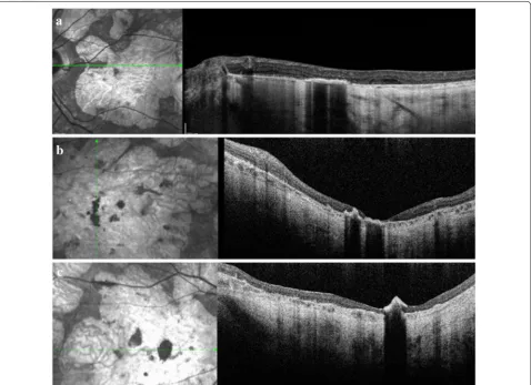
hyperpigmented lesions seen on CFP were not clearly identified on infra-red image on SD-OCT. Hyperpig-mentation appeared as hyperreflective and highly back-scattering lesions. In 31 eyes, hyperpigmentation was observed in the outer retina (comprising the IS/OS and RPE), while in 8 eyes, it was observed in the INL, ONL, ganglion cell layer (GCL) or retinal nerve fiber layer (RFNL) in addition to the IS/OS and RPE. Subretinal hyperpigmentation was observed in 2 eyes. SD-OCT images are shown in Fig. 2.
Progression
Seventeen (56.6%) patients had CFP follow-up images. Eleven patients (64.7%) demonstrated increases (for both distribution and density) of the hyperpigmented lesions, along with progression of the underlying RPE atro-phy, between the baseline visit and the most recent visit (mean follow up of 6.58 years). In 8 eyes, the pigment observed in the outer retina expanded to the inner lay-ers, including the INL, ONL, GCL or RNFL on SD-OCT.
VA remained stable. There was no statistically significant difference between the mean VA at the baseline (0.935 logMar) and the mean VA at the most recent visit (1.29 logMar) (p= 0.21).
Discussion
In this study, it has been demonstrated that patients with STGD with hyperpigmented lesions in the posterior pole had a long disease duration and severe phenotype. This was confirmed by a poor VA, widespread disease on FAF, and photopic and scotopic dysfunction on ERG in those with MH. In addition, over time, hyperpigmented lesions increased in size, spread across the retina, and migrated to different retinal layers, as seen by SD-OCT. Macular hyperpigmentation may be a marker of advanced stage of the disease, when diffuse RPE atrophy leads to pathologic remodeling and clumping of the remaining RPE cells. Similarly, the development of peripheral pigmentation in patients with STGD has been previously associated with a severe phenotype and advanced disease stage. Previous
studies have demonstrated the presence of peripheral pigmented retinal lesions resembling CHRPE lesions in a subset of patients with STGD disease who also had a more severe phenotype [24]. Therefore hyperpigmen-tation, whether in the macula or periphery, may be a marker of end-stage pathologic remodeling of the retina after many years of degeneration.
In patients with STGD, hyperpigmentation has been described following human embryonic stem cell-derived RPE (hESC-RPE) transplantation. In most eyes, hyper-pigmentation was observed as subretinal hyperreflective foci at the border of atrophic areas, consistent with loca-tion of hyperpigmentaloca-tion in the RPE-transplanted cells [25, 26]. In a few eyes that underwent hESC-RPE trans-plantation, a patch of epiretinal pigmentation also devel-oped, which likely resulted from a reflux of the implanted material from the subretinal space or from injection into the pre-retinal space [25, 26]. In retinal dystrophies over-all, RPE proliferation seems to occur in response to loss of surrounding RPE cells, which in turn leads to further retinal cell damage and loss [27, 28]. In our study, hyper-pigmented lesions increased in size, spread across the retina, and migrated to different retinal layers over time. Therefore, it is possible that, similar to the pathogenesis of hyperpigmentation in patients with AMD and other retinal dystrophies, the hyperpigmentation observed in patients with STGD may also occur in response to loss of surrounding RPE cells, which in turn leads to further RPE damage and loss [27, 28], resulting in RPE degeneration, displacement and migration [19–21].
In AMD, macular hyperpigmentation occurs after extensive loss of the RPE and increases the risk of progression to either geographic atrophy or choroi-dal neovascularization [22]. Comparisons have been made between patients with AMD and STGD in part due to the similar pathogenesis that characterizes these degenerative diseases [4–6]. Interestingly, the pres-ence of a single heterozygous mutation in ABCA4 has been associated with an increased risk for AMD [29]. Considering that the use of RPE stem cell therapies are being investigated for both of these diseases, and that pathologic RPE remodeling is an important disease mechanism in both diseases, a robust understanding of RPE remodeling is necessary to anticipate possible outcomes for patients with STGD who undergo a thera-peutic intervention. In STGD, macular hyperpigmen-tation may not be predictive or causative of a severe phenotype, but it may be a finding associated with an advanced stage of disease, after extensive photore-ceptor degeneration and RPE atrophy have occurred. The relationship between RPE atrophy, photorecep-tor degeneration, and RPE remodeling in both of these macular diseases may elucidate more about the etiology
of macular hyperpigmentation and lead to potential treatments that might reverse the pathologic remod-eling seen in these degenerative retinal diseases.
The limitations of this study include its retrospective design and limited number of visual functional tests. The absence of change in VA between baseline and fol-low up visits may have occurred because patients with STGD are known to have oscillation of the preferred retinal locus and have also been shown to reach a rela-tive plateau in visual acuity at 20/200 [30–33]. In addi-tion, in many cases, the areas of hyperpigmentation were outside the fovea, and, therefore, the effect of the lesions on visual function could not be evaluated only by measuring the central VA. The areas of geographic atrophy were also subjectively quantified, and, there-fore, the association between progression of geographic atrophy and development of hyperpigmentary changes cannot be established.
Since cell and gene therapies are being developed for inherited retinal dystrophies, we need to be able to stratify patients by severity and progression of disease in order to optimize the ability to elicit successful responses to therapy. Clinical trials evaluating efficacy of novel therapies for patients with STGD may wish to document the presence of hyperpigmentation at baseline as well as after therapy. Furthermore, studies assessing other functional testing, such as microperimetry and contrast sensitivity, may provide insight into how these hyperpig-mentary changes are associated with other visual func-tion measures.
Authors’ contributions
MFA and AAO have contributed equally to this manuscript (data collection, study design and writing); DS and NK: data collection and review; TJ: study design, review. All authors read and approved the final manuscript.
Author details
1 Department of Ophthalmology and Visual Sciences, W. K. Kellogg Eye Center,
University of Michigan, 1000 Wall Street, Ann Arbor, MI 48150, USA. 2
Depart-ment of Ophthalmology and Otolaryngology, University of Sao Paulo Medical School, Sao Paulo, São Paulo, Brazil.
Competing interests
The authors declare that they have no competing interests.
Availability of data and materials
Additional data may be presented upon request.
Consent for publication
This manuscript has not been published elsewhere and has not been submit-ted simultaneously for publication elsewhere. All authors have approved the manuscript for submission.
Ethics approval and consent to participate
The study was approved by the University of Michigan Institutional Review Board and conducted accordingly to the Declaration of Helsinki.
Funding
Publisher’s Note
Springer Nature remains neutral with regard to jurisdictional claims in pub-lished maps and institutional affiliations.
Received: 23 November 2018 Accepted: 25 March 2019
References
1. Saksens NT, Fleckenstein M, Schmitz-Valckenberg S, Holz FG, den Hol-lander AI, Keunen JE, et al. Macular dystrophies mimicking age-related macular degeneration. Prog Retinal Eye Res. 2014;39:23–57.
2. Allikmets R, Singh N, Sun H, Shroyer NF, Hutchinson A, Chidambaram A, et al. A photoreceptor cell-specific ATP-binding transporter gene (ABCR) is mutated in recessive Stargardt macular dystrophy. Nat Genet. 1997;15(3):236–46.
3. Lim LS, Mitchell P, Seddon JM, Holz FG, Wong TY. Age-related macular degeneration. Lancet (London, England). 2012;379(9827):1728–38. 4. Cideciyan AV, Aleman TS, Swider M, Schwartz SB, Steinberg JD, Brucker
AJ, et al. Mutations in ABCA4 result in accumulation of lipofuscin before slowing of the retinoid cycle: a reappraisal of the human disease sequence. Hum Mol Genet. 2004;13(5):525–34.
5. Molday RS. Insights into the molecular properties of ABCA4 and its role in the visual cycle and Stargardt disease. Prog Mol Biol Transl Sci. 2015;134:415–31.
6. Washington I, Saad L. The rate of vitamin A dimerization in lipofuscino-genesis, fundus autofluorescence, retinal senescence and degeneration. Adv Exp Med Biol. 2016;854:347–53.
7. Paavo M, Lee W, Allikmets R, Tsang S, Sparrow JR. Photoreceptor cells as a source of fundus autofluorescence in recessive Stargardt disease. J Neurosci Res. 2018;97:98–106.
8. Westeneng-van Haaften SC, Boon CJ, Cremers FP, Hoefsloot LH, den Hollander AI, Hoyng CB. Clinical and genetic characteristics of late-onset Stargardt’s disease. Ophthalmology. 2012;119(6):1199–210.
9. Fujinami K, Zernant J, Chana RK, Wright GA, Tsunoda K, Ozawa Y, et al. Clinical and molecular characteristics of childhood-onset Stargardt disease. Ophthalmology. 2015;122(2):326–34.
10. McBain VA, Townend J, Lois N. Progression of retinal pigment epithelial atrophy in Stargardt disease. Am J Ophthalmol. 2012;154(1):146–54. 11. Fujinami K, Lois N, Mukherjee R, McBain VA, Tsunoda K, Tsubota K, et al. A
longitudinal study of Stargardt disease: quantitative assessment of fun-dus autofluorescence, progression, and genotype correlations. Investig Ophthalmol Vis Sci. 2013;54(13):8181–90.
12. Wang JJ, Foran S, Smith W, Mitchell P. Risk of age-related macular degeneration in eyes with macular drusen or hyperpigmentation: the Blue Mountains Eye Study cohort. Arch Ophthalmol (Chicago, Ill: 1960). 2003;121(5):658–63.
13. Abdelfattah NS, Zhang H, Boyer DS, Rosenfeld PJ, Feuer WJ, Gregori G, et al. Drusen volume as a predictor of disease progression in patients with late age-related macular degeneration in the fellow eye. Investig Ophthalmol Vis Sci. 2016;57(4):1839–46.
14. Klein ML, Ferris FL 3rd, Armstrong J, Hwang TS, Chew EY, Bressler SB, et al. Retinal precursors and the development of geographic atrophy in age-related macular degeneration. Ophthalmology. 2008;115(6):1026–31. 15. Pauleikhoff D, Barondes MJ, Minassian D, Chisholm IH, Bird AC. Drusen
as risk factors in age-related macular disease. Am J Ophthalmol. 1990;109(1):38–43.
16. Klein R, Klein BE, Knudtson MD, Meuer SM, Swift M, Gangnon RE. Fifteen-year cumulative incidence of age-related macular degeneration: the Beaver Dam Eye Study. Ophthalmology. 2007;114(2):253–62.
17. Schmitz-Valckenberg S, Sahel JA, Danis R, Fleckenstein M, Jaffe GJ, Wolf S, et al. Natural history of geographic atrophy progression secondary to age-related macular degeneration (Geographic Atrophy Progression Study). Ophthalmology. 2016;123(2):361–8.
18. Ferris FL, Davis MD, Clemons TE, Lee LY, Chew EY, Lindblad AS, et al. A simplified severity scale for age-related macular degeneration: AREDS Report No. 18. Arch Ophthalmol (Chicago, Ill: 1960). 2005;123(11):1570–4. 19. Hageman GS, Mullins RF. Molecular composition of drusen as related to
substructural phenotype. Mol Vis. 1999;5:28.
20. Pieroni CG, Witkin AJ, Ko TH, Fujimoto JG, Chan A, Schuman JS, et al. Ultrahigh resolution optical coherence tomography in non-exudative age related macular degeneration. Br J Ophthalmol. 2006;90(2):191–7. 21. Anderson DH, Mullins RF, Hageman GS, Johnson LV. A role for local
inflammation in the formation of drusen in the aging eye. Am J Ophthal-mol. 2002;134(3):411–31.
22. Folgar FA, Chow JH, Farsiu S, Wong WT, Schuman SG, O’Connell RV, et al. Spatial correlation between hyperpigmentary changes on color fundus photography and hyperreflective foci on SDOCT in intermediate AMD. Investig Ophthalmol Vis Sci. 2012;53(8):4626–33.
23. Ho J, Witkin AJ, Liu J, Chen Y, Fujimoto JG, Schuman JS, et al. Documenta-tion of intraretinal retinal pigment epithelium migraDocumenta-tion via high-speed ultrahigh-resolution optical coherence tomography. Ophthalmology. 2011;118(4):687–93.
24. Zhao PY, Abalem MF, Nadelman D, Qian CX, Branham K, Schlegel D, et al. Peripheral pigmented retinal lesions in Stargardt disease. Am J Ophthal-mol. 2017;188:104–10.
25. Schwartz SD, Tan G, Hosseini H, Nagiel A. Subretinal transplantation of embryonic stem cell-derived retinal pigment epithelium for the treatment of macular degeneration: an assessment at 4 years. Investig Ophthalmol Vis Sci. 2016;57(5):ORSFc1-9.
26. Schwartz SD, Regillo CD, Lam BL, Eliott D, Rosenfeld PJ, Gregori NZ, et al. Human embryonic stem cell-derived retinal pigment epithelium in patients with age-related macular degeneration and Stargardt’s macular dystrophy: follow-up of two open-label phase 1/2 studies. Lancet (Lon-don, England). 2015;385(9967):509–16.
27. Longbottom R, Fruttiger M, Douglas RH, Martinez-Barbera JP, Greenwood J, Moss SE. Genetic ablation of retinal pigment epithelial cells reveals the adaptive response of the epithelium and impact on photoreceptors. Proc Natl Acad Sci USA. 2009;106(44):18728–33.
28. Kampik D, Basche M, Luhmann UFO, Nishiguchi KM, Williams JAE, Green-wood J, et al. In situ regeneration of retinal pigment epithelium by gene transfer of E2F2: a potential strategy for treatment of macular degenera-tions. Gene Ther. 2017;24:810.
29. Allikmets R, Shroyer NF, Singh N, Seddon JM, Lewis RA, Bernstein PS, et al. Mutation of the Stargardt disease gene (ABCR) in age-related macular degeneration. Science. 1997;277(5333):1805–7.
30. Testa F, Melillo P, Di Iorio V, Orrico A, Attanasio M, Rossi S, et al. Macular function and morphologic features in juvenile Stargardt disease: longitu-dinal study. Ophthalmology. 2014;121(12):2399–405.
31. Strauss RW, Ho A, Munoz B, Cideciyan AV, Sahel JA, Sunness JS, et al. The natural history of the progression of atrophy secondary to Stargardt disease (ProgStar) studies: design and baseline characteristics: ProgStar Report No. 1. Ophthalmology. 2016;123(4):817–28.
32. Bernstein A, Sunness JS, Applegate CA, Tegins EO. Mapping the dense scotoma and its enlargement in stargardt disease. Retina (Philadelphia, Pa). 2016;36(9):1741–50.