Open Access
R E S E A R C H A R T I C L E
© 2010 Nakajima et al; licensee BioMed Central Ltd. This is an Open Access article distributed under the terms of the Creative Commons Attribution License (http://creativecommons.org/licenses/by/2.0), which permits unrestricted use, distribution, and reproduction in any medium, provided the original work is properly cited.
Research article
Visual field defects of optic neuritis in
neuromyelitis optica compared with multiple
sclerosis
Hideto Nakajima*
†1,2, Takafumi Hosokawa
†1, Masakazu Sugino
1, Fumiharu Kimura
1, Jun Sugasawa
3,
Toshiaki Hanafusa
1and Toshiyuki Takahashi
4Abstract
Background: Neuromyelitis optica (NMO) is an inflammatory demyelinating disease that predominantly affects the optic nerves and the spinal cord, and is possibly mediated by an immune mechanism distinct from that of multiple sclerosis (MS). Central scotoma is recognized as a characteristic visual field defect pattern of optic neuritis (ON), however, the differing pathogenic mechanisms of NMO and MS may result in different patterns of visual field defects for ON.
Methods: Medical records of 15 patients with NMO and 20 patients with MS having ON were retrospectively analyzed. A thorough systemic and neurological examination was performed for evaluating ON. The total number of relapses of ON and visual fields was investigated. Visual fields were obtained by Goldmann perimeter with each ON relapse.
Results: All MS patients experienced central scotoma, with 90% of them showing central scotoma with every ON relapse. However, 53% of NMO patients showed central scotoma with every ON relapse (p = 0.022), and the remaining 47% of patients experienced non-central scotoma (altitudinal, quadrant, three quadrant, hemianopia, and bitemporal hemianopia). Thirteen percent of NMO patients did not experience central scotoma during their disease course. Altitudinal hemianopia was the most frequent non-central scotoma pattern in NMO.
Conclusions: NMO patients showed higher incidence of non-central scotoma than MS, and altitudinal hemianopia may be characteristic of ON occurring in NMO. As altitudinal hemianopia is highly characteristic of ischemic optic neuropathy, we suggest that an ischemic mechanism mediated by anti-aquaporin-4 antibody may play a role in ON in NMO patients.
Background
Neuromyelitis optica (NMO; Devic's disease) is an idio-pathic inflammatory disease of the central nervous sys-tem (CNS) that mainly affects the optic nerve and spinal cord. Traditionally, NMO is believed to differ from multi-ple sclerosis (MS) by causing very severe, often bilateral, optic neuritis (ON) and longitudinally extensive MRI spi-nal cord lesions but no MRI brain lesions or aggressive progression to disability and death [1]. Recent studies have reported a high frequency of brain MRI abnormali-ties in NMO patients. However, most were nonspecific
and were not considered typical of MS, and hypothalamic involvement has been emphasized [2]. NMO has a more negative outcome than MS, with frequent and early relapses. Within 5 years of onset, 50% of patients have become blind in both eyes and cannot walk unassisted, and 20% die of respiratory failure due to cervical myelitis [3]. Although no controlled therapeutic trials have been specifically performed in NMO, case series and observa-tional studies suggest that azathioprine in combination with oral steroid reduces the frequency of attacks [4,5], and rituximab and plasmapheresis can induce clinical remission of NMO [6-8]. Immuno-suppression rather than interferon β is the preferred treatment. Thus, distin-guishing NMO from MS is very important for the thera-peutic strategy of these disorders. Recently, clinical,
* Correspondence: [email protected]
1 Department of Internal Medicine I, Osaka Medical College, Takatsuki, Osaka, Japan
† Contributed equally
Nakajima et al.BMC Neurology 2010, 10:45 http://www.biomedcentral.com/1471-2377/10/45
Page 2 of 6
neuroimaging, laboratory, and pathological features have been established showing that NMO is distinct from MS. Histopathological and serological findings strongly sug-gest the involvement of the humoral immune system [9]. In particular, detection of serum anti-aquaporin-4 (AQP4) antibody can be used to distinguish NMO from MS [10,11]
ON is the most common and often initial symptom in both NMO and MS. In acute ON, the cardinal field defect is a widespread depression of sensitivity, and visual field testing typically reveals a central scotoma, although other visual field changes such as color blindness, bitemporal hemianopia, paracentral scotoma, and altitudinal deficits have also been reported. ON in NMO tends to be more severe and recovery is less complete compared with attacks of ON in the context of MS [3]. Clinical features such as ocular pain, visual field deficits, and positive phe-nomena, i.e. movement-induced phosphenes, have been thought not to differ from those found in MS-associated attacks [3]. Unlike patients with MS, those with NMO experience more severe disease symptoms due to myelitis characterized by centrally located spinal cord lesions that are longer than three vertebral segments and frequent early attacks. In NMO, the pathophysiology of spinal cord lesions and relation with seropositivity for anti-AQP4 antibody are well investigated [12,13]. However, the dif-ference of clinical symptoms or pathophysiologic findings for ON between NMO and MS have rarely been evalu-ated. We hypothesized that the differing pathogenic mechanisms of NMO and MS may result in different pat-terns of visual field defects as findings of ON. In this study, we evaluated the features of visual field defects in patients with NMO.
Method
We retrospectively analyzed the medical records of 15 patients with NMO (all women, mean age of onset: 36 ± 11, mean ± SD) and 20 patients with MS (5 men and 15 women, 29 ± 9), all of whom had ON. NMO patients ful-filled Wingerchuk's revised diagnostic criteria [14], except for NMO-IgG seropositive status. MS patients included in this study had definitive MS according to McDonald's criteria [15]. A thorough systemic and neu-rological examination was performed to evaluate ON. Visual field tests were performed on the Goldmann perimeter whenever visual acuity permitted. MRI was performed where deemed necessary and for those who could afford the investigation. This study received institu-tional review board approval and informed consent was obtained according to the Declaration of Helsinki.
Serum samples were stored at - 80°C until testing for anti-AQP4 antibody. Anti-AQP4 antibody was assessed as described previously [12,16]. Briefly, human
embry-onic kidney cells (HEK-293) were stably transfected with either a vector containing AQP4-cDNA or empty vector, and specimens were tested by indirect immunofluores-cence using these two cell lines (with or without AQP4). Specimens were incubated with the cells for 1 h, washed in phosphate-buffered saline (PBS), incubated with Alexa Fluor 488 goat anti-human IgG (Invitrogen, Eugene, Ore-gon, USA) for 30 min, and washed in PBS. The cells were then fixed in 4% paraformaldehyde and mounted in Per-mafluor aqueous mounting media (Beckman Coulter, Marseille, France).
Statistics
All data in this study are presented as mean ± SD. Cate-gorical variables were compared using the Mann-Whit-ney's U test and the Fisher's exact probability test. Significance levels were set at P < 0.05.
Results
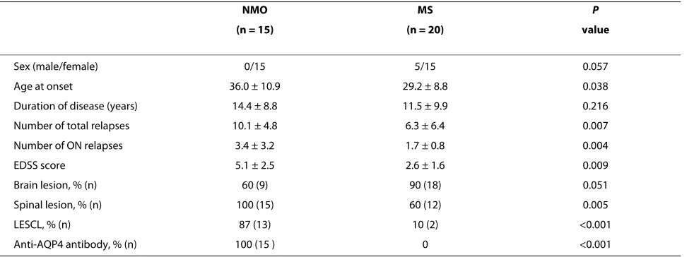
A total of 35 patients were included in this study: 15 NMO and 20 MS (Table 1). There were no differences between the 2 groups in gender and disease duration. Patients with NMO were older at disease onset, exhibited an increased number of total and ON relapses, and had a higher expanded disability status scale (EDSS) score.
When comparing visual field defect patterns of ON between the 2 groups, central scotoma was present in 31 out of 33 ON episodes in MS (94%) and 39 out of 51 epi-sodes in NMO (76%) (p = 0.041, Table 2). In 51 epiepi-sodes of ON, NMO patients exhibited 12 episodes of non-cen-tral scotoma (24%). Of the visual field defect patterns other than central scotoma, NMO patients showed 5 for altitudinal, 3 for quadrant, 2 for three quadrant, 1 for hemianopia, and 1 for bitemporal hemianopia. MS patients showed 1 each for three quadrant and hemiano-pia (Table 2).
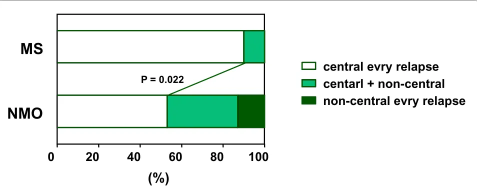
During the course of the disease, 90% of MS patients (18/20) showed central scotoma with every episode; how-ever, central scotoma with every episode was present in 54% of NMO patients (8/15) (p = 0.022, Figure 1). In the remaining 7 NMO patients, 5 showed both central and non-central scotoma, and 2 patients showed non-central scotoma with every episode. In 7 NMO patients showing non-central scotoma, altitudinal hemianopia was most frequent (5/7), and the location of the altitudinal hemi-anopia was inferior in 3 of 5 relapses and superior in 2 relapses. Notably, all altitudinal hemianopia occurred at the initial attack of each eye (Table 3).
Discussion
recognized as a typical visual field defect pattern of ON in MS [17]. In this study, all MS patients experienced central scotoma, with 90% showing central scotoma with every ON attack. On the contrary, 53% of NMO patients showed central scotoma with every ON attack, and the remaining 47% of patients experienced non-central sco-toma. Moreover, 13% of NMO patients did not experi-ence central scotoma during the course of their disease. Of the non-central scotoma patterns, altitudinal hemi-anopia was most frequent. Since altitudinal hemihemi-anopia was not recognized in MS patients, this visual field defect may be characteristic of ON for patients with NMO.
ON is the initial manifestation of NMO in 77% of patients. In 30% of NMO patients, the initial attack of ON led to blindness, with only 43% of patients completely recovering from the first attack. Compared with MS patients, NMO patients had a significantly higher rate of bilateral ON (70% versus 19%) [18]. Although the optic
nerve is mainly affected in both NMO and MS, the patho-genesis of ON in NMO might differ from that of MS. Compared to MS, the study using optical coherence tomography indicated a thinner overall average retinal nerve fiber layer, suggesting widespread axonal injury in the affected optic nerves in NMO [19].
Pathologically, NMO shares with MS a pattern of focal demyelination, inflammation, scar formation, and axonal destruction, but NMO also has an intense perivascular response, prominent necrosis, and cavitation, which are not seen in MS [9]. IgG, IgM, and products of comple-ment activation are deposited in a perivascular pattern in NMO, suggesting a pathogenic role involving autoanti-bodies [9]. Blood vessels within demyelination spinal lesions of NMO are thickened and hyalinized [20]. Active lesions exhibit tissue swelling, infiltrating polymorpho-nuclear macrophages, active microglia, demyelination, axonal loss, prominent necrosis, and variable degrees of Table 1: Demography and ocular findings of NMO and MS patients
NMO MS P
(n = 15) (n = 20) value
Sex (male/female) 0/15 5/15 0.057
Age at onset 36.0 ± 10.9 29.2 ± 8.8 0.038
Duration of disease (years) 14.4 ± 8.8 11.5 ± 9.9 0.216
Number of total relapses 10.1 ± 4.8 6.3 ± 6.4 0.007
Number of ON relapses 3.4 ± 3.2 1.7 ± 0.8 0.004
EDSS score 5.1 ± 2.5 2.6 ± 1.6 0.009
Brain lesion, % (n) 60 (9) 90 (18) 0.051
Spinal lesion, % (n) 100 (15) 60 (12) 0.005
LESCL, % (n) 87 (13) 10 (2) <0.001
Anti-AQP4 antibody, % (n) 100 (15 ) 0 <0.001
NMO = neuromyelitis optica; MS = multiple sclerosis; ON = optic neuritis; EDSS = expanded disability status scale; LESCL = longitudinally extensive spinal cord lesions.
Table 2: Visual field defect patterns of NMO and MS patients
NMO MS P value
Total number of ON relapses 51 33
Visual field defects
Central scotoma (%) 39 (76) 31 (94) 0.041
Non-central scotoma
Altitudinal (%) 5 (10) 0 (0) 0.151
Quadrant (%) 3 (6) 0 (0) 0.276
Three quadrant (%) 2 (4) 1 (3) 1
Hemianopia (%) 1 (2) 1 (3) 1
Bitemporal hemianopia (%) 1 (2) 0 (0) 1
Nakajima et al.BMC Neurology 2010, 10:45 http://www.biomedcentral.com/1471-2377/10/45
Page 4 of 6
perivascular inflammation with prominent eosinophils and products of their exocytosis. Chronic lesions show gliosis, cystic degeneration, cavitation, and atrophy [21]. These findings suggest that a humoral effector mecha-nism is initiated by binding of the NMO antibody at the blood-brain barrier (BBB).
Several studies have reported that areas of CNS inflam-mation correlate with expression pattern of AQP4 in NMO [22,23]. Expression of AQP4 in the brain and spinal cord is associated with astrocyte membranes that appose endothelial cell basal membranes. Astrocytes interact extensively with endothelial cells to maintain the CNS BBB, which normally limits the access of immune system effectors unless localized or distant events disrupt the BBB, thus allowing access of cellular or soluble immune effectors. AQP4 is also expressed by astrocytes that sur-round the optic nerve [24]. Since the optic nerve head is an area of the CNS where the BBB is more permissive, as evidenced by immunostaining for markers of intact BBB [25,26], tissues of the optic nerve might be more sensitive to AQP4 dysfunction mediated by anti-AQP4 antibodies [27]. Thus, in NMO, optic nerve lesions would have demyelination, axonal loss, and perivascular response, as seen in spinal cord lesions.
Central scotoma is recognized to be a typical visual field defect pattern of ON in MS. In this study, NMO patients showed higher incidence of non-central scotoma than MS patients (p = 0.022, Figure 1); altitudinal hemi-anopia was more common in non-central scotoma. An altitudinal visual field defect is suggestive of ischemic optic neuropathy, which occasionally is the result of pos-terior ciliary artery occlusion [28,29]. We suggest that
ischemic mechanism mediated by anti-AQP4 antibody may play a role in ON for NMO patients. Pathological study demonstrated that vascular degeneration, such as thickened or hyalinized vessels, existed in the spinal cord lesions [21]. Recent study indicated that NMO patients showed more vascular changes, including attenuation of the peripapillary vascular tree and focal arteriolar nar-rowing as the retinal features of ON than MS patients [30]. These vascular changes may results from direct vas-cular inflammation mediated by anti-AQP4 antibody [30,31]. Therefore, the tissue organization of optic nerve cells, such as the vascular structures associated with the optic nerves, is thought to express AQP4, resulting in non-central scotoma, especially altitudinal hemianopia.
Although NMO is often fulminant and has a more neg-ative outcome than MS [32], NMO responds to glucocor-ticoids, immunosuppressive agents, or plasmapheresis. Since monosymptomatic ON is often seen as being the first indication of an attack of NMO and MS, ophthalmo-scopic examination, especially the visual field test, is helpful for diagnosis of NMO, and anti-AQP4 antibody should be checked to decide the most effective treatment [33].
Conclusion
NMO patients showed higher incidence of non-central scotoma than MS, and altitudinal hemianopia may be characteristic of ON occurring in NMO. As altitudinal hemianopia is highly characteristic of ischemic optic neu-ropathy, we suggest that an ischemic mechanism medi-ated by anti-aquaporin-4 antibody may play a role in ON in NMO patients.
Figure 1 Comparison of visual field defects during the disorders. Ninety percent of MS patients showed central scotoma every time, but 54% in NMO (p = 0.022). In NMO patients, 33% of patients showed both central scotoma and non-central scotoma, and 13% of patients showed non-central scotoma every time. MS = multiple sclerosis; NMO = neuromyelitis optica.
central evry relapse
centarl + non-central
non-central evry relapse
0
20
40
60
80
100
NMO
MS
P = 0.022
Nakaj
ima
et
a
l.
BMC N
eurology
20
10,
10
:4
5
htt
p
:/
/ww
w
.biom
e
dcentr
al.
com/147
1-23
77/1
0
/45
Sex/Age at onset F/48 F/21 F/47 F/34 F/32 F/32 F/54
Duration of disease (years) 9 11 24 4 15 18 12
EDSS score 2.5 1 7 3.5 7 7.5 4
Number of total relapses 5 5 12 6 11 18 23
Number of ON relapses 3 2 2 2 3 4 12
Ocular pain moderate none none mild mild none moderate
Optic disk in acute phase normal NE NE normal normal normal normal
Course of ON lt-altitudinal lt-three quadrant rt-altitudinal lt-altitudinal rt-altitudinal bil-central rt-central
(inferior) rt-altitudinal (superior) (inferior) (superior) lt-guadrant lt-central: 2nd-3rd
rt-central (inferior) rt-central rt-quadrant lt-central rt-hemianopia rt-quadrant
lt-central lt-central lt-three quadrant
rt-central
lt-central
rt-central: 8th-11th
bitemporal
Outcome of ON rt-recover rt-recover rt-recover rt-recover rt-light perception rt-recover rt-light perception
lt-recover lt-recover lt-recover lt-light perception lt-recover lt-light perception
Nakajima et al.BMC Neurology 2010, 10:45 http://www.biomedcentral.com/1471-2377/10/45
Page 6 of 6
Competing interests
The authors declare that they have no competing interests.
Authors' contributions
HN performed analyses, collected data and wrote the manuscript. TH helped to draft the manuscript and collected data. MS, FK, JS, and TH helped to draft the manuscript. TT performed anti-AQP4 antibody assay. All authors read and approved the final manuscript.
Acknowledgements
This study was supported by the Osaka Medical Research Foundation, Osaka, Japan.
Author Details
1Department of Internal Medicine I, Osaka Medical College, Takatsuki, Osaka, Japan, 2Department of Internal Medicine, Seikeikai Hospital, Sakai, Osaka, Japan, 3Department of Ophthalmology, Osaka Medical College, Takatsuki, Osaka, Japan and 4Department of Neurology, Tohoku University Graduate School of Medicine, Sendai, Miyagi, Japan
References
1. Wingerchuk DM, Lennon VA, Lucchinetti CF, Pittock SJ, Weinshenker BG:
The spectrum of neuromyelitis optica. Lancet Neurol 2007, 6:805-815. 2. Pittock SJ: Neuromyelitis optica: A new perspective. Seminars in
neurology 2008, 1:95-104.
3. Wingerchuk DM, Hogancamp WF, O'Brien PC, Weinshenker BG: The clinical course of neuromyelitis optica (Devic's syndrome). Neurology
1999, 53:1107-1114.
4. Papeix C, Vidal JS, de Seze J, Pierrot-Deseilligny C, Tourbah A, Stankoff B, Lebrun C, Moreau T, Vermersch P, Fontaine B, Lyon-Caen O, Gout O:
Immunosuppressive therapy is more effective than interferon in neuromyelitis optica. Mult Scler 2007, 13:256-259.
5. Watanabe S, Misu T, Miyazawa I, Nakashima I, Shiga Y, Fujihara K, Itoyama Y: Low-dose corticosteroids reduce relapses in neuromyelitis optica: a retrospective analysis. Mult Scler 2007, 13:968-974.
6. Watanabe S, Nakashima I, Misu T, Miyazawa I, Shiga Y, Fujihara K, Itoyama Y: Therapeutic efficacy of plasma exchange in NMO-IgG-positive patients with neuromyelitis optica. Mult Scler 2007, 13:128-132. 7. Cree BA, Lamb S, Morgan K, Chen A, Waubant E, Genain C: An open label
study of the effects of rituximab in neuromyelitis optica. Neurology
2005, 64:1270-1272.
8. Jacob A, Weinshenker BG, Violich I, McLinskey N, Krupp L, Fox RJ, Wingerchuk DM, Boggild M, Constantinescu CS, Miller A, De Angelis T, Matiello M, Cree BA: Treatment of neuromyelitis optica with rituximab: retrospective analysis of 25 patients. Arch Neurol 2008, 65:1443-1448. 9. Lucchinetti CF, Mandler RN, McGavern D, Bruck W, Gleich G, Ransohoff RM, Trebst C, Weinshenker B, Wingerchuk D, Parisi JE, Lassmann H: A role for humoral mechanisms in the pathogenesis of Devic's neuromyelitis optica. Brain 2002, 125:1450-1461.
10. Lennon VA, Wingerchuk DM, Kryzer TJ, Pittock SJ, Lucchinetti CF, Fujihara K, Nakashima I, Weinshenker BG: A serum autoantibody marker of neuromyelitis optica: distinction from multiple sclerosis. Lancet 2004,
364:2106-2112.
11. Jarius S, Aboul-Enein F, Waters P, Kuenz B, Hauser A, Berger T, Lang W, Reindl M, Vincent A, Kristoferitsch W: Antibody to aquaporin-4 in the long-term course of neuromyelitis optica. Brain 2008, 131:3072-3080. 12. Takahashi T, Fujihara K, Nakashima I, Misu T, Miyazawa I, Nakamura M,
Watanabe S, Shiga Y, Kanaoka C, Fujimori J, Sato S, Itoyama Y: Anti-aquaporin-4 antibody is involved in the pathogenesis of NMO: a study on antibody titre. Brain 2007, 130:1235-1243.
13. Matsuoka T, Matsushita T, Kawano Y, Osoegawa M, Ochi H, Ishizu T, Minohara M, Kikuchi H, Mihara F, Ohyagi Y, Kira J: Heterogeneity of aquaporin-4 autoimmunity and spinal cord lesions in multiple sclerosis in Japanese. Brain 2007, 130:1206-1223.
14. Wingerchuk DM, Lennon VA, Pittock SJ, Lucchinetti CF, Weinshenker BG:
Revised diagnostic criteria for neuromyelitis optica. Neurology 2006,
66:1485-1489.
15. McDonald WI, Compston A, Edan G, Goodkin D, Hartung HP, Lublin FD, McFarland HF, Paty DW, Polman CH, Reingold SC, Sandberg-Wollheim M,
Sibley W, Thompson A, van den Noort S, Weinshenker BY, Wolinsky JS:
Recommended diagnostic criteria for multiple sclerosis: guidelines from the International Panel on the diagnosis of multiple sclerosis.
Ann Neurol 2001, 50:121-127.
16. Takahashi T, Fujihara K, Nakashima I, Misu T, Miyazawa I, Nakamura M, Watanabe S, Ishii N, Itoyama Y: Establishment of a new sensitive assay for anti-human aquaporin-4 antibody in neuromyelitis optica. Tohoku J Exp Med 2006, 210:307-313.
17. Warner J, Lessell S: Neuro-ophthalmology of multiple sclerosis. Clin Neurosci 1994, 2:180-188.
18. Merle H, Olindo S, Bonnan M, Donnio A, Richer R, Smadja D, Cabre P:
Natural history of the visual impairment of relapsing neuromyelitis optica. Ophthalmology 2007, 114:810-815.
19. Naismith RT, Tutlam NT, Xu J, Klawiter EC, Shepherd J, Trinkaus K, Song SK, Cross AH: Optical coherence tomography differs in neuromyelitis optica compared with multiple sclerosis. Neurology 2009, 72:1077-1082. 20. Mandler RN, Davis LE, Jeffery DR, Kornfeld M: Devic's neuromyelitis
optica: a clinicopathological study of 8 patients. Ann Neurol 1993,
34:162-168.
21. Lucchinetti CF, Parisi J, Bruck W: The pathology of multiple sclerosis.
Neurol Clin 2005, 23:77-105.
22. Misu T, Fujihara K, Kakita A, Konno H, Nakamura M, Watanabe S, Takahashi T, Nakashima I, Takahashi H, Itoyama Y: Loss of aquaporin 4 in lesions of neuromyelitis optica: distinction from multiple sclerosis. Brain 2007,
130:1224-1234.
23. Roemer SF, Parisi JE, Lennon VA, Benarroch EE, Lassmann H, Bruck W, Mandler RN, Weinshenker BG, Pittock SJ, Wingerchuk DM, Lucchinetti CF:
Pattern-specific loss of aquaporin-4 immunoreactivity distinguishes neuromyelitis optica from multiple sclerosis. Brain 2007,
130:1194-1205.
24. Nagelhus EA, Veruki ML, Torp R, Haug FM, Laake JH, Nielsen S, Agre P, Ottersen OP: Aquaporin-4 water channel protein in the rat retina and optic nerve: polarized expression in Müller cells and fibrous astrocytes.
J Neurosci 1998, 18:2506-2519.
25. Guy J, Rao NA: Acute and chronic experimental optic neuritis. Alteration in the blood-optic nerve barrier. Arch Ophthalmol 1984, 102:450-454. 26. Hofman P, Hoyng P, vanderWerf F, Vrensen GF, Schlingemann RO: Lack of
blood-brain barrier properties in microvessels of the prelaminar optic nerve head. Invest Ophthalmol Vis Sci 2001, 42:895-901.
27. Graber DJ, Levy M, Kerr D, Wade WF: Neuromyelitis optica pathogenesis and aquaporin 4. J Neuroinflammation 2008, 5:22.
28. Gerling J, Meyer JH, Kommerell G: Visual field defects in optic neuritis and anterior ischemic optic neuropathy: distinctive features. Graefes Arch Clin Exp Ophthalmol 1998, 236:188-192.
29. Levin LA, Rizzo JF, Lessell S: Neural network differentiation of optic neuritis and anterior ischaemic optic neuropathy. Br J Ophthalmol
1996, 80:835-839.
30. Green AJ, Cree BAC: Distinctive retinal nerve fibre layer and vascular changes in neuromyelitis optica following optic neuritis. J Neurol Neurosurg Psychiatry 2009, 80:1002-1005.
31. Roemer SF, Parisi JE, Lennon VA, Benarroch EE, Lassmann H, Bruck W, Mandler RN, Weinshenker BG, Pittock SJ, Wingerchuk DM, Lucchinetti CF:
Pattern-specific loss of aquaporin-4 immunoreactivity distinguishes neuromyelitis optica from multiple sclerosis. Brain 2007,
130:1194-1205.
32. Pirko I, Blauwet LK, Lesnick TG, Weinshenker BG: The natural history of recurrent optic neuritis. Arch Neurol 2004, 61:1401-1405.
33. Matiello M, Lennon VA, Jacob A, Pittock SJ, Lucchinetti CF, Wingerchuk DM, Weinshenker BG: NMO-IgG predicts the outcome of recurrent optic neuritis. Neurology 2008, 70:2197-2200.
Pre-publication history
The pre-publication history for this paper can be accessed here: http://www.biomedcentral.com/1471-2377/10/45/prepub
doi: 10.1186/1471-2377-10-45
Cite this article as: Nakajima et al., Visual field defects of optic neuritis in neuromyelitis optica compared with multiple sclerosis BMC Neurology 2010,
10:45
Received: 25 September 2009 Accepted: 18 June 2010 Published: 18 June 2010
This article is available from: http://www.biomedcentral.com/1471-2377/10/45 © 2010 Nakajima et al; licensee BioMed Central Ltd.
This is an Open Access article distributed under the terms of the Creative Commons Attribution License (http://creativecommons.org/licenses/by/2.0), which permits unrestricted use, distribution, and reproduction in any medium, provided the original work is properly cited.