Characteristics, Management and Survival Rate of
Ovarian Germ Cell Tumor
Karakteristik, Manajmen, dan Tingkat Kesintasan
Ovarian Germ Cell Tumor
Laila Nuranna1 , Zakiah Tourik2
1Division of Oncology Department of Obstetrics and Gynecology 2Department of Obstetrics and Gynecology
Faculty of Medicine Universitas Indonesia, Dr. Cipto Mangunkusumo National General Hospital
Jakarta
Correspondence author: Laila Nuranna. [email protected]
Research Article
Abstract
Objective : To determine the prevalence of malignant ovarian germ cell tumour in term of characteristics, management, and 3-year survival rate in Dr. Cipto Mangunkusumo Hospital Jakarta from 2011 to 2013.
Methods : This is a cross-sectional study. Secondary data were collected from medical record as well as interviewing patients through phone call or home visit.
Results : We collected data from 24 subjects. As many as 54.2% of subjects were between 20 to 40 year old, and 58.3% was single. Around 83.3% of the subjects came with chief complaint of abdominal enlargement. Histopathology fi nding confi rmed dysgerminoma in 50% subjects, mixed ovarian germ cell tumour in 25%, endodermal sinus tumour or yolk sac tumour in 16.7%, and immature teratoma in 8.3%. Half of the cases were found in stage I. The primary therapy was conservative surgical staging and adjuvant chemotherapy.In 2 subjects with dysgerminoma, neoadjuvant chemotherapy (bleomycin, etoposide, cisplatin, and cyclophosphamide-cisplatin regimen) resulted in a good response. The 3-year survival rate was 83.3% in dysgerminoma, 100% in mixed ovarian germ cell tumour, and 50% in immature teratoma.
Conclusions : In malignant ovarian germ cell tumour, conservative surgical staging followed by a complete course of chemotherapy is the treatment of choice with 3-year survival rate exceeding 70%.
Keywords : dysgerminoma, non-epithelial ovarian tumour,ovarian germ cell tumour, survival.
Abstrak
Tujuan : Mengetahui sebaran meliputi karateristik, penatalaksanaan dan kesintasan 3 tahun pasien tumor ganas sel germinal ovarium di RSCM tahun 2011 – 2013.
Metode : Penelitian ini menggunakan studi potong lintang dengan mengambil data sekunder dari rekam medis dan mewawancarai pasien atau keluarga pasien via telepon atau kunjugan rumah.
Hasil : Pada penelitian ini, dari 24 subjek penelitian, 54,2% ditemukan pada usia 20-40 tahun dan 58,3% subjek belum menikah. Sebanyak 83,3% datang dengan keluhan perut membesar. Secara histopatologi didapatkan jenis disgeminoma, tumor sel germinal campuran, sinus endodermal (yolk sac) dan teratoma imatur dengan proporsi masing-masing 50%, 25%, 16,7% dan 8,3%, sebagian besar kasus (50%) ditemukan pada stadium I. Conservative surgical staging dan kemoterapi adjuvan tatalaksana pilihan. Terdapat 2 subjek jenis disgerminoma yang diberikan dengan kemoterapi neoadjuvan (regimenbleomycin, etoposide, cisplatin dan cyclophosmide-cisplatin) memberikan respon yang baik. Kesintasan ≥ 3 tahun pada jenis disgerminoma mencapai 83,3%, pada tumor sel germinal campuran 100% dan pada teratoma imatur mencapai 50%.
Kesimpulan : Pada tumor ganas sel germinal ovarium conservative surgical staging diikuti kemoterapi lengkap merupakan pilihan terapi dengan kesintasan ≥ 3 tahun mencapai > 70%.
INTRODUCTION
Around 70% of ovarian tumour that affects teenager and young adult is ovarian germ cell tumour and one-third of the total cases is malignant.1Malignant ovarian germ cell tumour
is a rare occurrence, accounts for 5% of total ovarian malignancy.2Unlike malignant neoplasm
from epithelial ovary cells, this type of tumour affects women in reproductive age, including teenager and young adult.
According to Surveillance, Epidemiology and Results (SEER) in 1973 to 2003, identifi cation in 1,262 patients with malignant ovarian germ cell tumour showed dysgerminoma in 32.4% cases, immature teratoma with malignant degeneration in 35.6% cases, and mixed germ cell tumour in 28.7% cases.3In Asian and African population, the
most common type of malignant ovarian germ cell tumour is dysgerminoma.4
Management in malignant ovarian germ cell tumour has improved dramatically, giving rise to improvement in survival rate. Survival rate reaches 100% in dysgerminoma type and 85% in non-dysgerminoma type.5 Treatment in ovarian
germ cell tumour used to result in impaired fertility.6In the 1980s, reserving fertility could only
be performed in stage 1 and 2 malignant ovarian germ cell tumour with normal contralateral ovary and uterus. Starting in 1989, fertility function could be well maintained in all stage of the tumour, even in patients with involvement of both ovaries.7
Compared to data on testicle germ cell tumour, data in Indonesia regarding ovarian germ cell tumour is scarce. Therefore, we collected and analyzed the data to determine the prevalence and management of malignant ovarian germ cell in Indonesia, especially in Jakarta.
METHODS
This is a cross-sectional study. Secondary data were collected from the medical record and from interviewing patients. The interview was performed via phone call or home visit to determine the 3-year survival rate. We recruited
all patients with malignant ovarian germ cell tumour who received a complete course of therapy in Dr. Cipto Mangunkusumo Hospital Jakarta from 2011 to 2013.
We calculated the sample size based on prior data on disease prevalence in Asia. The result was 49 subjects. Subjects were recruited with total sampling from cancer registration in the oncology division of Obstetrics and Gynecology Department. Collected data were analyzed with the Statistical Program for Social Sciences (SPSS) 20. Ethical clearance was obtained from the Health Research Ethics Committee, Faculty of Medicine, Universitas Indonesia, in August 2017.
RESULTS
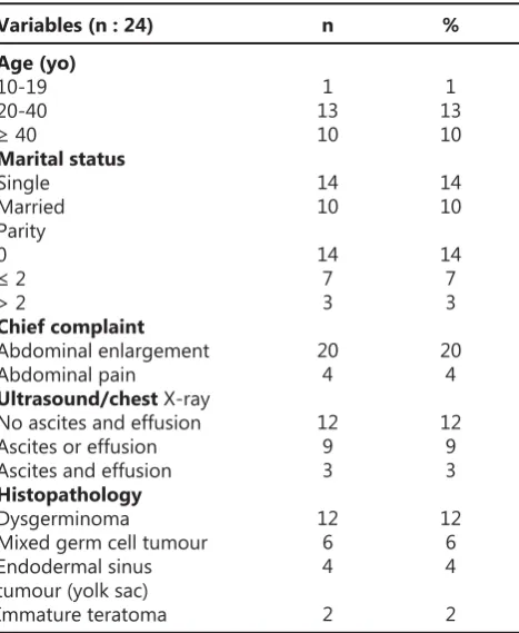
There were 57 patients with malignant ovarian germ cell tumour in Cipto Mangunkusumo Hospital Jakarta from 2011 to 2013. However, only 24 subjects fulfi lled the inclusion criteria of having a complete medical record. Baseline characteristics of the subjects are presented in table 1.
Table 1.Subjects Baseline Characteristics and Histo-pathology Finding
Variables (n : 24) Age (yo)
10-19 20-40
≥ 40
Marital status
Single Married Parity 0
≤ 2 > 2
Chief complaint
Abdominal enlargement Abdominal pain
Ultrasound/chest X-ray No ascites and effusion Ascites or effusion Ascites and effusion
Histopathology
Dysgerminoma
Mixed germ cell tumour Endodermal sinus tumour (yolk sac) Immature teratoma
1 13 10
14 10
14 7 3
20 4
12 9 3
12 6 4
2
n %
1 13 10
14 10
14 7 3
20 4
12 9 3
12 6 4
Table 2. Tumor Marker Findings
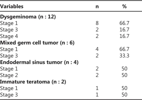
Table 3. Clinical Staging Grouped by Histopathology Type.
Variables
Variables
Dysgerminoma (n : 12)
Elevated LDH Elevated Ca125 Elevated bHCG Elevated LDH, Ca125 Elevated LDH, AFP, bHCG
Mixed germ cell tumor
Elevated LDH Elevated Ca125
Endodermal sinus tumor
Elevated AFP Elevated AFP, Ca125 Elevated AFP, LDH, Ca125
Immature teratoma (n : 2)
Elevated LDH
Dysgeminoma (n : 12)
Stage 1 Stage 3 Stage 4
Mixed germ cell tumor (n : 6)
Stage 1 Stage 3
Endodermal sinus tumor (n : 4)
Stage 1 Stage 2
Immature teratoma (n : 2)
Stage 1 Stage 3
6 2 1 2 1
3 3
2 1 1
2
8 2 2
4 2
2 2
1 1
50 16.7
8.3 16.7
8.3
50 50
50 25 25
100
66.7 16.7 16.7
66.7 33.3
50 50
50 50
n
n
%
%
We also collected data on tumour marker examination. Most of the subjects with dysgerminoma had raised the level of lactate dehydrogenase (LDH). We found two (16.7%) subjects with cancer antigen 125 (CA125) elevation, which is uncommon as Ca125 elevation is usually associated with the ovarian epithelial tumour. In three (25%) subjects, elevation in LDH was accompanied with other tumour marker elevation such as alpha-fetoprotein (AFP), human Chorionic Gonadotrophin (bHCG) and CA125. Another signifi cant elevation of a tumour marker is presented in Table 2.
Furthermore, we noted clinical staging from each subject grouped by type of histopathology
fi ndings, as shown in Table 3.
In term of case management, unilateral salphingo-oophorectomy (USO) was performed in half of the total subjects with or without omentectomy, appendectomy, and lymphadenectomy.
Options for chemotherapy were grouped based on histopathology type of the tumour. Half of the subjects with dysgerminoma underwent complete series of cisplatin, vinblastine, and bleomycin (PVB); bleomycin, etoposide, and cisplatin (BEP); and cyclophosphamide-cisplatin (CP) with the proportion of 8.3%, 25%, and 16.7% consecutively. The rest of the subjects did not undergo chemotherapy.
In term of 3-year survival rate, most of the patients (70.8%) were alive, and 83.3% were asymptomatic.
We analyzed survival functions with Kaplan Meier curve and found that all patients with stage 3 and 4 tumour despite their histopathology type had a 3-year survival rate of 100% with treatment. Similarly, the 3-year survival rate for patients with neoadjuvant chemotherapy is 100%. We also found that 3-year survival rate in dysgerminoma with complete treatment is higher than endodermal sinus tumour, immature teratoma, and mixed germ cell tumour.
DISCUSSION
The highest prevalence of malignant ovarian germ cell tumour was found in age 20 to 40 year old, followed by those older than 40, and younger than 20. This fi nding agrees with prior epidemiological data in which the highest incidence of malignant ovarian germ cell tumour occurs in young females aged 15 to 30-year-old.8However, one study
stated that the highest incidence is within 15 to 19 year old.3 Up until 20 years old, almost 70% of ovarian tumour originates from germ cells, and one-third of this number proves to be malignant.6 The tendency of malignancy in younger age may be caused by a hormonal factor as in the high level of exogenous and endogenous estrogen that disturbs the development of germ cell from primitive to mature cells. These abnormal germ cells are retained until puberty in which gonadotropin further initiates proliferation and results in tumour with various histopathology.9
We found that most of our subjects had dysgerminoma. This is consistent with epidemiology data from that stated dysgerminoma as the most frequent type of malignant ovarian germ cell tumour.10Another
important fi nding we need to highlight is tumour marker test. Carcinoembryonic antigen (CEA) is associated with gastrointestinal tract tumour. However, 4 of our subjects underwent this test that we think is unnecessary. Elevation in CA125 may be caused by various factors since this tumour marker is usually associated with ovary epithelial cell tumour. Each of our subjects underwent a test for multiple types of tumour markers, and therefore, we found overlapping results. However, we only presented results with signifi cant elevation.
Study on tumour marker found that there were 7 signifi cant tumour markers associated with various type of germ cell tumour in ovary. CA125 was found in more than 50% of the subjects. AFP was found positive mostly in endodermal sinus tumour (100%), immature teratoma (61.9%), and dysgerminoma (11.8%). Signifi cant elevation of LDH was found in 95% of dysgerminoma cases and 83.3% of endodermal sinus tumour. CEA is considered not associated with germ cell ovary tumor.12
Half of our subjects did not present with ascites or effusion. This condition may be caused by the fact that ascites or peritonitis due to effusion is a secondary manifestation from torsion, infection, or ovarian tumour rupture.13
Unilateral Salphingo-Oophorectomy (USO) was performed in 50% subjects in which 5 of them underwent additional procedures (omentectomy, appendectomy, and / or lymphadenectomy) due to the younger age and marital status of most subjects. One subject underwent bilateral salphingo-oophorectomy (BSO), and 11 subjects underwent total hysterectomy with BSO. In a study by Weinberg et al in 2011, USO was performed in 67.5% patients while total hysterectomy with BSO in 25% of patients. This study only examined subjects with surgery and preserved fertility.5
In term of histopathology, dysgerminoma, mixed germ cell tumour, endodermal sinus tumour, and immature teratoma, more than 50% were found in stage I. This fi nding agrees with
epidemiology data which stated that 60-70% cases were FIGO stage I or II.11
Malignant ovarian germ cell tumour is sensitive to chemotherapy. The standard regimen for non-dysgerminoma cases is BEP.14 In a study,
75% of patients with adjuvant chemotherapy postoperatively, 77% received BEP regimen. One patient with immature teratoma stage IIIc grade 2-3 was given additional VAC regimen when the tumour recurred.5 In our study, only 10 subjects
completed 6 series of chemotherapy, 8 of them had adjuvant chemotherapy (6 with PEB regimen, 1 with PVB regimen, and 1 with CP regimen) while the remaining 2 had neoadjuvant chemotherapy with BEP and CP regimen. Both groups presented a satisfying result.
We did not fi nd any VAC regimen in our data. There are no recurrent cases, and therefore we never used VAC regimen. Three patients with dysgerminoma had total abdominal radiotherapy, and 10 patients (5 with dysgerminoma and 5 with immature teratoma) did not receive postoperative adjuvant therapy unless 1 patient with stage Ia dysgerminoma with recurrence.5 In our study, we did not examine radiotherapy treatment, and we did adjuvant chemotherapy for dysgerminoma, mixed germ cell tumour, endodermal sinus tumour, and immature teratoma patients although chemotherapy was only performed in 42% subjects.
Overall survival rate was 100%. Based on epidemiology data, the majority of patients with ovarian germ cell malignancy has high cured rate with a small proportion experiencing recurrence within 24 months after primary diagnosis.14As
many as 71% of our subjects survived for at least 3 years after surgery. Based on histopathology type, dysgerminoma, mixed germ cell tumour, and immature teratoma had a 3-year survival rate of 83.3%, 100%, and 50% consecutively. Endodermal sinus tumour cases had a less-than-3-year survival rate of 100%.
We had 7 subjects who had survival duration of less than 3 years. This event may be caused by a certain tendency to refuse chemotherapy. We did not examine the recurrence or progression of the disease.
characteristic, management, and survival rate of patients with malignant germ cell tumour of the ovary, especially in Dr. Cipto Mangunkusumo Hospital Jakarta. Furthermore, there is no standard management guideline for such cases. However, we are also aware of a weakness in this study. In particular, we had inadequate subjects. The sample size was calculated with a minimal of 49 subjects. We only managed to recruit 24 subjects due to incomplete medical record data. We were unable to contact patients to learn about their current condition. Patients who live out of town presented as a challenge because we cannot perform the necessary physical and supporting examination to detect recurrence. Without routine follow-up, we lost contact from most of our patients.
CONCLUSION
Malignant ovarian germ cell tumour occurs mostly in teenagers, and young adult (20 to 40-year-old) and the most common type is dysgerminoma. Tumour marker elevation may differ based on histopathology. In dysgerminoma, mixed germ cell tumour, and immature teratoma, we found elevation in LDH while elevated AFP was found in endodermal sinus tumour.
All type of malignant ovarian germ cell tumour is mostly detected in stage I. Unilateral salphingo-oophorectomy (conservative surgical staging) followed with adjuvant chemotherapy (BEP regimen) is still the treatment of choice, especially for nulliparous patients. With adequate treatment (surgery and chemotherapy), patients with malignant ovarian germ cell tumour had 71% 3-year survival rate.
REFERENCES
Zalel Y, Piura B, Elchalal U, et al. Diagnosis and 1.
management of malignant germ cell ovarian tumours in young females. Int J Gynecol Obstet 1996; 55:1. Quirk JT, Natarajan N, Mettlin CJ. Age-Specifi c Ovarian 2.
cancer Incidence Rate Patterns in The United States. Gynecol Oncol. 2005;99(1):248–50.
Smith HO, Berwick M, Verschraegen CF, Wiggins C, 3.
Lansing L, Muller CY, et al. Incidence and survival rates for female malignant germ cell tumours. Obstet Gynecol. 2006;107(5):1075–85.
Pectasides D, Pectasides E, Kassanos D. Germ cell 4.
tumours of the ovary. Cancer Treat Rev. 2008 ;34(5):427– 41.
Weinberg LE, Lurain JR, Singh DK, Schink JC. Survival 5.
and reproductive outcomes in women treated for malignant ovarian germ cell tumours. Gynecol Oncol. 2011;121(2):285–9.
Low JJH, Ilancheran A, Ng JS. Malignant ovarian germ-6.
cell tumours. Best Pract Res Clin Obstet Gynecol. 2012 ;26(3):347–55.
Lai C-H, Chang T-C, Hsueh S, Wu T-I, Chao A, Chou H-H, 7.
et al. Outcome and prognostic factors in ovarian germ cell malignancies. Gynecol Oncol. 2005 ;96(3):784–91. DiSaia P, Creasman W. Epithelial ovarian cancer. Clinical 8.
Gynecologic Oncology, 6th ed. Mosby: 2002; 9:289-350.
Walker AH, Ross RK, Haile RW, Henderson BE. Hormonal 9.
factors and risk of ovarian germ cell cancer in young women. Bri J Cancer 1988;57:418-22.
Koshy M, Vijayananthan A, Vadiveloo V. Malignant 10.
ovarian mixed germ cell tumour: a rare combination. Biomed Imaging Interv J 2005;1(2). Available from: http://www.biij.org/2005/2/e10
Gershenson DM. Management of Ovarian Germ Cell 11.
Tumors. J Clin Oncol. 2007 ;25(20):2938–43.
Kawai M, Kano T, Kikkawa F, Morikawa Y, Oguchi H, 12.
Nakashima N, et al. Seven tumor markers in benign and malignant germ cell tumors of the ovary. Gynecol Oncol 1992;45:248-53.
Tewari K, Cappuccini F, Disaia PJ, Berman ML, Manetta 13.
A, Kohler MF. Malignant germ cell tumors of the ovary. Obstet Gynecol 2000;95(1):128–33
Williams SD, Blessing JA, DiSaia PJ, et al. Second-14.