Umesh D. Shivhare
Sharad Pawar College of Pharmacy, Nagpur (M.S.) E-mail: [email protected]
Address for correspondence
Access this article online www.japer.in
Preparation of Microbeads by different Techniques and
Study of their influence on Evaluation Parameters
INTRODUCTION
The goal of any drug delivery system is to provide a therapeutic amount of drug to the proper site in the body to achieve promptly and then maintain the desired concentration. That is drug delivery system should deliver the drug at a rate dictated by needs of the body over a specific period of treatment. [1] The design of effective drug delivery systems has recently become an integral part of the development of new medicines. Hence, research continuously keeps on searching for ways to deliver drugs over an extended period of time, with a well-controlled release profile. Oral drug delivery is the most desirable and preferred method of administering therapeutic agents for their systemic effects. In addition, the oral medication is generally considered as the first avenue investigated in the discovery and
acceptance, convenience, and cost effective development of new drug entities and pharmaceutical formulations, mainly because of patient manufacturing process. [2]
For many decades treatments of an acute disease or a chronic illness has been mostly accomplished by delivery of drugs to patients using various conventional pharmaceutical dosage like tablets, capsules, pills, suppositories, creams, ointments, liquids, aerosols and injectables as drug carriers. This type of drug delivery system is known to provide a prompt release of drug. So to achieve and maintain the drug concentration within therapeutically effective range needed for treatment, it is often essential to take this type of drug delivery system several times a day which results in a significant fluctuation in drug levels. For many drug substances, conventional immediate release formulations provide clinically effective therapy while maintaining the required balance of pharmacokinetic and pharmacodynamic profiles with acceptable level of safety to the patient. [3]
Microparticulate drug delivery systems have various well-known advantages over single unit dosage form. One of the most exploited techniques to formulate microparticulate drug delivery is microencapsulation. Although it offers many significant advantages it is only at the sake of some drawbacks. Some of important drawbacks of these techniques include the use of more or less harsh conditions in the formulation process which limits the many substances such as protein, enzyme and live cells etc. as core material for encapsulation. Preparation of microbeads drug delivery system is one of the alternatives to overcome above problem which involves neither use of harsh chemical nor elevated temperature. The conventional techniques involve the use of- Ionotropic gelation method, Emulsion gelation method, Polyelectrolyte complexation method. Majority of work has been done on preparation of microbeads by ionotropic gelation method rather than other methods owing to its ease of preparation for the treatment of various diseases it will be interesting in assess the release pattern of the drug from microbeads using different preparation techniques. Hence, the objectives of the present study is formulation and development of microbeads by different techniques using the water soluble drug and comparing the drug release pattern of prepared microbeads so as to obtain the ideal method among various techniques.
Key words: Microbeads, Ionotropic gelation, Emulsion gelation, Polyelectrolyte complexation.
ABSTRACT ABSTRACT ABSTRACT ABSTRACT
Umesh D. Shivhare*, Vijay B. Mathur, Chandrashekhar G. Shrivastava, Vivek I. Ramteke
Sharad Pawar College of Pharmacy, Wanadongri, Hingna Road, Nagpur-441 110 (M.S.) India
Multiple unit dosage form includes-
• Microgranules/spheroids- Drug wet granulated alone or incorporated into inert granules, and then coated to controlled the release pattern. • Pellets- Pellets are prepared by coating inert drug
pellets with film forming polymers. The release depends upon coating composition of polymer and amount of coatings.
• Microcapsules- Microcapsules are prepared by applying relatively thin coating to small particles of solids, droplet of liquid and dispersion. • Beads – Microbeads, as the name suggests they are
nearly spherical, small with diameter of 0.5-1000μm in size, solid and free flowing particulate carriers containing dispersed drug particles either in solution or crystalline form that allow a sustained release or multiple release profiles of treatment with various active agents without major side effects. Additionally, the beads maintain functionality under physiological conditions, can incorporate drug to deliver locally at high concentration ensuring that therapeutic levels are reached at the target site while reducing the side effects by keeping systemic concentration low. The microbeads are produced from several polymers such as cationic polymers e.g. chitosan, anionic polymers e.g. sodium alginate, and binding components e.g. gelatin, chondroitin sulfate, avidin in predetermined ratio.[4-5]
The techniques which are used for formulation of sustained release beads are as follows:
Ionotropic Gelation Method- It involves simply the interaction of an ionic polymer with oppositely charge ion to initiate cross linking. Unlike simple monomeric ions, the interaction of polyanion with cations cannot be completely explained by the electro-neutrality principle. The three dimensional structure and presence of other groups influence the ability of cations to conjugate with anionic functionalities or vice-versa. [6]
There are two sub-methods by which beads can be generated using ionotropic gelation technique. The methods differ from each other in the source of the linking ion. In one of the methods, the cross-linker ion is positioned externally, whereas in the other method, the cross-linker ion is incorporated within the polymer solution in inactive form. Ionortopic gelation method is classified into two types:-
a. External Gelation Method
The external gelation method involves the use of a metal ion solution as a source of the cross linking ion. The polymer solution containing drug is extruded through a needle into this solution with mild agitation. As soon as the polymeric drop comes in contact with the metal ion solution, instant gelation occurs, resulting into self sustained bead formation. The beads are cured for a specified time period into the gelation medium following which, they are removed and dried. The external gelation occurs as a result of rapid diffusion of the cross-linker ions into the partially gelled beads.
b. Internal Gelation Method
The internal gelation method involves the generation of the cross-linker ion ‘in situ’. This method involves the use of an insoluble metal salt (such as calcium carbonate and barium carbonate) as a source of cross-linking cation. The cation is released, in situ, by lowering the pH of the solution, thereby solubilizing the metal salt and releasing the metal ion.
Emulsion Gelation Method
Another method of Microbeads preparation is emulsion gelation techniques. The sodium alginate solution was prepared by dispersing the weighed quantity of sodium alginate in deionized water. Accurately weighed quantity of drug was added to polymeric solution of Sodium alginate and drug stirred magnetically with gentle heat to get a homogenous drug-polymeric mixture. Specific volume of cross-linking agent were added to form a viscous dispersion which was then extruded through a syringe
with a flat tipped needle of size no. 23 in to oil containing span 80 and 0.2% glacial acetic acid being kept under magnetic stirring at 1500 rpm. The microbeads are retained in the oil for 30 min to produce rigid discrete particles. They were collected by decantation and the products thus separated was washed with chloroform to remove the traces of oil the microbeads were dried at 400ºC for 12 h.
Polyelectrolyte Complexation Method
Another method of microbeads preparation is the complex coacervation of oppositely charges polyelectrolytes, polycation and polyanion materials, alginate–chitosan microcapsules with biocompatibility and biodegradability may be prepared under mild conditions, even physiological conditions, so they are suitable for the application in biomedical fields.[7] In recent years, there has been increasing interest in the study of the use of alginate– chitosan microcapsules as the drug-delivery systems of proteins and polypeptides. With this method, specific conditions of polyion concentration, pH and ionic strength, the mixture will separate into a dense coacertive phase containing the microbeads and a dilute equilibrium phase [8]. For example, complex coacervation between alginic acid and chitosan was achieved by spraying the sodium alginate solution into the chitosan solution, producing strong microbeads that remained stable over a large range of pH. For the best yield with coacervative bead preparation conditions should be set to a pH of 3.9, an ionic strength of 1 mM, and a 0.15% w/v total polyion concentration. [9]
MATERIAL AND METHODS
Diclofenac sodium was Gift sample from Zim Laboratories, Kalmeshwar, Nagpur. Sodium alginate was obtained from S.D. Fine Chemicals, Mumbai. Chitosan was Gift sample from Nitta Gelatin India Ltd., Cochin. All other ingredients used throughout the study were of analytical grade and were used as received.
Preparation of microbeads:
Ionotropic Gelation Method
Microbeads containing Diclofenac sodium were prepared by ionotropic gelation technique. The sodium alginate solution was prepared by dispersing the weighed quantity of sodium alginate in deionized water. Accurately weighed quantity (1 g) of Diclofenac sodium was added to 100 ml polymeric solution of Sodium alginate and drug were thoroughly mixed with help of homogenizer at 1500 rpm to get a homogenous drug-polymeric mixture. The formed mixture allowed to stand for 1 h to make it bubble free. By following the same procedure the alginate beads of different ratios of drug: polymer were prepared. The resulted homogenous dispersion was extruded into 100 ml of 6% cross-linker solution (CaCl2) through hypodermic syringe with flat tip needle (18 G) and stirred at 100 rpm. The formed microbeads were allowed to cure for 30 min in the cross-linker solution to complete the gelation. The beads were removed after the gelation period and washed with ethanol to harden the beads surface and finally with distilled water repeatedly to make free from un-reacted ion. The microbeads were then filtered and dried in hot air oven at 400ºC for 18 h.
Emulsion Gelation Method
30 min to produce rigid discrete particles. They were collected by decantation and the products thus separated was washed with chloroform to remove the traces of olive oil the microbeads were dried at 400ºC for 12 h. The composition of the microbeads formulation is listed in table.
Polyelectrolyte Complexation Method
Polyelectrolyte complexation techniques for mocrobeads preparation are based on complexation of oppositely charged polymers. 2.5% w/v carrageenan solution is prepared by using deionized water at 700ºC, a constant 1g drug was added. After
the drug was thoroughly dissolved, the solution of 2% (w/v) chitosan in 2% v/v acetic acid was added to the mixture of carrageenan and drug solution at the specific Carrageenan: Chitosan ratio. Then the volume was adjusted to 100 mL for each formulation. The mixtures were further stirred until becoming homogenous. 100 mL of the mixture was extruded in the form of droplet, using 18-gauge needle, into 100 mL of 0.3 M KCl/5.0%w/v NaOH as coagulant solution. Solutions were maintained at 100ºC for 5 hours to let the beads hardened. Then, the beads were filtered and washed with cold deionized water to remove excess NaOH and potassium ion, finally, the beads were freeze-dried for 24 h.
Table 1: Composition of microbeads formulation of ionotropic gelation method
Sr. No. Batch code Drug (%w/v) Polymer (% w/v) Cross-linker (%w/v)
1 C1 1 0.5 6
2 C2 1 1.0 6
3 C3 1 1.5 6
4 C4 1 2.0 6
5 C5 1 2.5 6
6 C6 1 3.0 6
Table 2: The composition of the microbeads formulation of emulsion gelation method Sr. No. Batch code Drug (%) Polymer (% ) Cross-linker (%) Surfactant (%) Olive oil (ml)
1 E1 1 0.5 6 1.5 300
2 E2 1 1.0 6 1.5 300
3 E3 1 1.5 6 1.5 300
4 E4 1 2.0 6 1.5 300
5 E5 1 2.5 6 1.5 300
6 E6 1 3.0 6 1.5 300
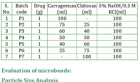
Table 3: The composition of the microbeads formulation of polyelectrolyte complexation method
S. No. Batch code Drug (g) Carrageenan (ml) Chitosan (ml)
5% NaOH/0.3 M KCl(ml)
1 P1 1 100 - 100
2 P2 1 75 25 100
3 P3 1 60 40 100
4 P4 1 50 50 100
5 P5 1 40 60 100
6 P6 1 25 75 100
7 P7 1 - 100 100
Evaluation of microbeads:
Particle Size Analysis
Particle size of microbeads was determined by using an optical microscope under regular polarized light, and the mean particle size was calculated by measuring 100 particles with the help of a calibrated ocular micrometer.
Swelling Index
The swelling index of the microbeads is an indication of the capacity of the microbeads to imbibe water and swell. For estimating swelling index, the microbeads (50 mg) were weighed initially then suspended in 25 ml of phosphate buffer pH 7.4. The beads were taken out at different time intervals using stainless steel grid and blotted carefully without pressing hard to remove the excess surface liquid. The swollen beads were weighed using electronic microbalance. The studies were performed in triplicate and average values were taken in data analysis.
Weight of wet microbeads Swelling Index = ×100 Weight of dry microbeads
Determination of Encapsulation Efficiency
The amount of Diclofenac sodium present in the microbeads was determined. The powdered microbeads were extracted in to 50 ml of phosphate buffer (pH 7.4) by magnetic stirring for a period of 2 h. The solution was filtered through Whatman filter paper no.5, suitably diluted and estimated for drug content spectrophotometrically at 282 nm using UV– Visible Spectrophotometer (UV – 1601). Encapsulation efficiency was calculated by the following formula:
Drug Content Estimation
Different batches of microbeads were checked for drug content uniformity. Accurately weighed (50 mg) amount of dried microbeads were taken in a pestle and mortar and powdered. The powdered microbeads were then separately dissolved in adequate quantity of 0.1 N HCl and 7.4 pH phosphate buffer and kept for 24 h. the solution was then filtered, scanned for absorbance was noted down at 282 nm using UV spectrophotometer (Shimadzu Model process was repeated in triplicate and average was calculated.
Morphology
Surface morphology of microbeads was investigated by Scanning Electron Microscopy (SEM) using JSM 6380A (JOEL, Japan). The microbeads, coated with Platinum by ion Auto fine coater JFC-1600 (JOEL, Japan), for 20 s at 1.1V under argon atmosphere were mounted onto metal stubs using double micrographs were taken.
In-vitro drug release studies
The in vitro drug release studies were performed using Dissolution test apparatus. The dissolution medium was hydrochloric acid buffer (pH 1.2) for first 2 h and 7.4) for subsequent h. The microbeads were efficiency was calculated by the following 1601, Japan). Each double-sided carbon adhesive tapze and the scanning electron phosphate buffer (pH allowed to sink in the vessel containing 900 ml of dissolution medium and the release of Diclofenac sodium was investigated at about 50 rpm at temp 37 ± 0.5°C. During dissolution 10 ml aliquot was withdrawn at interval of 1 h and same was replaced with equal volume of fresh medium. The withdrawn samples were filtered through Whatmann filter paper no.42 and diluted with the same buffer to 10 ml. Absorbance was measured at 282 nm using UV-Visible Spectrophotometer. Cumulative percent drug released was found out at each time interval and graph was plotted between cumulative % drug release v/s time.
RESULTS AND DISCUSSION
Table 4: Various Evaluation Parameters Of Batches Prepared By Ionotropic Gelation Method
Formulation Code Mean Particle size (µm) % swelling index % Encapsulation
Efficiency Drug content
pH 1.2 pH 7.4
C1 578.59 ± 3.58 295.23 ±3.78 532.34 ±6.26 41.34 24.64
C2 592.43 ± 5.35 318.67 ±5.02 555.60 ± 4.67 50.29 22.73
C3 616.52 ± 6.12 353.43 ±4.98 585.49±5.02 56.16 20.54
C4 667.30 ± 4.46 385.54 ±5.22 616.03 ±5.12 66.62 18.07
C5 787.14 ± 5.38 423.44 ±4.53 685.42 ± 4.25 72.28 17.11
C6 804.28 ± 6.54 456.20 ±3.23 705.32 ± 4.34 79.87 16.50 Mean + SD, n = 3
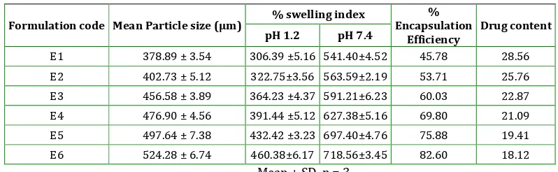
Table 5:Various Evaluation Parameters Of Batches Prepared By Emulsion Gelation Method
Formulation code Mean Particle size (µm) % swelling index
% Encapsulation
Efficiency
Drug content
pH 1.2 pH 7.4
E1 378.89 ± 3.54 306.39 ±5.16 541.40±4.52 45.78 28.56
E2 402.73 ± 5.12 322.75±3.56 563.59±2.19 53.71 25.76
E3 456.58 ± 3.89 364.23 ±4.37 591.21±6.23 60.03 22.87
E4 476.90 ± 4.56 391.44 ±5.12 627.38±5.16 69.80 21.09
E5 497.64 ± 7.38 432.42 ±3.23 697.40±4.76 75.88 19.41 E6 524.28 ± 6.74 460.38±6.17 718.56±3.45 82.60 18.12
Table 6: Various evaluation parameters of formulation batches p1 to p6
Formulation code Mean Particle size (µm) % swelling index
% Encapsulation
Efficiency
Drug Content
pH 1.2 pH 7.4
P1 742.29±3.65 406.45±2.41 558.71±3.23 59.42 16.87
P2 778.81±2.90 387.24±5.32 667.15±6.43 66.29 18.52
P3 780.14 ± 4.31 366.56 ± 3.76 686.56 ± 3.76 74.28 21.40
P4 784.30 ± 7.65 319.26 ± 4.68 754.26 ± 4.68 85.62 25.22
P5 776.52 ± 4.45 282.69 ± 3.23 705.69 ± 3.23 76.16 21.91
P6 752.43 ± 3.33 245.88 ± 4.85 559.88 ± 4.85 63.29 19.41
P7 738.59 ± 5.12 236.13 ± 5.92 543.40 ± 5.12 56.34 15.44 Mean + SD, n = 3
Table 7: Cumulative percentage drug release from formulation batches C1 to C6
Time (h)
Cumulative % Drug Release
C1 C2 C3 C4 C5 C6
1 22.46 20.78 17.31 15.14 12.73 10.16
2 41.73 38.80 30.75 28.98 21.27 16.98
3 66.41 56.17 46.52 45.46 40.36 30.28
4 91.31 75.37 60.37 56.11 52.12 41.85
5 97.17 90.87 85.52 68.89 60.89 49.93
6 - 96.18 91.61 87.21 71.64 56.16
7 - - 96.26 92.32 86.75 67.47
8 - - - 97.75 90.48 75.39
9 - - - - 94.91 85.40
10 - - - - 96.52 89.30
11 - - - 92.76
12 - - - 97.31
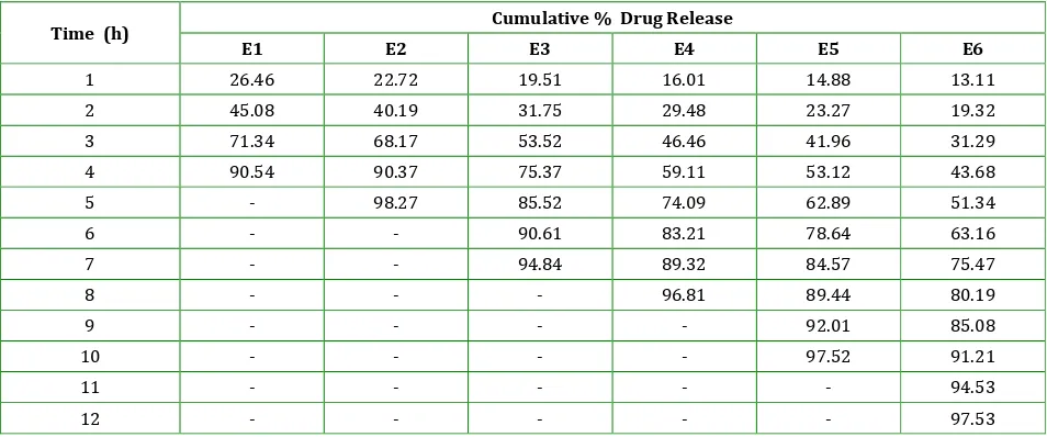
Table 8: Cumulative percentage drug release from formulation batches E1 to E6.
Time (h) Cumulative % Drug Release
E1 E2 E3 E4 E5 E6
1 26.46 22.72 19.51 16.01 14.88 13.11
2 45.08 40.19 31.75 29.48 23.27 19.32
3 71.34 68.17 53.52 46.46 41.96 31.29
4 90.54 90.37 75.37 59.11 53.12 43.68
5 - 98.27 85.52 74.09 62.89 51.34
6 - - 90.61 83.21 78.64 63.16
7 - - 94.84 89.32 84.57 75.47
8 - - - 96.81 89.44 80.19
9 - - - - 92.01 85.08
10 - - - - 97.52 91.21
11 - - - 94.53
12 - - - 97.53
Table 9: Cumulative Percentage Drug Release from Formulation Batches P1 to P7
Time (h) Cumulative % drug release
P1 P2 P3 P4 P5 P6 P7
1 17.38 9.33 8.86 5.24 4.67 3.06 2.19
2 28.30 17.73 15.93 9.65 8.88 6.18 3.78
3 41.17 36.66 32.17 26.02 28.78 33.23 21.20
4 63.37 54.31 50.56 38.37 45.70 51.50 38.87
5 78.17 65.24 63.17 48.52 60.76 65.63 51.67
6 86.38 73.19 70.18 53.61 68.47 72.36 68.19
7 97.46 78.69 75.56 60.26 72.40 76.47 78.89
8 - 83.17 79.99 64.96 76.03 81.39 85.71
9 - 88.33 86.40 68.78 81.87 87.40 96.52
10 - 94.71 89.47 71.06 86.78 90.3 -
11 - 98.88 94.51 74.14 89.05 94.76 -
12 - - 96.71 76.21 91.64 97.53 -
Selection of optimized batches:
From each method one optimized batch was selected based on their encapsulation efficiency and drug release.
Table 10: Optimized batch of various methods
Method Optimized batch
Encapsulation efficiency
% Cumulative drug release
(12 h)
Ionotropic gelation
method C6 79.87 97.53
Emulsion gelation
method E6 82.60 96.31
Polyelectrolyte complexation
method
P4 85.62 76.21
In ionotropic gelation method mean particle size of microbeads for batch C1 to C6 ranges from 578.59 µm to 804.28 µm. It was observed that as the concentration of sodium alginate increased (0.5% to 3.0%) size of microbeads also increased. Increasing concentration of polymer causes increasing viscosity of solution which in turn increases the droplet size during extrusion of the polymer dispersion to the harvesting medium which results formation of larger size beads.
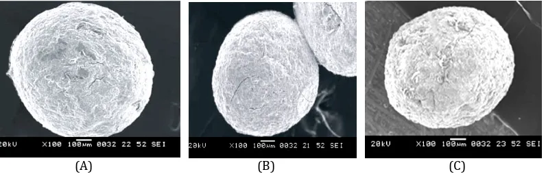
SEM of microbeads showed that formulations were found discrete and spherical as shown in figure 1. In ionotropic gelation method batches (C1 to C6) %swelling in pH 1.2 and pH 7.4 ranges from 295.23 to 456.20 and 532.34 to 705.32 respectively. All Batches showed low swelling in pH 1.2 than phosphate buffer pH 7.4. At acidic pH, alginate is protonated into insoluble form of the alginic acid this displays low swelling and in intestinal pH, at pH 7.4 carboxyl groups of alginate ionize, which weakens the electrostatic interactions, thus making the bead structure loose resulting in increased swelling. In ionotropic gelation method, % drug encapsulation efficiency for batches (C1-C6) ranges from 41.34% to 79.87%. The higher encapsulation efficiency was observed as the concentration of alginate increased. This is due to the greater availability of active calcium binding sites in the polymeric chains and consequently the greater degree of cross linking. Drug content of batches C1 to C6 ranges from 24.64 to 16.50%.
(A) (B) (C) Figure 1: SEM photograph A. Microbeads of ionotropic gelation method (Batch C6)
Drug release from batches C1, C2, C3, C4, C5 and C6 containing 0.5, 1.0, 1.5, 2.0, 2.5, 3.0% sodium alginate respectively showed 97.17% in 5 h, 96.18% in 6 h, 96.26% in 7 h, 97.75% in 8 h, 96.52% in 10 h and 97.31% in 12 h respectively.
It was observed from the swelling study that alginate beads had swollen in phosphate buffer pH 7.4 more than in 0.1 N HCl (pH 1.2). The release will depend on diffusion of Diclofenac sodium through the insoluble matrix of alginate polymer in pH 1.2 HCl buffer. On the other hand, rapid swelling and erosion of beads prepared form alginate were observed at pH 7.4 because at this pH exchange of Na+ ion and Ca2+ takes place and Ca-alginate is converted into Na-alginate which is more soluble.
From ionotropic gelation method, batch C6 showed 79.89% encapsulation efficiency and 97.31% drug in 12 h, hence considered as optimized batch.
In emulsion gelation method mean particle size of microbeads for batch E1 to E6 ranges from 378.89 µm to 524.28 µm. It was observed that as the concentration of sodium alginate increased (0.5% to 3.0%) size of microbeads also increased. Increasing concentration of polymer causes increasing viscosity of solution which in turn formation of larger size microbeads.
In emulsion gelation method batches E1 to E6 showed %swelling in pH 1.2 and pH 7.4 ranges from 306.39 to 460.38 and 541.40 to 718.56 respectively. All Batches showed low swelling in pH 1.2 than phosphate buffer pH 7.4. At acidic pH, alginate is protonated into insoluble form of the alginic acid this displays low swelling and in intestinal pH, at pH 7.4 carboxyl groups of alginate ionize, which weakens the electrostatic interactions, thus making the bead structure loose resulting in increased swelling. Percent drug encapsulation efficiency for batches (E1-E6) ranges from 45.78% to 82.60%. The higher encapsulation efficiency was observed as the concentration of alginate increased. This is due to the greater availability of active calcium binding sites in
the polymeric chains and consequently the greater degree of cross linking.
Drug content of batches E1 to E6 ranges from 28.56 to 18.12%. Drug content is higher as compared to ionotropic gelation method (24.64-16.50) because external oil gelation medium prevents diffusion of the drug.
Formulations were found smooth, discrete and spherical as shown in figure 1.
Batches E1, E2, E3, E4, E5 and E6 containing 0.5, 1.0, 1.5, 2.0, 2.5, 3.0% sodium alginate respectively showed 90.54% in 4 h, 98.27% in 5 h, 94.84% in 7 h, 96.81% in 8 h, 997.52% in 10 h and 97.53% drug release in 12 h respectively.
For emulsion gelation method it was observed from the swelling study that alginate beads had swollen in phosphate buffer pH 7.4 more than in 0.1 N Hcl (pH 1.2). The release will depend on diffusion of Diclofenac sodium through the insoluble matrix of alginate polymer in pH 1.2 Hcl buffer. On the other hand, rapid swelling and erosion of beads prepared form alginate were observed at pH 7.4. Ionization of cross-linked calcium salt increase and the process of exchange of Ca2+ for sodium start. As Ca2+ ions are replaced by Na+ ions, Ca-alginate is converted into Na-alginate which is more soluble.
Batch E6 showed 82.60% encapsulation efficiency and 97.53% drug in 12 h, hence considered as optimized batch.
In polyelectrolyte complexation method mean particle size of microbeads for batch P1, P2, P3, P4, P5, P6 to P7 was found 742.29 µm,778.81 µm, 780.14 µm, 784.30 µm, 776.52µm, 752.43µm and 738.59 µm respectively. From the results it was observed that increasing concentration of chitosan causes increasing viscosity of solution which in turn formation of larger size microbeads. Further increase in chitosan concentration with decrease in carrageenan lower the viscosity and size of microbeads reduced.
In polyelectrolyte complexation method batches P1 to P7 showed % swelling index in pH 1.2 and pH 7.4 ranges from 406.45 to 236.13 and 558-754
respectively. It was found that the beads shrink in acidic pH, this could be well justified due to the fact that, at acidic pH strong interaction occurs between ammonium groups of Chitosan and sulphate group of carrageenan which is due to the formation of intermolecular and intramolecular hydrogen bond (polyelectrolyte complex) between the two polymers. Additionally, a repulsive force within the microbeads is created due to protonation of primary ammonium group (-NH3+) of Chitosan. But because the force of H-bond is greater than the repulsive force, the beads are kept in a shrunken state in acidic medium. The increased swelling of beads in pH 7.4 phosphate buffer was due to, firstly, the breakage of H-bond, which reduces the interaction between the polyelectrolytes and ionization of sulphate group of carrageenan results in swelling of hydrogel network (beads) with subsequent imbibitions of fluid.
The% drug encapsulation efficiency was increased with the increase in concentration of chitosan from 0 to 50 ml for batches P1 to P4 ranges from 59.42% to 85.62%. Further increase in concentration of chitosan form 60 to 100 ml (P5 to P7) showed that decrease in percent encapsulation efficiency from 76.16% to 56.345 due to less availability of SO42- group of carrageenan for electrostatic interaction with –NH3+ group of chitosan. This interaction causes formation of network like structure for entrapment of drug. Drug content was increased with the increase in concentration of chitosan from 0 to 50 mL for batches P1 to P5 ranges from 16.87% to 25.22%. Further increase in concentration of chitosan form 60 to 100 ml (P5 to P7) results decreased in drug content from 21.91% to 15.44% due to less availability of SO4 2-group of carrageenan for electrostatic interaction with –NH3+ group of chitosan which results formation of loose network structure for entrapment of drug. SEM photomicrograph of the beads with polyelectrolyte complexation techniques revealed that the beads were not completely spherical, irregular in shape, rough moon-like surface and folded, shown in figure 1.
It was observed that as concentration of chitosan increased (0 to 100 ml) cumulative % release of drug in acetic medium extended from 28.30% to 3.78% in 2h. The release rate of drug in simulated intestinal fluid (pH 7.4) wasrelatively higher than in simulated gastric fluid (pH 1.2). Low release in acidic medium was due to strong interaction between ammonium groups of Chitosan and sulphate group of carrageenan which is due to the formation of intermolecular and intra molecular hydrogen bond between the two polymers. Additionally, a repulsive force within the microbeads is created due to the protonation of primary ammonium groups (-NH3+) of Chitosan. But because the force of H-bond is greater than the repulsive force, the beads are kept in a shrunken state in acidic medium and the drug is released slowly. However, under alkaline condition there was breakage of H-bond which reduces the interaction between the polyelectrolyte and ionization of sulphate group of carrageenan results in swelling of microbeads network with subsequent imbibitions of fluid and dissolution of drug followed by drug release by diffusion.
Batch P4 showed 85.62% encapsulation efficiency and 76.21% drug in 12 h, hence considered as optimized batch.
Among three methods polyelectrolyte complexation method was selected as optimized method as it showed comparatively higher encapsulation efficiency and extended drug release pattern. Optimized batch P4 of polyelectrolyte complexation method showed higher encapsulation efficiency (85.62%) and sustained drug release pattern (76.21% in 12 h) than optimized batch (C6) of ionotropic gelation method (encapsulation efficiency 79.89% and 97.31% drug release in 12 h) and (E6) of emulsion gelation method (encapsulation efficiency 82.60% and 97.53% drug release in 12 h).
CONCLUSION
emulsion gelation method and polyelectrolyte complexation method. The former two methods gave small, discrete, spherical microbeads. Polyelectrolyte complexation method produced comparatively large, rough and less spherical microbeads. Encapsulation efficiency was higher in polyelectrolyte complexation method than ionotropic gelation and emulsion gelation method. The swelling index and drug release pattern of microbeads depend upon polymer concentration and extent of complex formation between two oppositely charged polymers. The drug releases from the microbeads were found to be slow and spread over extended period of time depending on concentrations of polymers and method of preparation. The wash-off was faster at simulated intestinal pH (7.4) than at simulated gastric pH (1.2). There was no significant change in drug content of drug loaded microbeads, and prepared capsules stored at different storage conditions after 90 days of study. Micromeritic parameters of three different methods were similar but polyelectrolyte complexation method showed better drug encapsulation efficiency and sustained drug release pattern than ionotropic gelation and emulsion gelation method. Hence it can be concluded that polyelectrolyte complexation method is best suited for microbeads preparation.
ACKNOWLEDGEMENTS
The authors are thankful to Zim Laboratories, Kalmeshwar, Nagpur for providing the Diclofenac sodium as a gift sample.
REFERENCES
1. LeeY, Robinson J, In: Remington. 20th ed., Vol II, The
Science and Practice of Pharmacy. Lippincott
Williams and Wilkins.Noida:B.I. Publications, 2000,
903-904.
2. Lachman L, Liberman H, Kanig J. The Theory and
Practice of Industrial Pharmacy. 3rd ed.,
Mumbai:Varghese Publishing House, 1986, p. 430.
3. Gibaldi M, Parrier D. Biopharmaceutics and clinical
Pharmacokinetics.Philadelphia:Lea and Febiger 3rd
ed., Vol 15, ,1984,64-82.
4. Belyaeva E, Valle D.D., Neufeld R.J. Ponceleta D. New
approach to the formulation of hydrogel beads by
emulsification/thermal gelation using a static mixer.
Chem Eng Sci 2004; 59(2): 2913–20.
5. Badarinath A.V., Reddy J.R. Mallikarjuna R.
K.,.Alagusundaram M, Gnanaprakash K., Chetty M.S.
Formulation and Characterization of Alginate
Microbeads of Flurbiprofen by Ionotropic Gelation
Techniques. Int J Chem Tech Res 2010;2(1):361-367.
6. Reddy T., Tammishetty S. Formulation of barium
chloride crosslinked beads of carboxymethyl guar
gum. J Applied Poly Sci 2001; 82(7): 3084-3090.
7. Bopanna R., Kulkarni R.V., Setty C.M.
Carboxymethylcellulose-aluminium hydrogel
microbeads for prolong release of Simvastatin. Acta
Pharm Sci 2010; 52(2): 137-143.
8. Elzatahry A.A., Soliman E.A. Evaluation of
alginate-chitosan bioadhesive beads as a drug delivery
system for the controlled release of Theophylline, J
Apppl Poly Sci 2009; 111(34): 2452-2459.
9. Gursoy A., Cevik S. Sustained release properties of
alginate microsphere and tableted microsphere of
diclofenac sodium, J Microencapsul 2000; 17(4):
565-575.
How to cite this article: Umesh D. Shivhare*, Vijay B. Mathur, Chandrashekhar G. Shrivastava, Vivek I. Ramteke; Preparation of Microbeads by different Techniques and Study of their influence on Evaluation Parameters; J. Adv. Pharm. Edu. & Res. 2013: 3(3): 279-288.