This is an open access journal, and articles are distributed under the terms of the Creative Commons Attribution-Non Commercial-ShareAlike 4.0 License, which allows others to remix, tweak, and build upon the work non-commercially, as long as appropriate credit is given and the new creations are licensed under the identical terms.
© 2019 Journal of Advanced Pharmacy Education & Research | Published by SPER Publication
88
Serum levels of Osteoprotegerin, Matrix Metalloproteinase-III
and C-reactive protein in patients with Psoriasis and Psoriatic
Arthritis and their correlation with Radiological findings
Maha Abdelhadi Ali
1*, Hala Mohamed Raslan
1, Mahmoud F. Abdelhamid
2, Marwa Mohamed
Fawzy
3, Hany Ahmed Shehata
2, Heba Mohamed Abdelraheem
3, Aza Ahmed Ali
4, Alshymaa Ahmed
Ibrahim
4, Mohamed S. El Hussieny
5, Rokia Abdel Shafy El Banna
5, Mohamed Hussein Abohadeed
2 1Internal Medicine Department, National Research Centre, Cairo, Egypt. 2Dermatology and Venereology Department, National Research Centre, Cairo, Egypt. 3Dermatology and Venereology Department Faculty of Medicine, Cairo University, Cairo, Egypt. 4Clinical Pathology Department, National Research Centre, Cairo, Egypt. 5Biological Anthropology Department, National Research Centre, Cairo, Egypt.Correspondence: Maha Abdelhadi Ali. Internal Medicine Department, National Research Centre, Cairo, Egypt. Email: [email protected].
ABSTRACT
Background: Psoriasis is a chronic inflammatory disease of the skin that affects the joints in up to 62% of cases. Psoriatic arthritis can present with both peripheral articular and axial skeletal manifestations. It can be diagnosed by clinical, radiological and serological parameters including serum osteoprotegerin, matrix metalloproteinase-III, C reactive protein and macrophage colony stimulating factor. Aim: Our aim was to assess the serum levels of HS-CRP, OPG, MMP-3 and M-CSF in patients with psoriasis and PsA, and to correlate them with different radiological findings. Methods: Sixty-one patients with psoriasis (including 40 patients with PsA) were subjected to clinical, radiological and serological testing. Results: Osteopenia and osteoporosis of the spine were significantly higher among patients with PsA when compared to controls (p=0.024). Serum MMP-3 was significantly higher among patients when compared to controls (p=0.005). Serum levels of HS-CRP were significantly higher in patients showing juxta-articular osteoporosis (P=0.02). Serum OPG levels were positively correlated with radiological scores (P=0.003, r=0.595) and negatively correlated with Dexa femur score (p=0.039, r=-0.434). PASI score of the patients was positively correlated to serum levels of MMP-3 and GM-CSF (p=0.032, r=0. 363 and p=0.003, r=0.485 respectively). MMP-3 levels and GM-CSF were positively correlated to each other (p=0.006, r=0.423). Conclusion: Serum levels of MMP-3, HS-CRP, GM-CSF and OPG can be used to assess the activity of arthritis among patients with PsA and should be utilized, in addition to the clinical and radiological findings, to determine the severity of the disease and can be also used to follow up patients.
Keywords:Psoriasis, psoriatic arthritis, OPG, MMP-3, CRP, GM-CSF.
Introduction
Psoriasis is a chronic inflammatory disease of the skin that is prevalent in the general population (2–4%).[1] The prevalence
of inflammatory arthritis among psoriatic patients is estimated
to be varying from 6% to 42%. Approximately, 67% of the patients develop arthritis after psoriasis, and in 16% arthritis and psoriasis occur within 12 months of each other.[2] Psoriatic
arthritis is typically associated with psoriasis and psoriatic nail disease and has both peripheral articular manifestations (including synovitis, dactylitis, and enthesitis) and axial skeletal involvement.[3] Bone loss can occur, either locally in the form
of bone erosion and osteolysis affecting the peripheral joints, or systemically with loss of skeletal bone mineral density (BMD).[4]
Currently, the diagnosis of PsA is based on clinical, radiologic, and immunologic features which are consistent with the diagnosis of PsA, rather than other inflammatory arthritides.[5]
In 2006, a large international study group developed a simple and highly specific classification known as CASPAR (classification criteria for psoriatic arthritis).[6]
Access this article online
Website: www.japer.in E-ISSN: 2249-3379
How to cite this article: Maha Abdelhadi Ali, Hala Mohamed Raslan,
Mahmoud F. Abdelhamid, Marwa Mohamed Fawzy, Hany Ahmed Shehata, Heba Mohamed Abdelraheem et al.
Serum levels of Osteoprotegerin, Matrix Metalloproteinase-III and C-reactive protein in patients with Psoriasis and Psoriatic Arthritis and their correlation with Radiological findings. J Adv Pharm Edu Res 2019;9(1):88-92.
Several studies have shown that high sensitive C reactive protein (HS-CRP), osteoprotegerin (OPG) and matrix metalloproteinase-3 (MMP-3) are biomarkers for PsA and could also be related to the diagnosis, pathogenesis, prognosis, therapeutic response, and comorbidities associated with PsA.[7]
Also, macrophage colony stimulating factor (M-CSF), which promotes macrophage survival and proliferation and is a key regulator of osteoclastogenesis, which has been strongly implicated in the pathogenesis of tumor necrosis factor (TNF)-induced osteolysis in animal models. Studies have shown that serum levels of M-CSF are elevated in patients with PsA and that they strongly correlate with the severity of peripheral erosive disease.[8]
The aim of this study was to assess the serum levels of HS-CRP, OPG, MMP-3and M-CSF in patients with psoriasis and PsA, and to correlate them with different radiological findings in order to determine their significance in assessing the activity of the disease.
Materials and Methods
This study was conducted on 61 patients with psoriasis recruited from the outpatient dermatology clinic of the National Research Centre and Kasr Al Aini hospital, Cairo, Egypt. Inclusion criteria included patients with psoriasis above 12 years of age. Exclusion criteria included patients with joint disease other than PsA, patients with skin disease other than psoriasis, patients with thyroid disease, malignancy, chronic liver or renal disease and patients taking hormone replacement therapy, thyroxine or vitamin D. The study was approved by the Medical Research Ethics Committee of the National Research Centre. Thirty healthy controls were included as a control group. Informed written consent was obtained from all participants in the study.
All patients were subjected to detailed history taking including onset, course, duration of the disease and medications received. A thorough clinical examination was done by a dermatologist and a rheumatologist. Joint examination included determining the pattern of affected joints as well as the number of deformed joints. Severity of psoriatic skin disease was assessed using the Psoriasis Area and Severity Index (PASI) score graded from 0-72. CASPAR criteria were used to diagnose PsA. It consists of established inflammatory articular disease with at least 3 points from the following features: presence of current psoriasis, a history of psoriasis, a family history of psoriasis, dactylitis, juxta-articular new bone formation, rheumatoid factor negativity, and nail dystrophy.[6]
Activity and severity of articular disease was measured using the Patient Global assessment score in which the Visual Analogue Scale (0-100 mm) was utilized,[9] Patient Pain
assessment score in which pain intensity, physical functioning, emotional functioning and the participants’ rating of overall improvement were assessed,[10] Likert scale for Patient global
assessment, as well as the Modified Health Assessment Questionnaire (MHAQ) which includes questions concerning
perceived patient satisfaction regarding the same activities of daily living, along with perceived change in degree of difficulty.[11]
Bone mineral density (BMD) of proximal femur and lumbar spine was measured in both patients and controls by DEXA using the Norland XR-46 machine. Radiologic examination for hands and lumbar spine was done for patients to assess juxta-articular osteoporosis, proliferative changes, joint space narrowing (JSN) and erosion according to the Sharp van der Heijde score modified for use in PsA [Simple Erosion Narrowing Score (SENS method)]. SENS gives a score of 1 if at least one joint erosion is noted or if there is any narrowing in the joint. In the hands, the maximal erosion score is 32, and 30 for narrowing/(sub)luxation. In the feet, the maximal erosion score is 12, and 12 for narrowing/(sub)luxation. As a consequence, the range of the SENS method is from 0 to 86.[12]
Five milliliters of blood was drawn from patients and controls. Blood was centrifuged and the serum refrigerated at -20˚C until time of assessment of HS-CRP, OPG, MMP-3 and M-CSF by Enzyme linked immunosorbent assay (ELISA). Osteoprotegerin was measured using BioVendor kit, MMP-3 was measured using EIAab Science kit, GM-CSF was measured using IDlabs Biotechnology kit and HSCRP was measured using HSCRP kits Monobind Inc., Lake Forest, USA.
Results
This study was conducted on a total of 61 patients with psoriasis; 31 females (50.8%) and 30 males (49.2%), their age ranged from 12 to 60 years with a mean of 40.4±12.7. Forty patients had PsA as diagnosed by CASPAR criteria. The duration of psoriasis ranged from 1 to 35 years, with a mean of 10.5±9 years. The PASI score ranged from 0 to 54 with a mean of 12.9±12.7. The CASPAR score was ≥3 in 40 individuals (65.5%).
The studied symptoms of bone, joint and nail affection among patients are summarized in Fig.1.
Figure 1.Percentages of bone, joint and nail findings in psoriatic patients
Patient global assessment and patients pain assessment scores ranged from 0 to 100 with a mean of 49.3±28 and 45.7±30.7 respectively, while the Likert scale for Patient global assessment ranged from 1 to 5 with a mean of 2.2±1.1. All disease activity scores were significantly higher among patients having high swollen and tender joint counts, dactylitis, enthesitis, radiological erosions and spine affection compared with other psoriatic patients having no peripheral joint involvement (P<0.05).
Osteopenia and osteoporosis of the spine were significantly higher among patients with PsA when compared to controls (p=0.024).
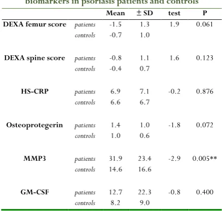
Regarding serum biomarkers, levels of MMP-3 were significantly higher among patients when compared to controls (p=0.005). The mean levels of DEXA scores and serum biomarkers are shown in Table 1. Also, serum levels of HS-CRP were significantly higher in patients showing juxta-articular osteoporosis (P=0.02).
Table 1. Mean levels of DEXA score and some biomarkers in psoriasis patients and controls
Mean ± SD test P
DEXA femur score patients -1.5 1.3 1.9 0.061
controls -0.7 1.0
DEXA spine score patients -0.8 1.1 1.6 0.123
controls -0.4 0.7
HS-CRP patients 6.9 7.1 -0.2 0.876
controls 6.6 6.7
Osteoprotegerin patients 1.4 1.0 -1.8 0.072
controls 1.0 0.6
MMP3 patients 31.9 23.4 -2.9 0.005**
controls 14.6 16.6
GM-CSF patients 12.7 22.3 -0.8 0.400
controls 8.2 9.0
Correlations were done between different clinical, radiological and serum parameters. DEXA femur score was negatively correlated with the radiological score (p=0.032, r=-0.448), positively correlated with DEXA spine score (p=0.005, r=0.489), and negatively correlated with serum levels of OPG (p=0.039, r=-0. 434).
Serum OPG levels were positively correlated with radiological scores (P=0.003, r=0.595). MMP-3 levels were positively correlated with GM-CSF (p=0.006, r=0.423).
Age of psoriatic patients was positively correlated to the radiological scores as well as serum levels of OPG (p=0.008, r=0.462 and p=0.000, r=0.610 respectively) and negatively related to DEXA femur score (p=0.03, r=-0.379).
PASI score of the patients was positively correlated to serum levels of MMP-3 and GM-CSF (p=0.032, r=0. 363 and p=0.003, r=0.485 respectively).
Radiological scores were positively correlated to MHAQ scores (p=0.047, r=0.360).
Discussion
Psoriatic arthritis is characterized by certain clinical features including the presence of axial involvement, distal interphalangeal joint involvement, dactylitis, and enthesitis.[13]
Several serological markers relating to the disease activity have been studied including serum OPG, MMP-3, CRP and GM-CSF.
In this study, we found a positive correlation between serum OPG levels and the radiological scores of patients with PsA, the same was reported by Nell-Duxneuner et al[14], who found
that psoriatic patients showing radiological findings of erosions had high serum OPG levels. Serum OPG levels were positively correlated with the age of psoriatic patients and negatively correlated with DEXA femur scores. Similarly, Fichna et al[15]detected that serum OPG level increased with age and was
negatively correlated with bone mineral density (BMD) at the lumbar spine and femoral neck. Mianini et al[16] also detected a
positive correlation between serum OPG levels with age, so did Nabipour et al[17]. The correlation between serum OPG
levels with age might reflect a compensatory response to enhanced bone resorption that occurs with aging and the increased osteoclastic bone resorption found in osteoporosis in order to minimize further bone loss.[18]
Osteoprotegerin is a glycoprotein belonging to the TNF-receptor superfamily. It is a part of the cytokine system responsible for osteoclast maturation, a process in which aRANK-Ligand (receptor activator of nuclear factor kappaB-ligand) on osteoblasts binds to RANK on osteoclast precursors, leading to their differentiation. Osteoprotegerin acts as a decoy receptor, it binds to RANK-Ligand instead of RANK, preventing the differentiation of precursor cells into osteoclasts, thus inhibiting bone resorption.[19]
In our study, serum levels of MMP-3 were found to be higher among patients than controls and they were positively correlated with PASI scores. Same findings were reported by Ribbenset al.[20]. The elevated MMP-3 levels reflect the
synovitis caused by articular inflammation, thus it can be a good method to determine the activity of synovial inflammatory diseases.
Serum levels of HS-CRP were higher among patients with JAO. Similar findings were reported by Maejima et al [21] who
detected that PASI scores, serum MMP-3 and CRP levels were higher among psoriatic patients especially those presenting with sacroiliitis. Also, Jadon and McHugh [22] detected elevated
levels of CRP in approximately 50% of PsA cases. C-reactive protein (CRP) is an acute phase reactant that increases in response to tissue damage, inflammation and infection and its elevation in PsA reflects a possible inflammatory mechanism for osteoporosis.[23]
IL-1 and TNF-α [24] and this might explain its positive
correlation with serum levels of MMP-3 in patients with PsA due to the presence of synovial inflammation.
Our results also showed that osteopenia and osteoporosis of the spine were significantly higher among psoriatic patients when compared to controls. Attia et al [25] found similar results
and concluded that psoriatic patients with or without arthritis may suffer from osteoporosis and have higher levels of serum OPG more than controls.
In conclusion, serum levels of MMP-3, HS-CRP, GM-CSF and OPG can be used to assess the activity of arthritis among patients with PsA and should be utilized, in addition to the clinical and radiological findings, to determine the severity of the disease and can be also used to follow up patients. Larger multi-center studies are needed for better estimation of the prevalence of PsA among Egyptian psoriatic patients.
References
1. Langley RG, Krueger GG, Griffiths CE. Psoriasis: epidemiology, clinical features, and quality of life. Ann Rheum Dis 2005; 64:18–23.
2. Gladman DD. Psoriatic arthritis. In: Gordon K, Ruderman E (eds). Psoriasis and Psoriatic Arthritis. Berlin: Springer-Verlag, 2005;57-65.
3. Taylor WJ, Porter GG, Helliwell PS. Operational definitions and observer reliability of the plain radiographic features of psoriatic arthritis. J Rheumatol 2003; 30:2645-58.
4. Frediani B, Allegri A, Falsetti P, et al. Bone mineral density in patients with psoriatic arthritis. J Rheumatol 2001;28:138-43.
5. Lee KH, son Mk, Ha YJ, et al. Inflammatory
Polyarthritis in a Patient with Psoriasis: Is It Psoriatic Arthritis or Rheumatoid Arthrirtis? Korean J Intern Med 2010; 25:224-6 .
6. Taylor W, Gladman D, Helliwell P, et al.
Classification criteria for psoriatic arthritis: development of new criteria from a large international study. Arthritis Rheum 2006; 54(8):2665–73. 7. Jadon Dr, Sengupta R, Nightingale A, et al. Serum
Bone-Turnover Biomarkers are Associated with the Occurrence of Peripheral and Axial Arthritis in Psoriatic Disease: A Prospective Cross-Sectional Comparative Study. Arthritis Resther 2017; 19:210. 8. Kitaura H, Zhou P, Kim HJ, et al. M-CSF mediates
TNF-induced inflammatory osteolysis. J Clin Invest 2005;115:3418-27.
9. Cauli A, Gladman DD, Mathieu A, et al. Patient global assessment in psoriatic arthritis, a multicenter GRAPPA and OMERACT study. J Rheumatol2011; 38:898-903.
10. Dworkin RH, Turk DC, Wyrwich KW, et al.
Interpreting the clinical importance of treatment
outcomes inchronic pain clinical trials: IMMPACT recommendations. J Pain 2008; 9:105–21.
11. Husted JA, Gladman DD, Long JA, et al. A modified version of the Health Assessment Questionnaire (HAQ) for psoriatic arthritis. Clin Exp Dermatol 1995;:13:439–43.
12. Van der Heijde D, Kavanaugh A, Gladman DD, et al. Infliximab inhibits progression of radiographic damage in patients with active psoriatic arthritis through one year of treatment: results from the induction and maintenance psoriatic arthritis clinical trial 2. Arthritis Rheum 2007;56:2698-707.
13. Alamanos Y, Papadopoulos NG, et al. Epidemiology of psoriatic arthritis in northwest Greece, 1982–2001. J Rheumatol 2003;30: 2641–4.
14. Nell-Duxneuner VP, Stamm TA, Machold KP, et al. Evaluation of the appropriateness of composite disease activity measures for assessment of psoriatic arthritis. Ann Rheum Dis 2010;69(3):546-49.
15. Fichna M, Zurawek M, Fichna P, et al. Increased serum osteoprotegerin in patients with primary adrenal insufficiency receiving conventional hydrocortisone substitution. J Physiol Pharmacol. 2012;63(6):677-82. 16. Mainini G, Incoronato M, Urso L, et al. Serum
osteoprotegerin correlates with age and bone mass in postmenopausal, but not in fertile age women.ClinExpObstetGynecol2011;38(4):355-9. 17. Nabipour I, Larijani B, Vahdat K, et al. Relationships
among serum receptor of nuclear factor-kappaB ligand, osteoprotegerin, high-sensitivity C-reactive protein, and bone mineral density in postmenopausal women:
osteoimmunity versus osteoinflammatory.Menopause2009; 16(5):950-5.
18. Grisar J, Bernecker PM, Aringer M. Ankylosing spondylitis, psoriatic arthritis and reactive arthritis show increased bone resorption, but differ with regard to bone formation. J Rheumatol 2002; 29:1430–6. 19. Aubin JE, Bonnelye E. Osteoprotegerin and its ligand:
a new paradigm for regulation of osteoclastogenesis and bone resorption. OsteoporosInt2000; 11:905–13. 20. Maejima H, Taniguchi T, Watarai A, et al. Analysis of
clinical, radiological and laboratory variables in psoriatic arthritis with 25 Japanese patients.J Dermatol 2010;37(7):647-56.
21. Jadon DR, McHugh NJ. Laboratory tests for psoriatic arthritis. In A. Adebajo, W-H. Boehncke, D. D. Gladman, & P. J. Mease (Eds.), Psoriatic Arthritis and Psoriasis: Pathology and Clinical Aspects 2016 Vol. Part V1, pp. 227-40.
23. Ribbens C, Martin M, Porras Y, et al. Increased matrix metalloproteinase-3 serum levels in rheumatic diseases: relationship with synovitis and steroid treatment. Ann Rheum Dis2002;61:161–6.
24. Ross F. M-CSF, c-Fms, and signaling in osteoclasts and their precursors.Ann N Y AcadSci2006;1068 (1):110– 6.
25. Attia EA, Khafagy A, Abdel-Raheem S, et al.