Rafał Małecki
1, A, B, D, Krzysztof Rosiński
2, B, D, Rajmund Adamiec
1, C, E, FEtiological Factors of Infections in Diabetic
Foot Syndrome – Attempt to Define
Optimal Empirical Therapy
1 Department of Angiology, Systemic Hypertension and Diabetology, Wroclaw Medical University, Poland 2 Students’ Scientific Group at the Department of Angiology, Systemic Hypertension and Diabetology, Wroclaw Medical University, Poland
A – research concept and design; B – collection and/or assembly of data; C – data analysis and interpretation; D – writing the article; E – critical revision of the article; F – final approval of article; G – other
Abstract
Background. Diabetic foot syndrome (DFS) represents one of the most frequent reasons for lower limb amputa-tion in developed countries. In most cases, it is associated with bacterial infecamputa-tion, requiring optimal antibiotic therapy.
Objectives. The aim of this study was to identify the most frequent pathogens responsible for infections associated with DFS, establish the optimal protocol of empirical therapy, and ascertain the clinical variables that may deter-mine the choice of the appropriate antibacterial agent.
Material and Methods. The analysis included hospital records of patients treated at the Department between 2008 and 2010. A total of 102 individuals were identified; their material was cultured and tested for antibiotic suscep-tibility.
Results. A total of 199 bacterial strains were isolated. There was a predominance of Gram-positive bacteria, par-ticularly Staphylococcus aureus, Staphylococcus coagulase-negative strains, and Enterococcus faecalis. Of note was the high percentage of E. faecalis infection (16.08%). One can speculate on the potential etiological factors in the case of some bacteria, e.g. patients infected with S. aureus were characterized by higher monocytosis and lympho-cytosis as compared to other patients. Analysis of drug susceptibility revealed that ciprofloxacin has the highest (but still only 44%) efficacy of all agents tested as monotherapy, and a combination of piperacillin and tazobactam or amoxicillin and clavulanate with aminoglycosides is particularly beneficial.
Conclusions. Staphylococcus spp. predominates amongst the etiological factors of DFS infection; however, the rate of E. faecalis infection is alarmingly high. Monotherapy enables effective treatment in a minority of cases; therefore, at least two-drug protocols should be implemented from the very beginning of the therapy (Adv Clin Exp Med 2014, 23, 1, 39–48).
Key words: antibiogram-based therapy, antibiotics, diabetic foot syndrome, Enterococcus faecalis.
Adv Clin Exp Med 2014, 23, 1, 39–48 ISSN 1899–5276
ORIGINAL PAPERS
© Copyright by Wroclaw Medical University
Diabetes affects more than 3 m patients in Po-land, and it is estimated that this number will in-crease to 3.4 m by 2030. The prevalence of diabetes slightly exceeds 10%. Impaired glucose tolerance (IGT) is detected in more than 5 m patients. The number of diabetes-related deaths is reaching 30,000 and the costs of treatment calculated per diabetic patient are estimated at USD 1143 per year in Poland [1]. Diabetic foot syndrome (DFS) is one of the chronic complications of diabetes. Ac-cording to the definition, this condition pertains to
the controls [4]. Antibiotic therapy, initially em-pirical and subsequently antibiogram-based, is the most important treatment modality in the case of DFS infection. The aim of this retrospective study was to determine the types of pathogens that are most frequently isolated from DFS patients treat-ed at the Department of Angiology, Systemic Hy-pertension and Diabetology of Wroclaw Medical University, analyze their drug resistance, propose the most potentially effective protocol of empirical antibiotic therapy, and verify any potential asso-ciations between the type of pathogen and clinical manifestation and laboratory findings.
Material and Methods
The analysis included 4500 hospital records of patients treated at the Department of Angiol-ogy between 2008 and 2010. The inclusion crite-ria for further analysis included the diagnosis of type 2 diabetes, co-existing DFS, and a bacterial culture with antibiotic susceptibility testing. The material for microbiological culture and antibiot-ic susceptibility testing (fragments of tissues, sur-face biopsies, aspirates) was obtained from deep-er laydeep-ers of the ulcdeep-eration, following disinfection of the skin surface. The susceptibility of microor-ganisms to antibiotics and chemotherapeutics was determined qualitatively by means of a diffusion method with paper discs impregnated with anti-biotic at a given concentration. Under such condi-tions, the susceptibility of the analyzed strain is de-termined based on the size of the inhibition zone around the disc impregnated with a given antibiot-ic. A detailed protocol of the testing can be found in relevant literature [5].
Aside from detected pathogens and their re-sistance, the analysis included the patient’s age, lo-cation of the ulceration, presence of obliterative atherosclerosis (abnormal values of the ankle-bra-chial index or abnormalities on a Doppler ultraso-nography), type of DFS, concentration of glycated hemoglobin, glomerular filtration rate (eGFR) es-timated on the basis of the MDRD (Modification of Diet in Renal Disease) formula, presence of mi-croalbuminuria or proteinuria, ketonuria, glycos-uria, anemia, concentration of C-reactive protein (CRP), peripheral monocyte, neutrophil, eosino-phil, basoeosino-phil, and lymphocyte count, and fasting glucose concentration.
The results were subjected to statistical anal-ysis. In the case of normally distributed variables (identified by the Shapiro-Wilk test) and homoge-neity of variance (confirmed by the Levene test), intergroup differences were analyzed by means of simple analysis of variance (ANOVA). Whenever
at least one of the abovementioned conditions was not satisfied, the non-parametric Kruskal-Wallis rank test (in the case of more than two groups, with subsequent post-hoc testing) or the Mann-Whit-ney U-test (in the case of two groups) was applied. Intergroup differences in the percentage distribu-tions of dichotomous variables were analyzed with the Pearson’s χ2 test. P value < 0.05 was considered statistically significant. Due to widely accepted rec-ommendations, 95% confidence intervals (CI) were calculated for each distribution aside from means and standard deviations. Additionally, logistic re-gression and cluster analysis were employed. Cut-off values characterized by maximal sensitivity and specificity were identified on the basis of a Receiv-er OpReceiv-erating CharactReceiv-eristic (ROC) curve analysis. All calculations were conducted with the Statistica 10 package (Stat Soft Inc.).
Results
The analysis included 4500 hospital records of patients treated at the Department of Angiol-ogy between 2008 and 2010. Overall, 102 individ-uals (67 men and 35 women) with an average age of 65 years were included. One-hundred and 99 bacterial strains were isolated, including 8 (4%) alert pathogens [6]: Enterobacter aerogenes ex-tended-spectrum beta-lactamase (ESBL) (n = 2),
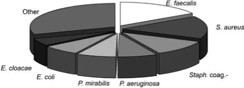
Escherichia coli ESBL (n = 2), methicillin-resis-tant Staphylococcus aureus (MRSA) (n = 2), Aci-netobacter baumannii (n = 1), and Enterobacter cloacae (n = 1). Only one strain was isolated from 38 individuals (37.5%), two strains – in 33 patients (32%), three strains – in 17 (16.3%), four strains – in 4 (4%), and five strains – in 9 (9%); in on-ly one case (1.2%) the culture was sterile. Gram-positive bacteria comprised 52.76% of all recov-ered strains, whereas the corresponding value was equal to 47.24% in the case of Gram-negative bac-teria. Facultative anaerobes were most prevalent (64.32%), followed by aerobes (29.14%), and an-aerobes (6.54%). Nearly two-thirds of infections were caused by one of the following 6 bacterial strains: Enterococcus faecalis, Staphylococcus au-reus, Staphylococcus coagulase-negative species,
Pseudomonas aeruginosa, Proteus mirabilis, or
Escherichia coli (Fig. 1). Staphylococcusspp. corre-sponded to 26.63% of all etiological factors; specifi-cally, coagulase-positive Staphylococcus aureus was recovered in the majority of cases. The percentages of various microorganisms isolated from the ana-lyzed patients are presented in Table 1.
Fig. 1. Percentage of various bacterial strains iso-lated from the infection of diabetic foot syndrome
Table 1. Percentage distribution of cultured microorganisms
Bacterium N % Comments
Enterococcus faecalis 32 16.08 10 HLAR
Staphylococcus aureus 31 15.58 27 MSSA, 2 MRSA (including 2 alert)
Staphylococci coagulase-negative 22 11.05 MRCNS, MSCNS,
Staphylococcusepidermidis, carnosus, simulans, lentus, haemoliticus, capitis
Pseudomonas aeruginosa 15 7.54
Proteus mirabilis 15 7.54
Escherichia coli 14 7.04 2 alert ESBL
Enterobacter cloacae 10 5.03 1 alert ESBL
Klebsiella oxytoca 7 3.52
Streptococcus pyogenes 6 3.02
Serratia marcescens 4 2.01
Klebsiella pneumoniae 4 2.01
Enterococcus faecium 4 2.01 2 HLAR
Enterobacter aerogenes 4 2.01 2 alert ESBL
Acinetobacter baumani 4 2.01 1 alert
Streptococcus agalactiae 3 1.51
Stenotrophomonas maltophilia 3 1.51
Peptostreptococcus spp. 3 1.51
Morganella morganii 3 1.51
Citrobacter freundii 3 1.51
Prevotella melaninogenica 2 1.01
Bifidobacterium spp. 2 1.01
Bacteroides fragilis 2 1.01
Providencia rettgeri 1 0.50
Kluyvera ascorbata 1 0.50
Eubacterium aerofaciens 1 0.50
Clostridium bifermentans 1 0.50
Citrobacter braakii 1 0.50
Alcaligenes faecalis 1 0.50
Total 199 100.00
other infections [mean 9.0% (95% CI: 7.96–10.05) vs. 7.47% (95% CI: 7.00–7.93), Mann-Whitney
U test p = 0.002] (Fig. 2). Logistic regression anal-ysis showed that an increase in the glycated hemo-globin fraction by one point was associated with a 1.38 odds ratio of infection with this bacteria (95% CI: 1.10–1.74), p < 0.01). Moreover, the frac-tion of glycated hemoglobin HbA1c ≥ 12.6% was associated with 98% specificity for E. faecalis infec-tion; however, the sensitivity was low and amount-ed to only 15% (likelihood ratio, LR = 9.23).
Compared to individuals infected with oth-er bactoth-eria, patients with S. aureus infection were characterized by a higher estimated glomeru-lar filtration rate (eGFR) determined by means of the MDRD method, and higher fasting glyce-mia [eGFR 68.97 mL/min, 95% CI: 61.90–76.03 vs.
57.08 mL/min, 95% CI: 51.43–62.72, Mann-Whit-ney U test p < 0.01; fasting glycemia 160.38 mg/dL, 95% CI: 140.44–180.31 vs. 133.25 mg/dL, 95% CI: 120.59–145.91, Mann-Whitney U test p < 0.01]. Additionally, infection with S. aureus was most frequent in patients with generalized atheroscle-rotic process (chi-square test; p < 0.01, ɸ = –0.27). In turn, patients infected with coagulase-nega-tive Staphylococci were characterized by an en-hanced immune response of the monocyte and lymphocyte system as compared to other bacteri-al infections (monocyte count: 0.96 G/l, 95% CI: 0.84–1.09 vs. 0.79 G/L, 95% CI: 0.73–0.85, Mann- -Whitney U test p < 0.05; lymphocyte count: 2.88 G/L, 95% CI: 2.06–3.70 vs. 1.71 G/L, 95% CI: 1.53– –1.88, Mann-Whitney U test p < 0.001; Fig. 3).
Lymphocyte count ≥ 3.11 G/l was characterized by 96% specificity (but only 40% sensitivity) with regards to the infections with coagulase-negative
Staphylococci (LR = 10).
Although the activation of the immune re-sponse was also observed in individuals with P. mi- rabilis infection, it involved monocytes and neu-trophils rather than lymphocytes (monocyte count: 0.99 G/L, 95% CI: 0.88–1.10 vs. 0.79 G/l, 95% CI: 0.73–0.85, Mann-Whitney U test p < 0.01; neutrophil count: 8.48 G/l, 95% CI: 7.32–9.63 vs.
5.36 G/L, 95% CI: 4.81–5.91, Fig. 4, Mann-Whit-ney U test p < 0.0001). Importantly, the neutro-phil count of one patient (7%) with this infec-tion fit within the normal range. Neutrophil count above 8.84 G/l was characterized by 57% sensitiv-ity and 91% specificsensitiv-ity with regards to P. mirabilis
infection (LR = 6.12), and monocyte count ≥ 2 G/L was 98% specific (but only 7% sensitive) for infec-tion with this bacteria (LR = 5.43). All infecinfec-tions with Proteus mirablis were associated with anemia; however, this relationship did not prove statistical-ly significant due to the high prevalence of anemia in the examined population (chi-square test with Yates’ correction p = 0.06; ɸ = 0.22).
No other significant relationships were identi-fied between infection with a given pathogen and laboratory or clinical characteristics.
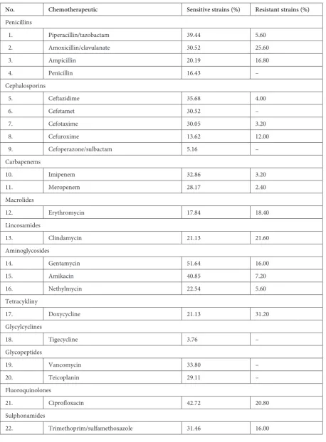
Data on the chemotherapeutic susceptibility of various strains is summarized in Table 2. An anal-ysis that was inclusive of all the pathogens revealed that the fraction of sensitive strains was the highest in the case of piperacillin/tazobactam, gentamycin,
and ciprofloxacin, while the lowest number of strains showed resistance to carbapenems, cefo-taxime, and piperacillin/tazobactam.
Taking into account the fraction of sensitive and resistant strains, a cluster analysis was con-ducted in order to significantly optimize empirical therapy. As a result, four principal groups of che-motherapeutics were identified (Fig. 5):
1) Nethylmycin, cefuroxime, trimethoprim/ sulfamethoxazole, clindamycin, erythromycin,
ampicillin – characterized by a relatively low per-centage of sensitivity and relatively high resistance rate; not recommended.
2) Doxycycline, amoxicillin/clavulanic acid – characterized by a relatively high percentage of sensitivity, but also high resistance rate; rather not recommended
3) Ciprofloxacin, gentamycin – antibiotics characterized by a high fraction of sensitivity and relatively low resistance rate; recommended.
Fig. 3. Monocyte and lym-phocyte count in individu-als with coagulase-negative staphylococcal infection compared to patients infected with other bacteria (mean, standard error, 95% CI)
4) Meropenem, cefotaxime, imipenem, cef-tazidime, amikacin, piperacillin/tazobactam – anti-biotics characterized by a high fraction of sensitive strains and extremely low percentage of resistant strains; definitely recommended.
The list of less and more recommended anti-biotic combinations (Table 3) was developed af-ter considering solely the in vitro efficacy, i.e. the percentage of strains sensitive to a given antibi-otic combination, estimated on the basis of the
Table 2. Sensitivity of all analyzed strains to various chemotherapeutics
No. Chemotherapeutic Sensitive strains (%) Resistant strains (%)
Penicillins
1. Piperacillin/tazobactam 39.44 5.60 2. Amoxicillin/clavulanate 30.52 25.60
3. Ampicillin 20.19 16.80
4. Penicillin 16.43 –
Cephalosporins
5. Ceftazidime 35.68 4.00
6. Cefetamet 30.52 –
7. Cefotaxime 30.05 3.20
8. Cefuroxime 13.62 12.00
9. Cefoperazone/sulbactam 5.16 – Carbapenems
10. Imipenem 32.86 3.20
11. Meropenem 28.17 2.40
Macrolides
12. Erythromycin 17.84 18.40
Lincosamides
13. Clindamycin 21.13 21.60
Aminoglycosides
14. Gentamycin 51.64 16.00
15. Amikacin 40.85 7.20
16. Nethylmycin 22.54 5.60
Tetracykliny
17. Doxycycline 21.13 31.20
Glycylcyclines
18. Tigecycline 3.76 –
Glycopeptides
19. Vancomycin 33.80 –
20. Teicoplanin 29.11 –
Fluoroquinolones
21. Ciprofloxacin 42.72 20.80
Sulphonamides
sensitivity to a single antibiotic determined by the disc method, and the loss, i.e. the percentage of strains sensitive to both antibiotics (in situations where application of one chemotherapeutic would be theoretically sufficient).
Discussion
A EURODIALE study revealed that bacteria can be recovered from 58% of newly-diagnosed cases of diabetes in which material was obtained from developing ulcerations [7]. The participa-tion of various etiological factors depends on geo-graphical latitude, as well as on the cultural and hygienic customs of a given region. Western coun-tries are predominated by Gram-positive bacte-ria, mostly Staphylococcus aureus and beta-he-molytic Streptococcus spp. [8, 9]. In contrast, the Gram-negative bacteria, mostly Pseudomonas ae-ruginosa and Escherichia coli, predominate in Eastern countries, particularly in India. The most prevalent Gram-positive bacteria observed in this region include Staphylococcus aureus and Entero-coccus spp. [10]. Taking the percentage distribu-tion of microorganisms into account, the situadistribu-tion in Poland approaches European trends. Howev-er, the high fraction of enterococci requires fur-ther research, mostly with regards to Enterococ-cus faecalis (16.8%), whose prevalence, together
with Enterococcus faecium, exceeds 18%, and thus is higher than in the case of Staphylococcus aureus
(slightly above 15%). During our search through the available literature, we did not find any report in which the prevalence of Enterococci would be higher than that of Staphylococcus aureus, which predominates amongst the Gram-positive micro-flora [10, 11]. There are several potential reasons behind such a high fraction of Enterococci, includ-ing severe dental caries [12], insufficient person-al hygiene, co-existing urinary tract infection, or contact with poultry/poultry meat [13]. Howev-er, the selection of strains resulting from empirical antibiotic therapy, started prior to hospitalization and based mostly on β-lactams, to which Entero-cocci show natural resistance, seems the most ra-tional explanation. About 30% of the enterococ-cal strains isolated from our patients were revealed as high level aminoglycoside resistant (HLAR); in contrast, there were no alarm strains resistant to glycopeptides (VRE – vancomycin-resistant En-terococcus). These values are lower than previous-ly presented in the available literature [14–17]. In most cases the material was obtained immediate-ly on admission to the clinic, and a relativeimmediate-ly low fraction of HLAR strains were recovered, both of which contradict the nosocomial source of entero-coccal infection.
The presented associations between the eti-ological factors and clinical data analyzed in this
No. Combination of antibiotics Efficacy (%) “Loss” (%)
β-lactam antibiotic and aminoglycoside
1. Amoxicillin/clavulanate + amikacin 61.5 9.9 2. Piperacillin/tazobactam + amikacin 52.6 27.7 3. Ceftazidime + amikacin 51.6 24.9 4. Cefetamet + amikacin 50.2 21.1 5. Cefotaxime + amikacin 50.1 20.2 6. Amoxicillin/clavulanate + gentamycin 73.7 8.5 7. Piperacillin/tazobactam + gentamycin 70.4 20.7 8. Ceftazidime + gentamycin 68.1 19.2 9. Cefetamet + gentamycin 64.3 17.8 10. Cefotaxime + gentamycin 66.2 15.5 β-lactam antibiotic and fluoroquinolone
11. Amoxicillin/clavulanate + ciprofloxacin 63.4 9.8 12. Piperacillin/tazobactam + ciprofloxacin 57.7 24.4 13. Ceftazidime + ciprofloxacin 53.4 24.4 14. Cefetamet + ciprofloxacin 51.2 22.1 15. Cefotaxime + ciprofloxacin 53.5 19.2 β-lactam antibiotic and macrolide
16. Amoxicillin/clavulanate + erythromycin 45.1 3.3 17. Piperacillin/tazobactam + erythromycin 57.2 0 18. Ceftazidime + erythromycin 53.5 0 19. Cefetamet + erythromycin 48.4 0 20. Cefotaxime + erythromycin 47.9 0 β-lactam antibiotic and lincosamide
21. Amoxicillin/clavulanate + clindamycin 44.6 7.0 22. Piperacillin/tazobactam + clindamycin 59.6 0.9 23. Ceftazidime + clindamycin 56.8 0 24. Cefetamet + clindamycin 51.2 0 25. Cefotaxime + clindamycin 51.6 0 Aminoglycoside and fluoroquinolone
26. Gentamycyna + ciprofloxacin 62.4% 31.9% 27. Amikacyna + ciprofloxacin 60.1% 23.5% Aminoglycoside and macrolide
28. Gentamycyna + erythromycin 56.9 12.7 29. Amikacyna + erythromycin 54.5 4.2 Aminoglycoside and lincosamide
30. Gentamycyna + clindamycin 59.6 13.1 31. Amikacyna + clindamycin 57.3 4.7 Macrolide and tetracycline
paper have purely practical applications, enabling the selection of a “better” protocol of antibiotic therapy when the results of a culture are unavail-able. Although we did not obtain sufficiently high sensitivity of the analyzed parameters, their high specificity and acceptable likelihood ratio (LR) are worth mentioning [18], particularly with regards to a ≥ 12.6% fraction of glycated hemoglobin in individuals with E. faecalis infection (LR = 9.23), lymphocytosis ≥ 3.11 G/L in the course of S. au-reus infection (LR = 10), and monocytosis ≥ 2 G/L in P. mirabilis infection (LR = 5.43). The complex and variable interaction between a given etiologi-cal factor and the host’s immune system, partic-ularly in diabetic patients, can be responsible for these clinically important findings [19, 20].
In view of the fact that even up to 34% of pa-tients with ulceration associated with DFS require amputation [5], early, appropriate antibiotic ther-apy plays an important role, aside from ratio-nal insulin therapy, potential revascularization, decompression of the foot, and local treatment. Therefore, we analyzed the antibiotic susceptibil-ity for all microorganisms isolated from our ma-terial rather than for individual strains. Such an approach is clinically justifiable since the empiri-cal antibiotic therapy should be started before the
etiological factors can be identified based on the culture results. Antibiotics that proved to be partic-ularly valuable in the monotherapy of our patients included ciprofloxacin, aminoglycosides (typical-ly not implemented as monotherapy), amoxicil-lin/clavulanate, and piperacillin/tazobactam; how-ever, it should be emphasized that the efficacy of any single chemotherapeutic did not exceed 43% of the cases. Therefore, it is quite obvious that combined therapy should be implemented at the very beginning of severe infection. Our analysis re-vealed that in such cases a combination of amox-icillin/clavulanate or piperacillin/tazobactam with aminoglycosides is most efficient; this is generally consistent with the recommendations of PDA and monographs dealing with antibiotic therapy [21]. Of note are the small benefits associated with com-bining ciprofloxacin with chemotherapeutics from other groups.
The fact that the data presented originates from a single center is unambiguously the princi-pal limitation of this study. Nevertheless, it is like-ly that the significance of at least some of the hereby identified problems, such as the high prevalence of
E. faecalis infection, laboratory abnormalities specif-ic to certain types of infection, and analysis of drug susceptibility spreads beyond the Wrocław center.
References
[1] IDF Atlas: http://www.idf.org/atlasmap/atlasmap, 2011.
[2] Polskie Towarzystwo Diabetologiczne: Zalecenia kliniczne dotyczące postępowania u chorych na cukrzycę 2011. Diabet Prakt 2011, 5 (supl. A), A33.
[3] Korzon-Burakowska A: Zespół stopy cukrzycowej – patogeneza i praktyczne sposoby postępowania. Choroby serca i naczyń 2007, 4, 93–98.
[4] Shah BR, Hux JE: Quantifying the risk of infectious diseases for people with diabetes. Diabetes Care 2003, 26, 510–513.
[5] Yekta Z, Pourali R, Nezhadrahim R, Ravanyar L, Ghasemi-Rad M: Clinical and behavioral factors associated with management outcome in hospitalized patients with diabetic foot ulcer. Diabetes Metab Syndr Obes 2011, 4, 371–375.
[6] Rozporządzenie Ministra Zdrowia z dnia 23 grudnia 2011 r. w sprawie listy czynników alarmowych, rejestrów zakażeń szpitalnych i czynników alarmowych oraz raportów o bieżącej sytuacji epidemiologicznej szpitala. Dziennik Ustaw 2011, 294, 17195–17205.
[7] Prompers L, Huijberts M, Apelqvist J: High prevalence of ischaemia, infection and serious comorbidity in patients with diabetic foot disease in Europe. Baseline results from the Eurodiale study. Diabetologia 2007, 50, 18–25. [8] Dang CN, Prasad YD, Boulton AJ, Jude EB: Methicillin resistant Staphylococcus aureus in the diabetic foot clinic:
a worsening problem. Diabet Med 2003, 20, 159–161.
[9] Citron DM, Goldstein EJC, Merriam CV, Lipsky BA, Abramson MA: Bacteriology of moderate to severe diabetic foot infections and in vitro activity of antimicrobial agents. J Clin Microbiol 2007, 45, 2819–2828.
[10] Ramakant P, Verma AK, Misra R: Changing microbiological profile of pathogenic bacteria in diabetic foot infec-tions: time for a rethink on which empirical therapy to choose? Diabetologia 2011, 54, 58–64.
[11] Crouzet J, Lavigne JP, Richard JL: Diabetic foot infection: a critical review of recent randomized clinical trials on antibiotic therapy. Int J Infect Dis 2011, 15, e601–e610.
[12] Sebeena M, Boopathy T: Enterococcus faecalis – an endodontic challenge. J Indian Acad Dent Spec 2010, 1, 46–48.
[13] Poulsen LL, Bisgaard M, Son NT, Trung NV, An HM, Dalsgaard A: Enterococcus faecalis Clones in Poultry and in Humans with Urinary Tract Infections, Vietnam. Emerg Infect Dis 2012, 18, 1096–1100.
[15] Vinodkumar CS, Srinivasa H, Basavarajappa KG, Geethalakshmi S, Bandekar N: Isolation of bacteriophages to multi-drug resistant Enterococci obtained from diabetic foot: A novel antimicrobial agent waiting in the shelf? Indian J Pathol Microbiol 2011, 54, 90–95.
[16] Mendiratta DK, Kaur H, Deotale V, Thamke DC, Narang R, Narang P: Status of high level aminoglycoside resistant Enterococcus faecium and Enterococcus faecalis in a rural hospital of central India. Indian J Med Microbiol 2008, 26, 369–371.
[17] Rudy M, Zientara M, Bek T, Martirosian G: Occurrence of Antibiotic Resistant Enterococci in Clinical Specimens from a Pediatric Hospital. Pol J Microbiol 2005, 54, 77–80.
[18] Bożek J, Jaeschke R, Leśniak W: Ocena informacji o metodzie diagnostycznej. In: Podstawy EBM, czyli medycyny opartej na danych naukowych dla lekarzy i studentów medycyny. Eds.: Gajewski P, Jaeschke R, Brożek J. Med Prakt, Kraków 2008, 1st ed., 102–104.
[19] Sava IG, Heikens E, Huebner J: Pathogenesis and immunity in enterococcal infections. Clin Microbiol Infect 2010, 16, 533–540.
[20] Takahashi H, Tsuda Y, Takeuchi D, Kobayashi M, Herndon DN, Suzuki F: Influence of systemic inflammatory response syndrome on host resistance against bacterial infections. Crit Care Med 2004, 32, 1879–1885.
[21] Dzierżanowska D, Dzierżanowska-Fangrat K: Przewodnik antybiotykoterapii 2011. Alfa-Medica Press, Bielsko- -Biała 2011.
Address for correspondence:
Rafał MałeckiDepartment of Angiology, Systemic Hypertension and Diabetology Wroclaw Medical University
Borowska 213 50–556 Wrocław Tel.: 71 733 22 00
E-mail: [email protected]
Conflict of interest: None declared