Mustafa Said Aydogan
1, A, C, D, Mehmet Ali Erdogan
1, D, E, Aladdin Polat
2, B,
Aytac Yücel
1, E, Ulkü Ozgül
1, E, Hakan Parlakpinar
3, B, Zeynep Rumeysa Duran
2, B,
Azibe Yildiz
4, C, Mahmut Durmus D.
1Protective Effects of Melatonin and β-d-Glucan Against
Liver Injury in Rats – a Comparative Study
Działanie ochronne melatoniny i β-d-glukanu
w uszkodzeniach wątroby u szczurów – studium porównawcze
1 Department of Anesthesiology and Reanimation 2 Department of Physiology
3 Department of Pharmacology
4 Department of Histology, Inonu University, Faculty of Medicine, Malatya, Turkey
A – research concept and design; B – collection and/or assembly of data; C – data analysis and interpretation; D – writing the article; E – critical revision of the article; F – final approval of article; G – other
Objectives. The aim of this study was to investigate the possible protective effects of melatonin and β-d-glucan against ischemia-reperfusion (IR) injury in rats.
Materials and Methods. Forty rats were randomly divided into 5 groups, each consisting of 8 animals, as follows. Sham group [S], IR group [C], IR + β-Glucan group [β], IR + melatonin group [MLT], IR + melatonin + β-Glucan group [MLT + β]. The rats in the C, β, MLT and MLT + β groups were subjected to IR for 60 min each. Melatonin (10 mg∙kg–1) was intraperitoneally injected for a single dose 30 min before IR. β-Glucan (50 mg∙kg–1∙day–1) was
orally administered for 10 days to rats. All of the rats were killed on day 11, and histological changes in the liver and tissue levels of oxidants and antioxidants were evaluated.
Results. Malondialdehyde [MDA] level were significantly higher in the C group compared to the S group (p = 0.007). MDA level were significantly higher in the β group compared to the MLT and MLT + β groups (p =0.007). Tissue antioxidant markers (superoxide dismut ase [SOD], glutathione-peroxidase [GPx], and catalase [CAT]) were sig-nificantly lower in the C group than the S group (p < 0.05). SOD levels were simply not significant in the β group compared to the MLT and MLT + β groups. CAT and GPx activities were significantly higher in the β group compared to the MLT and MLT + β groups (p = 0.004).The histological damage ameliorated in β, MLT and MLT + β groups compared to C group.
Conclusion. Our results suggest that melatonin and β-glucan combination pretreatment suppressed oxidative stress and increased antioxidant levels in an experimental rat model of liver IR injury (Adv Clin Exp Med 2013, 22, 5, 621–627).
Key words: liver, ischemia reperfusion injury, melatonin, β-glucan, oxidative stress.
Słowa kluczowe: wątroba, szkodzenie niedokrwienno-reperfuzyjne, melatonina, β-glukan, stres oksydacyjny.
Adv Clin Exp Med 2013, 22, 5, 621–627 ISSN 1899–5276
ORIGINAL PAPERS
© Copyright by Wroclaw Medical University
Liver ischemia-reperfusion injury (IRI) oc-curs during transplantation and major hepatic surgery [1]. IRI may contribute to postoperative liver dysfunction and negatively affect graft func-tion in liver transplant patients following vascular
clamping [2]. Antioxidant enzymes such as super-oxide dismutase (SOD), catalase (CAT), and glu-tathione peroxidase (GPx) are protective against IRI. For these reasons, treatment with exogenous antioxidants, particularly in the early stages of
reperfusion, markedly reduces the severity of liv-er IRI [3].
β-glucans have shown beneficial effects, in-cluding anti-inflammatory, antioxidant properties
and enhanced immune response [4, 5].
Melato-nin (N-acetyl-5-methoxytryptamine) is an indole-amine produced by the pineal gland in a circadian rhythm, and it is one of the most powerful stimula-tion of antioxidative enzymes, including glutathi-one reductase and superoxide dismutase [6, 7].
Liver IRI is a persistent problem that can-not be prevented during liver surgery and trans-plant [8, 9]. Multi pharmacologic preconditioning is a new strategy to reduce IRI. Because different pathways in various cell types are involved in I/R-injury, it is conceivable to use multiple drugs to prevent IRI [10]. In the present study, we aimed to investigate if a combination of β-d-glucan and melatoninwould be better than drugs used alone against liver IRI in rats by using biochemical and histological analyses for the evaluation of the ex-tent of oxidative damage.
Material and Methods
Animals
The authors obtained 40 Spraque-Dawley male rats weighing 210–240 g from Inonu Univer-sity Laboratory Animals Research Center, Malatya, Turkey. The rats were kept in a room that was 21 ± 2°C with relative humidity of 60% ± 5% and a 12-h light/dark cycle. The animals were housed in plastic cages (50 × 35 × 20 cm, 8 animals per cage). Experiments were carried out according to the standards of animal research issued by the Nation-al HeNation-alth Research Institute and with the approvNation-al of the Inonu University Ethical Committee.
Experimental Design
Rats were randomly divided into 5 groups, each consisting of 8 animals, as follows. (1) Sham group [S]: sham-operated animals only a mid-line laparotomy was performed, and the abdomen
was closed without any further procedure. (2) IR
group [C]: animals were subjected to 60 min of ischemia followed by 60 min of reperfusion. (3)
IR + β-Glucan group [β]: animals received oral β-glucan and were subjected to 60 min of ischemia
followed by 60 min of reperfusion. (4) IR +
mela-tonin group [MLT]: animals received i.p. melato-nin and were subjected to 60 min of ischemia fol-lowed by 60 min of reperfusion. (5) IR + melatonin + β-Glucan group [MLT + β]: animals received i.p.
melatonin and oral β-glucan and were subject-ed to 60 min of ischemia followsubject-ed by 60 min of reperfusion.
Glucan Administration
The micro particulate form of β-glucan
was prepared from Saccharomyces cerevisiae
yeast (Mustafa Nevzat Drug Co., Turkey). β-d- -Glucan was suspended in saline and a dose of 50 mg/kg/day was administered by intragastric gavage for 10 days prior to, and 30 min after, IR procedure [11].
Melatonin Administration
Melatonin was obtained from Sigma Chemi-cal, St Louis, MO, USA. It was dissolved in etha-nol and diluted in saline to a final concentration of 5% ethanol. A single dose of melatonin (10 mg/kg, i.p.) was injected 30 min before IR procedure. The authors determined the melatonin dose on the ba-sis of the results from recent studies [12].
Surgical Procedure
Determination
of Enzyme Activities
Determination of Superoxide
Dismutase Activity
Total SOD activity was determined according to the method of Sun et al. [13]. The principle of the method is the inhibition of nitroblue tetrazoli-um (NBT) reduction by the xanthine–xanthine ox-idase system as a superoxide generator. One unit of SOD was defined as the enzyme amount caus-ing 50% inhibition in the NBT reduction rate. SOD activity was given as units per gram protein (U/g protein).
Determination of Catalase Activity
Catalase activity was determined according to Aebi’s method [14]. The principle of the as-say is based on the determination of the rate con-stant (k, s−1) or the H2O2 decomposition rate at
240 nm. Results are given as k per gram protein (k/g protein).
Determination of Glutathione
Peroxidase Activity
Glutathione peroxidase activity was measured by the Paglia and Valentine method [15]. An en-zymatic reaction in a tube containing NADPH, re-duced glutathione (GSH), sodium azide, and gluta-thione reductase, was initiated by addition of H2O2,
and the change in absorbance at 340 nm was ob-served by a spectrophotometer. Activity was given in units per gram protein (U/g protein).
Determination
of Malondialdehyde Activity
The MDA contents of homogenates were de-termined spectrophotometrically [16], by measur-ing the presence of thiobarbituric acid reactive sub-stances. 3 mL of 1% phosphoric acid and 1 mL 0.6% thiobarbituric acid solution were added to 0.5 mL of
homogenate pipetted into a tube. The mixture was heated in boiling water for 45 min. After the mix-ture had cooled, the colored part was extracted into 4 mL of n-butanol. The absorbance was measured by a spectrophotometer (UV-1601; Shimadzu, Kyo-to, Japan) at 532 and 520 nm. The amount of lipid peroxides was calculated as thiobarbituric acid re-active substances of lipid peroxidation. The results were given in nanomoles per gram tissue (nmol/g tissue) according to a prepared standard graph.
Determination of Protein Content
Protein contents were measured according to the method of Lowry et al. [17].
Histological Analysis
Liver tissues fixed in 10% formaldehyde solu-tion were embedded in paraffin. The 4-μm secsolu-tions were stained with hematoxylin-eosin (H-E). Score of liver damage severity was semi-quantitatively assessed as follows: disruption in radial arrange-ment around central vein, sinusoidal dilatation, congestion, intracellular vacuolization and nucle-ar changes. The sections were examined by a his-tologist who was unaware of the treatment group by using a Leica DFC 280 light microscope and the Leica QWin Plus analysis system (Leica Micros Imaging Solution Ltd., Cambridge, UK).
Statistical Analysis
The data is expressed as median (min–max). Normal distribution was confirmed using the Sha-piro-Wilk test. Statistical analyses were performed using the Kruskal-Wallis H test or two-tailed un-paired Student’s t-test with SPSS for Windows (SPSS Inc., Chicago, IL, United States), as appro-priate. If the results of the Kruskal-Wallis H test were significant, multiple comparisons were evalu-ated by the Conover test. A difference was consid-ered significant at p < 0.05.
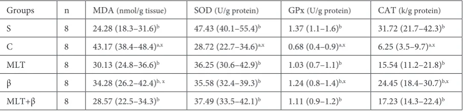
Table 1. Enzyme activities in liver tissue
Groups n MDA (nmol/g tissue) SOD (U/g protein) GPx (U/g protein) CAT (k/g protein)
S 8 24.28 (18.3–31.6)b 47.43 (40.1–55.4)b 1.37 (1.1–1.6)b 31.72 (21.7–42.3)b
C 8 43.17 (38.4–48.4)a,x 28.72 (22.7–34.6)a,x 0.68 (0.4–0.9)a,x 6.25 (3.5–9.7)a,x
MLT 8 30.13 (24.8–36.6)b 36.25 (30.6–42.9)b 1.03 (0.7–1.1)b 15.54 (11.2–21.8)b
β 8 34.28 (26.2–42.4)b, x 35.58 (32.4–39.3)b 1.24 (0.8–1.4)b,x 24.45 (18.4–30.7)b,x
MLT+β 8 28.57 (22.5–34.3)b 37.49 (33.5–42.1)b 1.11 (0.9–1.2)b 17.23 (14.3–22.4)b
The data were presented as median (min–max).S – sham; C – ischemia/reperfusion; MLT – ischemia/reperfusion + Melatonin intraperitoneal; β – ischemia/reperfusion + β-glucan orally; MLT + β – ischemia/reperfusion + Melatonin + β-glucan; MDA – malondialdehyde; SOD – superoxide dismutase; GPx – glutathione peroxidase; CAT = catalase; k = rate constant (k, s−1).
Results
As shown in table 1, MDA level were significant-ly higher in the C group compared to the S group (p = 0.007). MDA level were significantly higher in the β group compared to the MLT and MLT + β groups (p = 0.007). Tissue antioxidant markers
[SOD, GPx and CAT] were significantly lower in the C group than the S group (p < 0.05). SOD levels were not significantly in the β group compared to the MLT and MLT + β groups. CAT and GPx activities were significantly higher in the β group compared to the MLT and MLT + β groups (p = 0.004).
cv
B A
cv
D C
cv
cv
F E
cv
Histological Changes
The histological characteristics of all groups are given in Figure 1A–F. The liver tissue in the S group showed a normal histological appearance, except light degenerative changes such as minimal sinusoi-dal dilatation (Fig. 1A). However, in the C group, si-nusoidal dilatation, congestion, hemorrage were ob-served when compared with regular S group (Fig. 1B and 1C). On the other hand, the histological damage ameliorated in β, MLT and MLT + β groups com-pared to C group (Fig. 1D, 1E and 1F).
Discussion
To the best of our knowledge, the effect of MLT + β combination on the liver IRI has not been pre-viously reported. Also, these results indicate that administration MLT + β have a combined stronger effect than MLT or β-glucanalone in reducing lip-id peroxlip-idation, which was apparent from the bio-chemical and histological findings.
IR is an inevitable event during liver surgery, including transplantation and tumor resection [18]. Because IRI is a complex process involving numer-ous intracellular signaling pathways, mediators, cells, and pathophysiological disturbances [19], the extent of IR-associated liver injury is a major factor directly affecting graft survival and post-transplan-tation function. During transplanpost-transplan-tation, both cold and warm ischemia occur. However, the authors used warm ischemia in our surgical protocol.
Many investigators have reported that lipid per-oxidation is thought to be closely associated with liver IRI pathogenesis [20]. MDA is a by-product of oxidant-induced lipid peroxidation and protein oxidation [21]. In the present study, we observed that MDA levels were increased after IRI. However, MDA levels almost returned to control levels with β-glucan and melatonin combination treatment.
GSH is a necessary component of the antioxi-dant system and cellular GSH is important for the maintenance of cellular redox states through the direct scavenging of radical species or participation in reactions catalyzed by antioxidant enzymes, such as GPx [22, 23]. In the present study, there was low-er GPx activity in rat livlow-ers by IRI than in livlow-ers that were not IRI. Pre-treatment with melatonin or β-d-glucan reduced the damage caused by IRI by reduc-ing oxidative stress and increasreduc-ing antioxidant ac-tivity. Because melatonin or β-d-glucan are known free-radical scavengers [8, 24] the idea that free radicals are involved in the pathogenesis of IRI is supported. Melatonin is a strong direct free radical scavenger and an indirect antioxidant through the induction of antioxidant enzymes [25, 26].
GPx further catalyzes the transformation of hydrogen peroxide to form water [27]. CAT is an antioxidant defense enzyme and a potent H2O2
scavenger. It prevents the formation of highly tox-ic hydroxyl radtox-icals when SOD is insufftox-icient to neutralize ROS [28]. Thus, the production of CAT provides additional antioxidative activity against oxidative stress during IR. CAT has been shown to have beneficial effects toward the end of the isch-emic period [29, 30]. The delivery of CAT protein successfully prevented liver IRI in mice [31]. The authors found that β-glucan pretreatment signifi-cantly increased CAT and GPx activities compared to the MLT + β G group, and this may explain the observed ROS decrease.
SOD has been widely studied among the en-dogenous antioxidant. SOD catalyzes the dysmu-tation of superoxide anion to hydrogen peroxide and oxygen, but hydrogen peroxide still produc-es liver oxidative injury. In many different reports, however, SOD activity was not affected, suggesting that this enzyme is a less sensitive predictor of oxi-dative stress [32, 33]. The authors found that MLT or β-glucan pretreatment were not significant-ly increased SOD activity compared to the MLT + β G group and this may result from MLT and β G combination treatment indirectly reducing ox-idative damage.
The histological investigation suggested that IR caused severe pathological alterations in the liver, including edema, vascular congestion, hem-orrhage, and leukocyte infiltration. Pretreatment with β, MLT and MLT + β combination ameliorat-ed the IR-inducameliorat-ed histological changes that were attributed to its antioxidant efficacy. As a result, we may explain these results due to the antioxidant effect of β-glucan and melatonin.
The limitations of the study were explained as follow: Firstly, the sample size used in this study was small. Secondly, we did not evaluate β-glucan and melatonin doses or longer time-dependent response to IRI. However, we established that β-glucan and melatonin attenuates IRI even if it is incomplete protection.
References
[1] Clavien PA, Selzner M, Rüdiger HA, Graf R, Kadry Z, Rousson V, Jochum W: A prospective randomized study in 100 consecutive patients undergoing major liver resection with versus without ischemic preconditioning. Ann Surg. 2003, 238, 843–852.
[2] Dogan S, Aslan M: Hepatic ischemia-reperfusion injury and therapeutic strategies to alleviate cellular damage. Hepatol Res 2011, 41, 103–117.
[3] Wang J, Kan Q, Li J, Zhang X, Qi Y: Effect of neferine on liverischemia-reperfusioninjury in rats. Transplant Proc 2011, 43, 2536–2539.
[4] Toklu HZ, Sener G, Jahovic N, Uslu B, Arbak S, Yegen BC: Beta-glucan protects against burn-induced oxidative organ damage in rats. IntImmunopharmacol 2006, 6, 156–169.
[5] Babincova M, Bacova Z, Machova E, Kogan G: Antioxidant properties of carboxymethylglucan: comparative analysis. J Med Food 2002, 5, 79–83.
[6] Kang JW, Koh EJ, Lee SM: Melatonin protects liver against ischemia and reperfusion injury through inhibition of toll-like receptor signaling pathway. J Pineal Res 2011, 50, 403–411.
[7] Rodriguez C, Mayo JC, Sainz RM, Antolín I, Herrera F, Martín V, Reiter RJ: Regulation of antioxidant enzymes: a significant role for melatonin. J Pineal Res 2004, 36, 1–9.
[8] Klune JR, Tsung A: Molecular biology of liver ischemia/repefusion injury: established mechanisms and recent advancements. Surg Clin North Am 2010, 90, 665–677.
[9] Czubkowski P, Socha P, Pawlowska J: Current status of oxidative stress in pediatric liver transplantation. Pediatr Transplant 2010, 14, 169–177.
[10] Schindler G, Kincius M, Liang R, Backhaus J, Zorn M, Flechtenmacher C, Gebhard MM, Büchler MW, Schemmer P: Fundamental efforts toward the development of a therapeutic cocktail with a manifold ameliorative effect on hepatic ischemia/reperfusion injury. Microcirculation 2009, 22, 1–10.
[11] Toklu HZ, Sehirli AO, Velioğlu Oğünç A, Cetinel S, Sener G: Acetaminophen-induced toxicity is prevented by beta-d-glucan treatment in mice. Eur J Pharmacol 2006, 14, 133–140.
[12] Sener G, Sehirli AO, Ayanoglu Dülger G: Protective effects of melatonin, vitamin E and N-acetylcysteine against acetaminophen toxicity in mice: a comparative study. J Pineal Res 2003, 35, 61–68.
[13] Sun Y, Oberley L, Li Y: A simple method for clinical assay of superoxide dismutase. Clin Chem 1988, 34, 497–500. [14] Aebi H: Catalase. [In:] Methods of enzymatic analysis. Ed.: Bergmeyer HU. Academic Press, New York 1974.
673–677.
[15] Paglia DE, Valentine WN: Studies on the quantitative and qualitative characterization of erythrocyte glutathione peroxidase. J Lab Clin Med 1967, 70, 158–170.
[16] Mihara M, Uchiyama M: Determination of malonaldehyde precursor in tissues by tiobarbituric acid test. Anal Biochem 1978, 86, 271–278.
[17] Lowry O, Rosenbraugh N, Farr L, Rondall R: Protein measurement with the folin-phenol reagent. J Biol Chem 1951, 193, 265–275.
[18] Hasselgren PO: Prevention and treatment of ischemia of the liver. Surg Gynecol Obstet 1987, 164, 187–196. [19] Sakon M, Ariyoshi H, Umeshita K, Monden M: Ischemia reperfusion injury of the liver with special reference to
calcium dependent mechanisms. Surg Today 2002, 32, 1–12.
[20] Dalle-Donne I, Rossi R, Colombo R, Giustarini D, Milzani A: Biomarkers of oxidative damage in human disease. Clin Chem 2006, 52, 601–623.
[21] García JJ, Reiter RJ, Guerrero JM, Escames G, Yu BP, Oh CS, Muñoz-Hoyos A:Melatonin prevents changes in microsomal membrane fluidity during induced lipid peroxidation. Febs Lett 1997, 408, 297–300
[22] Budziński G, Suszka-Świtek A, Caban A, Oczkowicz G, Heitzman M, Wystrychowski W, Dolińska B, Ryszka F, Cierpka L: Evaluation of cysteine effect on redox potential of porcine liver preserved by simple hypothermia. Transplant Proc. 2011, 43, 2897–2899.
[23] Deleve LD, Kaplowitz N: Glutathione metabolism and its role in hepatotoxicity. Pharmacol Ther 1991, 52, 287–305. [24] Noyan T, Kömüroğlu U, Bayram I, Sekeroğlu MR: Comparison of the effects of melatonin and pentoxifylline on
carbon tetrachloride-induced liver toxicity in mice. Cell Biol Toxicol 2006, 22, 381–391.
[25] Reiter RJ, Tan DX, Mayo JC, Sainz RM, Leon J, Czarnocki Z: Melatonin as an antioxidant: biochemical mecha-nisms and pathophysiological implications in humans. Acta Biochim Pol 2003, 50, 1129–1146.
[26] Reiter JR, Tan DX, Sainz RM, Mayo JC, Lopez-Burillo S: Melatonin: in reducing the toxicity and increasing the efficacy of drugs. J Pharm Pharmacol 2002, 54, 1299–1321.
[27] Galecka E, Jacewicz R, Mrowicka M, Florkowski A, Gałecki P: Antioxidative enzymes-structure, properties, func-tions. Pol Merk Lek 2008, 25, 266–268.
[28] Yabe Y, Kobayashi N, Nishihashi T, Takahashi R, Nishikawa M, Takakura Y, Hashida M: Prevention of neutro-phil – mediated hepatic ischemia/reperfusion injury by superoxide dismutase and catalase derivatives. J Pharmacol Exp Ther 2001, 298, 894–899.
[29] Koolj A, Schiller HJ, Schlins M, Van Noorden CJ, Frederiks WM: Conversion of xanthine dehydrogenase into xan-thine oxidase in rat liver and plasma at the onset of reperfusion after ischemia. Hepatology 1994, 19, 1488–1495. [30] McKillen MN: Respiratory burst oxidase of the neutrophil. Biochem Soc Trans 1990, 19, 49–78.
[32] Ozansoy G, Akin B, Aktan F, Karasu C: Short-term gemfibrozil treatment reverses lipid profile and peroxidation but does not alter blood glucose and tissue antioxidant enzymes in chronically diabetic rats. Mol Cell Biochem 2001, 216, 59–63.
[33] Ceylan A, Karasu C, Aktan F, Güven C, Can B, Ozansoy G: Effects of simvastatin treatment on oxidant/antioxi-dant state and ultrastructure of diabetic rat myocardium. Gen Physiol Biophys 2003, 22, 535–547.
Address to correspondence:
Mustafa Said AydoganInonu University School of Medicine
Department of Anesthesiology and Reanimation Malatya
Turkey
Tel: +90 422 341 06 60 31 60 Fax: +90 422 341 07 28
E-mail: [email protected]
Conflict of interest: None declared