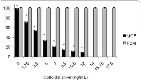
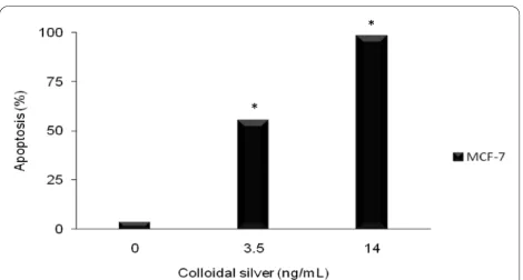
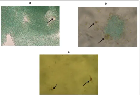
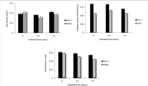
Antitumor activity of colloidal silver on MCF-7 human breast cancer cells
Full text
Figure
Related documents
De placer des mines automatiques de contact amarrees qui ne deviennent pas inoffensives des qu'elles auront rompu leurs amarres.- Institut, 1913 .... MEANS AND METHODS OF
REGULATIONS RELATING TO FOREIGN VESSELS OF WAR IN 'VATERS UNDER THE ,JURISDICTION OF THE UNITED STATES. In time of peace. In general, foreign vessels of 'var need
Cultural Assessment: A Study of Midwives’ Knowledge, Attitude and Self-reported Practice in Uganda.. Journal of
oracle (depending on the attack model atk ). As a result, it outputs some state information s for its second phase. Then a fair coin is tossed and depending on the result, either
IVSd: Interventricular septum thickness at end diastole; IVSs: Interventricular septum thickness at end systole; LA: Left atrium; LAV: Left atrial volume; LAVI: Left atrial
Specif- ically, the number of references pertaining to chiropractic geriatric education has increased from 3 to 11, the number of demographic and epidemiological studies has
As the signal-to-excess noise ratio will be independent o f received optical power level (for constant source conditions), this noise term puts a maximum limit on system SNR (and
However, it was found that this type of resin has a large amount of free formaldehyde (about 20%), which is unacceptable from environmental and human exposure point
