doi:10.4236/nm.2011.22019 Published Online June 2011 (http://www.SciRP.org/journal/nm)
Fas Receptor Modulates Lineage Commitment and
Stemness of Mouse Neural Stem Cells
Julia Knight1, Charles Hackett2, John Soltys2, Yang Mao-Draayer2
1Neuroscience Graduate Program, University of Vermont Burlington, USA; 2Neurology Department, Fletcher Allen Health Care, Burlington, USA.
Email: [email protected]
Received February 1st, 2011; revised February 25th, 2011; accepted March 15th, 2011.
ABSTRACT
Although transplanted neural stem/progenitor cells (NPCs) can ameliorate disease course in animal models of central nervous system inflammatory and neurodegenerative diseases, little is known about the regulation of NPC differentia-tion and proliferadifferentia-tion. The Fas receptor, a member of the tumor necrosis factor (TNF) superfamily, has recently been shown to be important in NPC survival and immunoregulatory functions. We were interested in further investigating this system utilizing NPCs isolated from Fas-deficient (lpr) mutant mice. We found that lpr NPCs have increased sur-vival and decreased proliferation. Additionally, RT-qPCR, confocal microscopy, and flow cytometry surface staining reveal that lpr NPCs have a significantly more robust differentiation to neuronal and oligoprogenitor cell lineages as compared to wild-type (wt) NPCs. These effects correlated with an upregulation of three of the major fate specification modulators in lpr NPCs: sonic hedgehog (Shh), slit homolog 2(Slit2), and noggin. These data indicate Fas plays an important role in determining the stemness and differentiation fate of NPCs. Additionally, our research reveals a novel connection between Fas and major modulators of NPC differentiation—Shh, Noggin, and Slit2. This is the first indica-tion of a possible link between Fas and these particular signaling molecules that control neuronal fate specificaindica-tion. Therefore, our results suggest Fas is a novel target for controlling the development of neurons versus mature oligoden-drocytes.
Keywords: Fas, Neural Stem/Progenitor Cell, Differentiation
1. Introduction
There is a critical need for developing direct neuroregen- erative therapies for demyelinating diseases, including multiple sclerosis (MS). Currently MS treatments are limited to peripheral immunoregulation and are ineffec- tive in later, progressively neurodegenerative stages of the disease. Neural stem/progenitor cell (NPC) therapies offer a potentially powerful treatment approach for the chronic neurodegeneration that occurs in these later stages [1]. The two main experimental modalities for NPC therapy are exogenous transplantation and activa- tion/recruitment of the endogenous adult NPC compart- ment [2]. In either method, differentiation and integration of NPCs to enhance central nervous system (CNS) repair is the most sought after outcome. Though exogenous application of NPC has shown significant recovery in animal models of MS (experimental autoimmune ence- phalomyelitis), the functional integration and terminal differentiation of transplanted cells is extremely rare [1,
3]. The signaling pathways that control NPC survival and differentiation are not well understood; therefore, eluci- dating these complex pathways is paramount for ad- vancing this therapeutic approach.
The Fas receptor (hereafter referred to as “Fas”)—a member of the TNF receptor superfamily—has emerged as a major modulator of NPC biology. Previous knowl-edge limited this receptor to cell-death related roles: in the immune system controlling homeostasis by inducing T-cell death [4], and in the CNS mediating programmed death during embryologic development [5,6]. However, Fas is now recognized as a mediator of diverse, cell-de- pendent mechanisms. Namely, activation of Fas via its cognant ligand (FasL) promotes survival in NPCs [7,8]. Furthermore, Fas activation induces neurogenesis and neurite outgrowth [8,9]. These recent findings suggest that Fas plays a complex role in mediating NPC fate, and modulation of this pathway may be a crucial target for future therapies.
of Fas-deficient (lpr) mutant mice to further investigate the role of Fas in mediating NPC fate. Here we report that lpr NPCs exhibit increased survival, increased neu- ronal specification, and higher populations of oligopro- genitor cells (OPCs) compared to wild type (wt) NPCs. Consistent with a more committed progenitor phenotype,
lpr NPCs show deficiency in mitogenic capacity. We have identified the signaling molecules sonic hedgehog (Shh), Slit2, Hes1, and Noggin as probable mediators of these phenomena.
2. Results
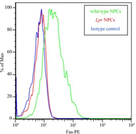
2.1. Lpr NPCs do not Express Fas
In order to confirm that lpr NPCs lack Fas, we stained NPCs with a Fas-specific phycoerythrin (PE)-conjugated antibody (or isotype control antibody). Flow cytometric analysis demonstrates high levels of Fas surface expres-sion in wt NPCs, and no Fas expresexpres-sion in lpr NPCs (Figure 1).
2.2. Fas-Deficient NPCs Have Stunted
Proliferation and Neurosphere Formation
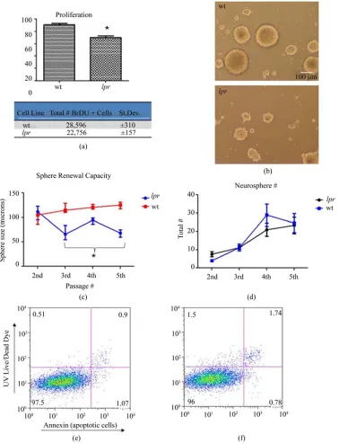
Proliferation in response to mitogens and the ability to self-renew are defining properties of NPCs. All prolifera-tion experiments were performed in the presence of epi-dermal growth factor (EGF) and fibroblast growth factor (FGF). A bromodeoxyuridine (BrDU) assay was used to determine the effects of Fas deficency on NPC prolifera-tion. Lpr NPCs showed significantly less BrDU incorpo-ration over 16vhours in complete media compared to wt NPCs. Lpr NPCs had 18% ± 1.6 less proliferation com-pared to wt (Figure 2(a)).
Additionally, neurosphere growth in lpr NPCs was abrogated compared to wt NPCs (Figure 2(b)). We per-formed the neurosphere assay to quantify this difference. In this assay, size of the neurosphere is an indicator of mitogenic or proliferative capacity, while neurosphere number is an estimate of the absolute total number of putative stem cells [10]. Over three subsequent passages,
lpr NPCs developed significantly smaller neurospheres than wt (Figure 2(c)). While, the total number of neuro-spheres remained the same (Figure 2(d)).
To be certain the abrogated proliferative capacity of
[image:2.595.311.536.87.308.2]lpr NPCs was not due to altered apoptosis profiles, we also performed UV Live-Dead dye (labeling dead cells) and annexin (labeling early apoptotic cells) double-stai- ning on NPCs growing in the presence of growth factors (EGF and FGF, similar to proliferation experiments). Four days post-cell plating, there was no difference in the amount of dead and dying cells in lpr and wt cultures (Figures 2(e) and (f)). Quantification of the data showed no significant difference between the percentage of live/
Figure 1. Lpr NPCs lacks Fas. Lpr mice have a large inser-tion in the Fas gene which results in improper splicing and absence of the protein. We confirmed absence of Fas in lpr
NPC using a phycoerythrin (PE)-conjugated Fas anti-body. Background staining was assessed using an isotype control IgG antibody (blue line). Unlike wild-type NPCs (green line), lpr NPCs (red line) do not express Fas (+peak completely overlaps with isotype control). Y-axis represents % of maximum staining intensity (arbitray units) while the X-axis represents the voltage of positive PE flurophore staining.
healthy cells in lpr (97% ± 2.3) and wt (95% ± 2.6) cells. Therefore, the decrease in proliferation cannot be attrib-uted to increased cell death.
In summary, Fas deficient NPCs are deficient in pro-liferative properties, evidenced by decreased BrDU in-corporation and smaller sphere size, but Fas expression has no impact on the total number of stem cells (number of neurospheres).
2.3. Enhanced Survival of Lpr NPCs in Growth Factor-Free Conditions
The ability of NPCs to survive in the absence of growth factors (EGF, FGF) is a fundamental issue for transplan-tation models. If NPCs are unable to survive after trans-plantation due to inadequate levels of growth factors, their beneficial effects are negated. Normally, upon growth factor withdrawal in vitro, a large percentage of NPCs die [7]. We observed significantly higher survival in lpr
Figure 2. Abrogated proliferation and sphere growth in lpr NPCs. (a-f) All experiments performed in complete media (pres-ence of EGF and FGF). (a) Cells growing in medium were pulsed overnight with BrDU. Proliferating cells were detected us-ing a FITC-conjugated anti-BrDU antibody (*p < 0.05, n = 4). Both percent positive (top) and average total number of BrDU positive cells (bottom) are shown. The total number of BrDU positive cells is out of 30,000 total events detected by the flow cytometer; (b) light microscope pictures of 7 day old tertiary neurospheres growing in complete media; (c) and (d) Neuro-sphere self-renewal assay: Neuro-sphere size and number was quantified over 4 passages starting with passage 2 cells. Cells were dissociated and re-plated at each passage (i.e. every 7 days). n = 6 separate wells from 2 independent experiments; (c) Neuro-sphere size quantification using Neurolucida® software. Lpr neurospheres were significantly smaller than wild-type at pas-sage number 3, 4, and 5 (*p < 0.05); (d) Quantification of neurosphere number. Shown is the average number within 20X phase microscope images (as shown in (b)); (e) and (f) representative dot plots of cells growing in complete media for four days. Y-axis represents dead cells (staining positive for UV Live/Dead dye) and X-axis represents apoptotic cells (staining positive for annexin-AlexaFluor647). The majority of cells are live/healthy (lower left quadrant), with no differences between
(Figures 3(a) and (b)).
2.4. Lpr NPCs Exhibit Enhanced Differentiation to Neuronal and Oligoprogenitor Cell Lineages
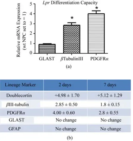
One distinguishing feature of NPCs is their capacity to differentiate into the three neuroectodermal lineages. We compared lpr and wt NPC lineage marker RNA expres-sion for astrocytes (glial fibrillary acidic protein (GFAP), glutamate aspartate transporter (GLAST)), OPCs (plate-let-derived growth factor receptor-α (PDGFRα)) and neurons (βIII-tubulin, Doublecortin (Dcx)) using RT- qPCR. After two days of differentiation, lpr NPCs ex-pressed significantly higher levels of OPC and neuronal markers compared to wt NPCs (Figure 4(a)). This trend maintained statistical significance after seven days in
differentiating media (Figure 4(b)).
We confirmed the enhanced OPC phenotype in lpr
NPCs by staining for A2B5, a putative OPC cell surface marker, after two days of differentiation. Flow cytomet-ric analysis revealed 60% of lpr NPCs were positive for A2B5, while only 20% of wt NPCs were A2B5-positive (Figures 5(a) and (b)).
NPC cell morphology was also confirmed at the pro-tein level using immunocytochemistry. Staining for βIII- tubulin and PDGFRα corroborated mRNA expression levels, as the percentage of neuronal cells (βIII-tubulin+) and OPCs (PDGFRα+) in lpr cultures was double that found in wt (Figures 5(c) and (d)).
[image:4.595.86.516.300.647.2]Altogether, these data indicate that the absence of Fas enhances NPC differentiation to the neuronal and OPC lineages at both the genomic and protein level.
Figure 4. lpr NPCs are more committed to oligo- and neu-ronal lineages. (a) Spontaneous differentiation of lpr cells compared to wt after 2 days in minimal media; (b) sum-mary of lineage marker results at 2 days (minimal media) and 7 days (0.1% FCS). Fold changes are lpr expression levels relative to wt. Each represented fold difference reached statistical significance (p < 0.05, n = 5 independent experiments). Fold changes calculated using 2-ΔΔCtmethod with β-actin as the endogenous control gene and wt NPC samples as the calibrator (mRNA expression levels set to equal 1).
2.5. Lpr NPCs Exhibit Altered Levels of
Neuronal Fate Specification Genes, Including Shh
In search of possible mechanisms and pathways control-ling the lpr phenotype, we ran a “Neurogenesis and Neural Stem Cell” (SA BiosciencesTM) PCR array using
RNA isolated from wt or lpr NPCs after 7 days of dif-ferentiation. Of the >80 genes tested, four were down- regulated (apolipoprotein E (Apoe), bone morphogenetic protein 2 (BMP2), FGF2, S100 calcium binding protein A6 (S100a6)) and 6 were up-regulated (α-filamin (Flna), hairy and enhancer of split 1 (Hes1), Neurod1, Noggin, neuronal pentraxin 1 (NptX1), Shh) in lpr compared to wt (Figure 6(a)). Neurod1, Noggin, and Nptx1 are all genes known to be involved in regulating neuronal fate specification. Shh is a major modulator of both oli-godendrocyte and neuronal differentiation pathways. The Shh antagonist BMP2 is down-regulated in lpr NPCs (–3.87 fold, p < 0.0012); further indicating Shh pathway alterations as a probable mechanism for the altered lpr
phenotype.
Upregulation of Hes1, Noggin, and Shh was confirmed
using RT-qPCR (Figure 6(b)). To further evaluate the involvement of Shh in the lpr phenotype, we also tested expression levels of Gli1, a major regulator and target of Shh signaling. Gli1 was up-regulated 3.86-fold (±0.55) in
lpr NPCs compared to wt.
3. Discussion
There are currently no efficacious treatments for devas-tating neurodegenerative diseases including Alzheimer’s and MS. Therefore, there is a pressing need to develop therapeutics that exhibit neuroprotective effects by acting directly on the CNS. NPC therapies represent a novel and promising avenue for developing neuroregenerative treat- ments [2,11]. However, all stem cell therapies currently in development ultimately fail in that they are unable to completely repair lost or damaged CNS tissue [12,13]. If endogenous or transplanted NPCs could be activated to terminally differentiate, CNS damage and consequent long-term disability could be minimized or even reversed. Therefore, determining modulators of NPC differentation and their mechanisms of action is critical to advancing therapies for neurogenenerative disease.
By utilizing a Fas-deficient mutant NPC cell line (lpr), we were able to determine the significance of Fas-sig- naling in NPC survival, self-renewal capacity, and dif-ferentiation into the three neuroectodermal lineages. Lpr
mice have a retroviral insertion of poly(A) adenylation signal repeats in the gene for Fas [4]. This large insertion results in improper splicing and truncation of Fas mRNA so that the cells are devoid of protein expression. Our study using NPCs isolated from lprmice is novel in that there has not been detailed characterization of how lack of Fas affects NPC stemness and differentiation ex vivo. We showed that lpr NPCs have dramatically decreased cell death upon withdrawal of growth factors—a condi-tion that normally induces both significant cell death and spontaneous differentiation to astrocytes, neurons, and sparse oligodendrocytes. The significance of this discov-ery is two-fold. First, this suggests that Fas manipulation may represent a feasible solution to promote NPC viabil-ity in harsh environments, including that introduced in exogenous stem cell transplantation paradigms. Second, enhanced survival of NPCs could allow for enhanced neuroprotective and immunoregulatory capacity in dis-ease models. We hypothesize that extended survival of
lpr NPCs in the absence of growth factors increases their capacity for terminal OPC and neuronal differentiation, as wild-type NPCs are more likely to die before activat-ing differentiation pathways.
Figure 5. LPR NPCs have enhanced differentiation into Neuronal and OPC lineages. (a) Representative fluorescence histo-gram for A2B5 positivity after 2 days in minimal media (red = isotype control [ISO]); (b) Quantification of A2B5 flow data (n
= 4 independent experiments, **p < 0.01). After 3 days of differentiation (right columns), lpr NPC still expressed a signifi-cantly higher amount of A2B5, though overall levels of this marker is decreased in both cell types. (c & d) Immunocytochem-istry (ICC) was performed on cells after seven days in differentiating media; (c) Differentiation of wt (top panels) and lpr
(bottom panels) into neurons (βIII-tubulin+ cells, left panels) and OPCs (PDGFRα+ cells, right panels); (d) Quantification of ICC data using Image J software. Percent positive = #lineage marker+/total # of Hoechst+. 12 - 14 serial photos were taken from 3 separate coverslips. *p < 0.05, n = 3.
OPC marker expression in lpr compared to wt NPCs was detected by analyzing mRNA levels using RT-qPCR and confirmed using both flow cytometry surface staining (A2B5) and confocal microscopy to confirm the OPC phenotype (PDGFRα).
Along with elevated levels of OPC markers, lpr NPCs have elevated levels of neuronal-specific lineage markers (BIII-tubulin, doublecortin) both at early (two days) and late (seven days) differentiation time points. In addition, the astrocyte-specific marker GFAP remains unchanged.
These data indicate a preferential neuronal-fate specifica-tion in NPCs lacking Fas. Corroborating these data, lpr
Figure 6. Upregulation of Shh and neuronal specification genes in lpr NPCs. (a) Shown are genes with significant mRNA fold-changes (out of >80 tested, SA BiosciencesTM
“Neurogenesis and Neural Stem Cell” PCR Array) in lpr
NPCs compared to wt NPCs after seven days in differentia-tion medium. Down-regulated genes (red font) have been converted to whole numbers via 1/[fractional fold change]; (b) RT-qPCR confirmation of gene upregulation in lpr
NPCs after one week in differentiation media. Data are based on 4 biological replicates (n = 4), all fold changes are significantly above levels in wt cells (p < 0.01).
dulate differentiation of ES cells to neuronal and meso-dermal cells and oscillating levels contribute to hetero-geneous responses from these cells [14]. Slit2 acts through the Roundabout (Robo) receptor and is involved in axon guidance [15,16] and neuronal migration [17]. Shh is a critical gene involved in patterning during em-bryogenesis and regulates NPC proliferation and neuro-genesis [18,19]. Since the absence of functional Fas in our culture system enhanced neuronal differentiation, it is possible that active Fas in wt NPCs provides inhibitory signals against this lineage development. Whether these inhibitory signals act upon the candidate genes identified here requires further study.
Previously, we [7] and others [8] have shown that ac-tivation of Fas via FasL enhances NPC survival. Addi-tionally, in vivo overexpression of FasL increases sub-ventricular zone neurogenesis. An explanation for these seemingly contradictory results is that exposure to FasL in NPCs enhances both survival and neurogenic path-ways [7,8]; however, the presence of a functional Fas system in the absence of activating signals (FasL) inhib-its these pathways. The latter is indicated by our current study since endogenous FasL levels are extremely low in our culture system [4] and wt NPCs have decreased sur-vival and differentiation compared to lpr NPCs—pre-sumably because unactived Fas serves an inhibitory role in these phenomena. This suggests a dynamic role for Fas in both NPC fate specification and survival that is dependent on the presence or absence of FasL.
Taken together, we propose the following paradigm for the role of Fas in NPCs. Fas targeting leads to two
unique physiological processes: first, Fas ablation pro-motes NPC survival in deprived-environments and sec-ond, Fas ablation promotes increased neuronal and oligo differentiation. Coupled together, Fas manipulation may produce a viable means of exogenous stem cell trans-plantation by overcoming the challenge of cell survival under harsh transplantation conditions, while promoting complete integration of NPCs into functional neuronal circuits by stimulation of NPC differentiation. The Shh signaling pathway, or other molecules identified here, may be a useful target in promoting this response. This paradigm offers novel candidates deserving further in-vestigation because of their potential to advance NPC transplantation therapies.
4. Conclusions
We have identified Fas as an important modulator of NPC survival, differentiation, and proliferation. This study is the first to demonstrate a putative mechanistic link between Fas and Shh signaling. Overall, we provide important insight into understanding the elusive mecha-nisms that dictate NPC fate-determination, an important step in improving NPC-based therapies for neurodegen-erative disorders.
5. Experimental Procedure
5.1. NPC Isolation and Culture5.2. Flow Cytometry
Cells were lifted using 0.05% trypsin/EDTA (Gibco) at 37˚C. Trypsin enzyme was stopped using 20% FCS/ neural basal media after 7 mins. Cells were placed in Falcon® tubes (#352058) and then rinsed with 1XPBS and spun at 1400 rpm before proceeding with staining. All stains were added to a 100 µL volume cell suspen-sion containing approximately 1 × 10⁶ cells.
Fas receptor stain: Samples blocked with hamster IgG (50 ug/mL). Samples stained with either anti-Fas or iso-type control (Hamster IgG2, λ1) phycoerythrin (PE)-con- jugated antibodies (1:30, BD Biosciences). All rinses and dilutions were done with 1%BSA/1XPBS. All incuba-tions performed at 4˚C (15 min for block, 30 min for PE-conjugated antibodies).
A2B5 stain: biotynlated anti-A2B5 (50 uL, 1:100, BD Pharm) added to each sample. Primary antibody incu-bated for 30 min at 4˚C. After rinsing with PBS, Stre-pavidin-Phycoerythrin (100 uL, undiluted, BD Pharm) added to each sample and incubated for 30 min at 4˚C. Samples rinsed twice before analyzing on flow. Controls included: no primary and a sample of NPCs growing in the presence of EGF and FGF (to serve as a low percent differentiation negative control).
Death & Apoptosis Assay (UV/Annexin stain): UV Live/Dead Dye® and Annexin Alexa Fluor-647 both used according to manufacturer instructions (Invitrogen). UV dye labels dead cells because of disruptions in the cell membrane. Viable cells to not stain positive for UV dye because of an intact cell membrane. Annexin binds strongly to phosphatidylserine residues that have flipped to the exterior surface of the cell, thus labeling early apoptotic cells. Briefly, 200 µL of diluted UV dye (2 µL per mL 1 × PBS) was added to cell suspension contain-ing 10,000 cells/µL. Cells were incubated on ice, in the dark, for 30 min. After cells with 1 × PBS and aspirating the supernatant, 100 uL of diluted Annexin (1 µL per 400 µL HEPES buffer) was added to cells. Cells grown in complete media (growth factors present) were used as a negative control because >90% of cells in these cultures are live/healthy. Cells briefly treated with 3% parafor-maldehyde were used as positive controls because >90% of cells are dead/dying.
Cell Proliferation: BrDU assay performed according to manufacturer instructions (FITC Detection BrdU Flow Cytometry Assay Kit, BD Biosciences). Briefly, cells were first pulsed overnight (16 hours) with BrDU and subsequently fixed and stained with a FITC-conjugated anti-BrDU antibody for flow analysis. Cells grown in minimal media (no growth factors) were used as a nega-tive control because <10% of cells grown in these condi-tions will stain positive for BrDU incorporation.
Analysis: flow cytometry analysis was performed by a BD LSRII flow cytometer equipped with 3 lasers (488, helium neon, and UV) and data were analyzed with FlowJo software, respectively.
5.3. Neurosphere Self-Renewal Assay
Single-cell suspensions were plated in uncoated 24-well plates (seeding density of 100,000 cells/mL) using NPC media containing EGF and FGF. After 7 days, 2-3 pic-tures of random frames from each well (3 wells per cell type) were taken at 20X using a phase-contrast micro-scope. After taking pictures, neurospheres were dissoci-ated using Neurocult® Chemical Dissociation Kit and then filtered through a 40 µm nylon cell strainer (BD FalconTM). Cells were then counted, plated as described
above, and grown for 7 days before the next passage time point was recorded (this same process completed four total times over four weeks). Exact cell circumferences were measured using Neurolucida software.
5.4. Immunocytochemistry
Cells were plated in 24-well plates with poly-D/Laminin coated coverslips and grown to confluency. At this point, cells were rinsed and allowed to differentiate 1 week in minimal media containing 0.2% FCS. For all differentia-tion markers, 3 coverslips were scored per cell line and repeated for at least 3 biological replicates.
Stains performed as follows: Cells fixed with Zam-boni’s fixative and blocked (10%HS/0.1% Triton/0.02% Azide in 1 × PBS) for 30 min. Block for PDGFRα stain did not include triton. Primaries incubated overnight at 4˚C, while secondary antibodies incubated for 1.5 hr at RT. Cells rinsed 3X between primary and secondary. All cells counterstained with Hoechst nuclear stain (1:2000).
The following antibodies were used: rabbit anti-BIII Tubulin (Sigma, 1:100), rabbit anti-PDGFRα (Santa Cruz, 1:50), rabbit anti-GFAP (Dako, 1:1000), goat anti-rabbit Cy3 (1:500, Jackson Immunoresearch).
lin or PDGFRα) by the total number cells (hoecsht stain-ing nuclei).
5.5. RT-qPCR and Gene Expression Arrays
Performed as previously described [7]. Briefly, RNA was extracted from cells using RNeasy® Mini Kit (Qiagen) according to manufacturer protocol. For RT-qPCR, cDNA was prepared using Superscript® III First-Strand Synthe-sis Supermix for qRT-PCR. RT-qPCR completed using Taqman® Master Mix and applied Biosystems 7500 Fast Software. Primers were purchased from Applied Biosys-tems (Taqman Assay on Demand).
Neurogenesis and Neural Stem Cell (PAMM-404) PCR arrays purchased from SA BiosciencesTM and RNA
sam-ples were processed by UVM Cancer Center DNA Facil-ity.
5.6. Statistics
Graphs were created and statistical analyses were run using GraphPad Prism software. Student’s t-test or ANOVA with Tukey’s post-test were used to determine significance (p-values).
6. Acknowledgements
We would like to thank: Dr. Rae Nishi for helpful dis-cussion and guidance; Dr. Jeffrey Spees and the UVM Stem Cell Core for resources and general support; Dr. Ralph Budd for graciously supplying lpr mice; Dr. Karen Fortner for expertise and assistance; Dr. Issei Shimada for technical assistance and guidance; John DeWitt and Anthony Pappas for technical assistance; Timothy Hunter and colleagues for services provided by the Ver-mont Cancer Center DNA Analysis Facility; UVM Neu-roscience Center of Biomedical Research Excellence (COBRE) for resources and technical support; and the UVM Microscopy and Imaging Center for resources and Marilyn Wadsworth for technical assistance and training. The project described was supported by NIH Grant Number P20 RR16435 from the COBRE Program of the National Center for Research Resources.
7. Authors’ Contributions
JK—study concept and design, manuscript preparation, data acquisition and analysis; CH—data acquisition and analysis; JS—data acquisition and analysis, manuscript revisions; YMD—study concept and design, manuscript revisions.
REFERENCES
[1] S. Pluchino and G. Martino, “The Therapeutic Use of Stem Cells for Myelin Repair in Autoimmune Demyeli-nating Disorders,” Journal of the Neurological Sciences, Vol. 233, No. 1-2, 2005, pp. 117-119.
doi:10.1016/j.jns.2005.03.026
[2] G. Martino and S. Pluchino, “The Therapeutic Potential of Neural Stem Cells,” Nature Reviews Neuroscience, Vol. 7, No. 5, 2006, pp. 395-406. doi:10.1038/nrn1908
[3] S. Pluchino, L. Zanotti, B. Rossi, E. Brambilla, L. Otto-boni, G. Salani, M. Martinello, A. Cattalini, A. Bergami, R. Furlan, et al., “Neurosphere-Derived Multipotent Pre-cursors Promote Neuroprotection by an Immunomodula-tory Mechanism,” Nature, Vol. 436, No. 7048, 2005, pp. 266-271. doi:10.1038/nature03889
[4] S. Nagata and P. Golstein, “The Fas Death Factor,” Sci-ence, Vol. 267, No. 5203, 1995, pp. 1449-1456.
doi:10.1126/science.7533326
[5] F. K. H. van Landeghem, U. Felderhoff-Mueser, A. Moysich, C. Stadelmann, M. Obladen, W. Brück and C. Bührer, “Fas (CD95/Apo-1)/Fas Ligand Expression in Neonates with Pontosubicular Neuron Necrosis,” Pediat-ric Research, Vol. 51, No. 2, 2002, pp. 129-135.
doi:10.1203/00006450-200202000-00003
[6] R. Nat, E. Radu, T. Regalia and L. M. Popescu, “Apop-tosis in Human Embryo Development: 3. Fas-Induced Apoptosis in Brain Primary Cultures,” Journal of Cellu-lar and MolecuCellu-lar Medicine, Vol. 5, No. 4, 2001, pp. 417- 428. doi:10.1111/j.1582-4934.2001.tb00177.x
[7] J. Knight, E. Scharf and Y. Mao-Draayer, “Fas Activation Increases Neural Progenitor Cell Survival,” Journal of Neuroscience Research, Vol. 88, No. 4, 2010, pp. 746- 757.
[8] N. S. Corsini, I. Sancho-Martinez, S. Laudenklos, D. Glagow, S. Kumar, E. Letellier, P. Koch, M. Teodorczyk, S. Kleber, S. Klussmann, et al., “The Death Receptor CD95 Activates Adult Neural Stem Cells for Working Memory Formation and Brain Repair,” Cell Stem Cell, Vol. 5, No. 2, 2009, pp. 178-190.
doi:10.1016/j.stem.2009.05.004
[9] J. Desbarats, R. B. Birge, M. Mimouni-Rongy, D. E. Weinstein, J.-S. Palerme and M. K. Newell, “Fas En-gagement Induces Neurite Growth through ERK Activa-tion and P35 UpregulaActiva-tion,” Nature Cell Biology, Vol. 5, No. 2, 2003, pp. 118-125. doi:10.1038/ncb916
[10] S. Pluchino, L. Muzio, J. Imitola, M. Deleidi, C. Al-faro-Cervello, G. Salani, C. Porcheri, E. Brambilla, F. Cavasinni, A. Bergamaschi, et al., “Persistent Inflamma-tion Alters the FuncInflamma-tion of the Endogenous Brain Stem Cell Compartment,” Brain, Vol. 131, No. 10, 2008, pp. 2564-2578. doi:10.1093/brain/awn198
[11] R. J. M. Franklin and C. Ffrench-Constant, “Remyelina-tion in the CNS: From Biology to Therapy,” Nature Re-views Neuroscience, Vol. 9, No. 11, 2008, pp. 839-855.
doi:10.1038/nrn2480
[12] M. Stangel, “Neuroprotection and Neuroregeneration in Multiple Sclerosis,” Journal of Neurology, Vol. 255, Supplement 6, 2008, pp. 77-81.
doi:10.1007/s00415-008-6014-x
1-4. doi:10.1016/j.jns.2007.03.006
[14] T. Kobayashi, H. Mizuno, I. Imayoshi, C. Furusawa, K. Shirahige and R. Kageyama, “The Cyclic Gene Hes1 Contributes to diverse Differentiation Responses of Em-bryonic Stem Cells,” Genes & Development, Vol. 23, No. 16, 2009, pp. 1870-1875. doi:10.1101/gad.1823109
[15] K. Patel, J. A. Nash, A. Itoh, Z. Liu, V. Sundaresan and A. Pini, “Slit Proteins Are Not Dominant Chemorepellents for Olfactory Tract and Spinal Motor Axons,” Develop-ment, Vol. 128, No. 24, 2001, pp. 5031-5037.
[16] L. Lin and O. Isacson, “Axonal Growth Regulation of Fetal and Embryonic Stem Cell-Derived Dopaminergic Neurons by Netrin-1 and Slits,” Stem Cells, Vol. 24, No. 11, 2006, pp. 2504-2513.
doi:10.1634/stemcells.2006-0119
[17] W. Wu, K. Wong, J. Chen, Z. Jiang, S. Dupuis, J. Y. Wu and Y. Rao, “Directional Guidance of Neuronal Migra-tion in the Olfactory System by the Protein Slit,” Nature, Vol. 400, No. 6742, 1999, pp. 331-336.
doi:10.1038/22477
[18] G. Zhu, M. F. Mehler, J. Zhao, Y. S. Yu and J. A. Kessler, “Sonic Hedgehog and BMP2 Exert Opposing Actions on Proliferation and Differentiation of Embryonic Neural Progenitor Cells,” Developmental Biology, Vol. 215, No. 1, 1999, pp. 118-129. doi:10.1006/dbio.1999.9431
[19] V. Palma, D. A. Lim, N. Dahmane, P. Sánchez, T. C. Brionne, C. D. Herzberg, Y. Gitton, A. Carleton, A. Al-varez-Buylla and A. Ruiz i Altaba, “Sonic Hedgehog Controls Stem Cell Behavior in the Postnatal and Adult Brain,” Development, Vol. 132, No. 2, 2005, pp. 335-344.
doi:10.1242/dev.01567
Abbreviations
NPC: neural stem/progenitor cell OPC: oligoprogenitor cell MS: multiple sclerosis
Lpr: Fas receptor deficient mice TNF: tumor necrosis family
GFAP: glial fibrillary acidic protein
GLAST: glial high affinity glutamate transporter PDGFRα: platelet-derived growth factor receptor-α
Dcx: Doublecortin GalC: galactosylceramidase Shh: sonic hedgehog

![Figure 5. LPR = 4 independent experiments, **gram for A2B5 positivity after 2 days in minimal media (red = isotype control [ISO]); (b) Quantification of A2B5 flow data (cantly higher amount of A2B5, though overall levels of this marker is decreased in both](https://thumb-us.123doks.com/thumbv2/123dok_us/9060513.402383/6.595.88.511.87.495/figure-independent-experiments-positivity-control-quantification-overall-decreased.webp)

