D. T. Rockwell, E. R. Melhem, and R. G. Bhatia
PURPOSE:To assess the clinical utility of GRASE (gradient- and spin-echo) MR imaging of the brain by comparing it with the T2-weighted turbo spin-echo technique.METHODS:Fifty-three consecutive patients referred for MR imaging of the brain were studied with T2-weighted turbo spin-echo and GRASE techniques, matched for effective echo time (110 milliseconds), echo train length (eight), and spatial resolution. The examinations were evaluated independently by two neuroradiologists for lesion detection (high- and low-signal-intensity lesions) and lesion conspi-cuity, and for susceptibility, motion, and chemical-shift artifacts.RESULTS:The GRASE technique provided greater detection of both high- and low-signal-intensity lesions and of low-signal-intensity lesions with paramagnetic susceptibility characteristics (ie, calcium and hemorrhage). Chemical-shift artifacts in the frequency-encoding direction were more prominent with the turbo spin-echo technique, whereas chemical-shift artifacts in the phase-encoding direction were more prominent with the GRASE technique. There was no significant difference in the degree of diamagnetic susceptibility artifacts at the base of the skull, or in motion artifacts.CONCLUSION:T2-weighted GRASE is a fast imaging technique with a potential for replacing turbo spin-echo in routine MR imaging of the brain. GRASE maintains the contrast resolution of turbo spin-echo imaging and is better at depicting lesions with paramagnetic susceptibility characteristics.
Index terms:Brain, magnetic resonance; Magnetic resonance, technique
AJNR Am J Neuroradiol18:1923–1928, November 1997
At many institutions, turbo spin-echo se-quences have replaced conventional spin-echo sequences in routine T2-weighted magnetic resonance (MR) imaging of the brain. Turbo spin-echo imaging is faster and thus decreases motion artifacts and allows for quicker patient throughput. One disadvantage of the turbo spin-echo method in brain imaging is that the multiple trains of radio-frequency (RF) pulses and the short echo spacing used with this tech-nique decrease the conspicuity of susceptibility effects (1, 2), making it more difficult to detect hemorrhagic or calcified lesions as compared with standard spin-echo imaging. Gradient- and spin-echo (GRASE) imaging is a fast multisec-tion MR technique that combines gradient and spin echoes. GRASE makes use of the speed of
gradient refocusing while overcoming chemi-cal-shift and field inhomogeneity artifacts (3, 4). We assessed the clinical utility of GRASE in MR imaging of the brain by comparing it with the T2-weighted turbo spin-echo technique. We hypothesized that the gradient-echo component of GRASE would improve the detection of hem-orrhagic or calcified lesions while the spin-echo component would maintain the contrast resolu-tion offered by the turbo spin-echo technique. Image artifacts related to both techniques were evaluated.
Materials and Methods
MR Technique
All studies were performed using a 1.5-T superconduct-ing magnet with a peak gradient of 15 mT/m and a quadrature head coil operating in receive mode. Fifty-three consecutive patients referred for brain MR were im-aged with axial T2-weighted turbo spin-echo and GRASE techniques, matched for effective echo time (110 millisec-onds), echo train length (eight), and spatial resolution. The field of view was 220 cm, the section thickness was 5 mm with a 0.5 mm gap, and the matrix was 2563256. For the
Received February 17, 1997; accepted after revision May 1. Supported in part by a research grant from Philips Medical Systems. From the Department of Radiology, Boston University Medical Center, 88 E Newton St, Boston, MA 02118. Address reprint requests to Elias R. Melhem, MD.
AJNR 18:1923–1928, Nov 1997 0195-6108/97/1810 –1923
©American Society of Neuroradiology
GRASE sequence, three gradient-recalled echoes were generated within successive 180° RF pulses. Image acqui-sition time for the turbo spin-echo sequence was 2 minutes 47 seconds; for the GRASE sequence, it was 1 minute 17 seconds. For both sequences, the number of signal aver-ages was two. Repetition time for the turbo spin-echo sequence was 3164 milliseconds; for the GRASE se-quence, it was 5503 milliseconds. Echo spacing for the turbo spin-echo technique was 15.1 milliseconds; for the GRASE sequence, it was 24.0 milliseconds. Minimum al-lowable bandwidth was used for both techniques.
Study Design
Two board-certified neuroradiologists, blinded to the patients’ clinical histories, reviewed the T2-weighted turbo spin-echo and GRASE images independently. Lesion de-tection and conspicuity, and susceptibility, chemical-shift, and motion artifacts were graded for each study as follows: Lesion Detection.—The number of lesions and their lo-cation were recorded. Lolo-cations were categorized as being either on the left or right side in one of the following areas: cerebellum, cerebral hemisphere, basal ganglia, brain stem, corpus callosum, or extraaxial. Enlarged Virchow-Robbins spaces were noted, but not included in the anal-ysis. Confluent periventricular white matter ischemic changes were also noted, but not graded as individual lesions.
Lesion Signal and Conspicuity.—The detected lesions were then divided into two groups as either low (dark) or high (bright) signal relative to brain parenchyma. Lesion conspicuity was scored on a three-point scale as subtle (0 points), average (1 point), or obvious (2 points).
Artifacts.—Chemical-shift, diamagnetic susceptibility and motion artifacts were graded on a four-point scale as none (0 points), mild (1 point), moderate (2 points), or marked (3 points). Chemical-shift artifacts in the frequen-cy- and phase-encoding directions were evaluated at the globe– orbital fat, skull– cerebrospinal fluid, and scalp-skull interfaces. The degree of diamagnetic susceptibility artifacts at the base of the skull was evaluated in the temporal lobes at the level of the petrous bone and in the frontal lobes just above the orbital roofs.
To standardize the grading of these subjective criteria, the reviewers met before evaluating the images to review the grading systems. An example of each of the grades for each criterion was selected (from images not included in this study) and reviewed by the panel.
Statistical Analysis
A pairedttest was used to compare GRASE and turbo spin-echo techniques with regard to each of the following seven criteria: the number of high-signal lesions identified in each patient, the number of dark signal lesions, the conspicuity of the dark lesions, the degree of diamagnetic susceptibility at the skull base, the degree of chemical-shift artifacts in the frequency- and phase-encoding direc-tions, and the degree of motion artifacts. The average
score of the two observers was calculated for each crite-rion, and these averages were then used to compare the GRASE and turbo spin-echo techniques.
Interobserver reliability was examined by comparing the results of the two observers for each of the seven criteria. This was done separately for each technique (GRASE and turbo spin-echo) using pairedttests.
A two-tailedPvalue of .05 or less was considered sig-nificant. No correction was made for multiple compari-sons. Because the assumption of a gaussian distribution of the differences in a pairedttest is questionable, the anal-yses were repeated with a nonparametric test of signifi-cance. The conclusions were identical.
Results
More high-signal (bright) lesions were de-tected on the T2-weighted GRASE sequences than on the turbo spin-echo sequences (P ,
.05), for a total of 103.5 on the GRASE quences and 81.5 on the turbo spin-echo se-quences (Fig 1).
More low-signal (dark) lesions were detected with the GRASE technique than with the turbo spin-echo technique (P,.01), for a total of 17 on the GRASE sequences and 10.5 on the turbo spin-echo sequences (Fig 2), (Table 1). The dark lesions were identified in 14 patients by at least one observer; in no case were more dark lesions identified on turbo spin-echo images than on GRASE images. In eight of these cases, dark lesions were detected on both sequences, and scores for conspicuity could thus be com-pared. Dark-signal lesions were more conspic-uous on GRASE studies than on turbo spin-echo studies (P ,.05) (Fig 3).
Chemical-shift artifacts in the frequency-encoding direction were more prominent with turbo spin-echo imaging (P , .0001); in the phase-encoding direction, they were more prom-inent with GRASE imaging (P,.0001) (Fig 4). There was no significant difference in the de-gree of diamagnetic susceptibility artifacts at the base of the skull (P . .10), or in motion artifacts (P. .10).
For both GRASE and turbo spin-echo
se-TABLE 1: Average number of lesions detected per patient on T2-weighted GRASE images versus turbo spin-echo images in 53 pa-tients
High-Signal-Lesions Low-Signal Lesions
GRASE 1.95 0.32 Turbo spin-echo 1.54 0.20
[image:2.587.307.546.676.729.2]Fig 1. A 75-year-old woman with a change in mental status, diabetes, and hy-pertension. A lesion of high signal intensity (arrow) in the left frontal subcortical white matter is well seen on T2-weighted GRASE image (5503/110/2 [repetition time/echo time/excitations]) (A); however, it is much more difficult to identify on T2-weighted turbo spin-echo image (3164/110/2) (B).
Fig 2. A 34-year-old woman with Sturge-Weber syndrome.
AandB, A low-signal, curvilinear calci-fication (arrow) in the right cerebellar hemi-sphere is well seen on T2-weighted GRASE image (5503/110/2) (A); however, it is much more difficult to identify on T2-weighted turbo spin-echo image (3164/ 110/2) (B).
[image:3.587.49.379.315.726.2]quences, there was no significant difference be-tween the observers with regard to bright- and dark-lesion detection, dark-lesion conspicuity, and motion artifacts (P . .50). With GRASE imaging, the only criterion for which a statisti-cally significant difference was noted was for diamagnetic susceptibility at the skull base (P5
.03). With the turbo spin-echo technique, the only criterion for which a statistically significant difference was noted was chemical-shift arti-facts in the frequency-encoding direction (P 5
.02) (Table 2).
Discussion
At many institutions, turbo spin-echo tech-niques are being used in place of conventional spin-echo for T2-weighted MR imaging of the
brain. At our institution, we rarely use conven-tional spin-echo techniques for routine brain imaging because of frequent problems with pa-tient motion and time constraints. Turbo echo offers contrast resolution similar to spin-echo techniques (5), with a much shorter imaging time. Turbo spin-echo decreases im-aging time by acquiring multiple spin echoes during each repetition time interval, resulting in more efficient filling of k-space relative to con-ventional spin-echo imaging. However, multiple 180° RF pulses and the short echo spacing im-plemented in turbo spin-echo cause diminished sensitivity to susceptibility effects (1, 2) and in turn reduce conspicuity of intracranial hemor-rhagic and calcified lesions as compared with conventional spin-echo imaging.
[image:4.587.218.549.83.494.2]GRASE is a fast multisection MR technique Fig 3. A 60-year-old woman with
bilat-eral posttraumatic subdural hematomas containing blood-fluid levels. The low-sig-nal in the dependent portion of the left-sided hematoma (arrow) is more conspicuous on T2-weighted GRASE image (5503/110/2) (A) than on turbo spin-echo image (3164/ 110/2) (B). Note also the reduced motion artifact on the GRASE image relative to the turbo spin-echo image, which is in part due to the faster technique.
combining gradient and spin echoes (3, 4). Be-tween successive 180° RF pulses, multiple short gradient-recalled echo trains (three or more) are generated. The total number of signals per repetition time interval is the product of the number of RF refocused pulses (in our case, eight) and the number of gradient-recalled ech-oes between each (in our case, three). The speed advantage of GRASE over turbo spin-echo is proportional to this latter factor. Thus, one advantage of GRASE imaging is that for a fixed number of RF refocusing pulses, there is decreased imaging time relative to turbo spin-echo. In our study, the image acquisition time for the GRASE sequence was 1 minute 30 sec-onds faster than for the turbo spin-echo se-quence. Another advantage of GRASE is that, at a fixed imaging time, there is less energy dep-osition to the patients’ bodies (3, 4, 6). As with turbo spin-echo, the effective echo time in GRASE is the time at which the center of k-space (zero-order phase encoding) is sampled. Because of the multiple gradient-recalled echo component of GRASE imaging, we
antic-ipated an increased sensitivity to diamagnetic, paramagnetic, and ferromagnetic susceptibility effects. Gradient-echo imaging is much more sensitive to susceptibility-induced frequency shifts than is spin-echo imaging, because of the lack of refocusing 180° RF pulses (which refo-cus dephased spins) (7–9). We found an im-proved detection of paramagnetic lesions, such as hemorrhage or calcium, with GRASE imag-ing. Not only did GRASE show more dark le-sions than turbo spin-echo (P , .01) but such lesions were significantly more conspicuous on GRASE sequences (P ,.05) (Figs 2 and 3).
Because there is a gradient component to GRASE imaging, there is the possibility of un-wanted diamagnetic susceptibility artifacts at the skull base. We therefore included this as one of the criteria to be graded in each study to determine whether this would pose a significant problem in clinical imaging. We found there were no greater diamagnetic susceptibility arti-facts than with turbo spin-echo imaging (P .
.10).
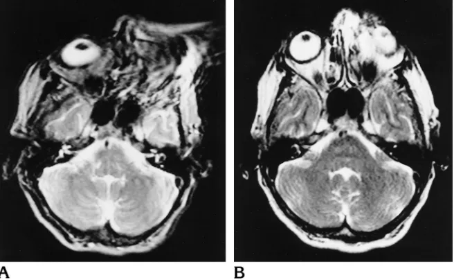
[image:5.587.50.547.96.187.2] [image:5.587.51.379.225.427.2]Although not specifically addressed in our Fig 5. A 68-year-old woman with tran-sient ischemic attacks and fixed metallic dental hardware (braces). The ferromag-netic susceptibility artifact is greater on the GRASE image (5503/110/2) (A) than on the turbo spin-echo image (3164/110/2) (B), completely obliterating the left orbit.
TABLE 2:Pvalues for interobserver variability for T2-weighted GRASE versus turbo spin-echo techniques
Average Number of High-Signal Lesions Detected
Average Number of Low-Signal Lesions Detected
Conspicuity of Dark Lesions
Artifacts
Diamagnetic Susceptibility
Chemical-Shift:
Frequency-Encoding Direction
Chemical-Shift: Phase-Encoding Direction
Motion
GRASE .80 .60 .42 .03* .26 .08 .13 Turbo spin-echo .24 .26 .56 .06 .02* .10 .42
study design, we noticed greater artifacts from ferromagnetic susceptibility with GRASE imag-ing than with turbo spin-echo imagimag-ing (Fig 5). This may pose a limitation to the utility of GRASE in patients with internal metallic hard-ware.
As in echo-planar imaging, chemical-shift ar-tifacts in GRASE sequences are more promi-nent along the phase axis of the image than along the frequency axis, because of frequency sensitivity through the echo train (4). However, in GRASE imaging, chemical-shift effects and field inhomogeneity evolve over the relatively short time period between 180° pulses, whereas in echo-planar imaging they have a longer time to develop (the time of the total echo train). Hence, GRASE sequences have relatively fewer of these artifacts than do echo-planar imaging sequences (4). Although there was significantly more chemical-shift artifacts in the phase-en-coding direction with GRASE imaging than with turbo spin-echo imaging, we do not think this was detrimental to image quality or interpreta-tion (Fig 4). Also, these artifacts could be re-duced by using frequency-selective fat satura-tion (4, 10 –12).
In our study, the GRASE technique main-tained the contrast resolution of turbo spin-echo imaging and afforded improved detection of high-signal lesions (P , .05). The reason the GRASE technique maintained the contrast res-olution provided by turbo spin-echo imaging is that the multiple spin-echo component was used to fill the center of k-space. The actual improvement in detection of high-signal lesions on GRASE sequences as compared with turbo spin-echo sequences is probably not intrinsic to the GRASE technique itself but is related to the longer repetition time used with the GRASE sequence (5503 milliseconds) rela-tive to the turbo spin-echo sequence (3164 milliseconds).
One other observed difference between the two techniques, which was not addressed in our study design, was related to fat signal. One well-recognized effect with T2-weighted turbo spin-echo imaging is that fat tends to have a high signal (13, 14). On GRASE images, fat was persistently lower in signal intensity than it was on the turbo spin-echo images (Fig 2).
Al-though this observation probably does not have a significant impact on brain imaging, it may prove useful for T2-weighted imaging of the neck and spine. Also, we noted decreased sig-nal intensity from fat in the bone marrow on GRASE images relative to turbo spin-echo im-ages. This may improve the ability to detect bone lesions (ie, metastases) on fast T2-weighted sequences.
In conclusion, T2-weighted GRASE is a fast imaging technique with a potential for replacing turbo spin-echo in routine MR imaging of the brain. GRASE maintains the contrast resolution of turbo spin-echo, is faster, and is better at depicting lesions with paramagnetic suscepti-bility characteristics, such as calcium and hem-orrhage.
References
1. Vinitski S, Mitchell DG, Einstein MS, et al. Conventional and fast spin-echo MR imaging: minimizing echo time. J Magn Reson Imaging1993;3:501–507
2. Jones KM, Mulkern RV, Mantello MT. Brain hemorrhage: evalua-tion with fast spin-echo and convenevalua-tional dual-echo sequences.
Radiology1992;182:53–58
3. Oshio K, Feinberg DA. GRASE imaging: a novel fast MRI tech-nique.Magn Reson Med1991;20:344 –349
4. Feinberg DA, Oshio K. GRASE MR imaging: a new fast clinical imaging technique.Radiology1991;181:597– 602
5. Melki PS, Mulkern RV, Panych LP, Jolesz FA. Comparing the FAISE method with conventional dual-echo sequences.J Magn Reson Imaging1991;1:319 –326
6. Hennig J, Nauerth A, Friedberg H. RARE imaging: a fast imaging method for clinical MR.Magn Reson Med1986;3:823– 833 7. Luedeke KM, Roeschmann P, Tischler R. Susceptibility artifacts in
NMR imaging.Magn Reson Imaging1985;3:329 –343
8. Elster AD. Sellar susceptibility artifacts: theory and implications.
AJNR Am J Neuroradiol1993;14:129 –136
9. Atlas SW, Grossman RI, Hackney DB, et al. Calcified intracranial lesions: detection with gradient-echo-acquisition rapid MR imag-ing.AJNR Am J Neuroradiol1988;9:953–954
10. Joseph PM, Shetty A. A comparison of selective saturation and selective echo chemical shift imaging techniques.Magn Reson Imaging1988;6:421– 430
11. Frahm J, Haase A, Hanicke W, Matthaei D, Bomsdorf H, Helzel T. Chemical shift selective MR imaging using a whole-body magnet.
Radiology1985;156:441– 444
12. Rosen BR, Wedeen VJ, Brady TJ. Selective saturation NMR im-aging.J Comput Assist Tomogr1984;8:813– 818
13. Henkelman RM, Hardy PA, Bishop JE et al. Why fat is bright in RARE and fast spin-echo imaging.J Magn Reson Imaging1992; 2:533–540