Printed in Great Britain
CHANGES IN HEART RATE AND RESPIRATORY
FREQUENCY DURING NATURAL BEHAVIOUR OF
DUCKS, WITH PARTICULAR REFERENCE TO
DIVING
BY P. J. BUTLER AND A. J. WOAKES
Department of Zoology and Comparative Physiology, University of Birmingham, Birmingham B15 zTT, U.K.
{Received 17 July 1978)
SUMMARY
1. Heart rate and respiratory frequency were recorded from free-range pochards and tufted ducks by using an implantable radio-transmitter. Particular attention was paid to the changes associated with natural diving behaviour which occurred on an outside pond (4 x 8 x 0-65 m depth).
2. Spontaneous dives, which occurred in the absence of any obvious external stimuli, often occurred in a series and there could be in excess of 40 dives in fairly quick succession. The first dive in a series was heralded by tachycardia and tachypnoea. Upon submersion there was transient brady-cardia, but heart rate then increased over a period of 6-8 s until it was close to the value recorded when the bird was swimming on the pond before the dive. This rate was maintained throughout the dive. Upon surfacing, heart rate increased when lung ventilation commenced, and then decreased steadily until a few seconds before the next dive when it increased. The average duration of all spontaneous dives of a series was 8-4 ± 0-2 (137) s for pochards and 9-9 + o-8 (95) s for tufted ducks. The birds also dived in response to food being thrown onto the water (feeding dives). Changes in heart rate associated with these dives were similar to those described for the spontaneous dives. Heart rate did, however, tend to be higher during all stages of the feeding dives, and it often increased at the end of a dive before the bird started to ventilate its lungs.
3. When being chased with a net, the birds had extremely high values of heart rate and respiratory frequency. Immediately they dived to escape, heart rate fell to a rate which was similar to the rate recorded when the un-stressed birds were swimming. This rate was maintained throughout the dive. Anticipatory increases in heart rate preceded lung ventilation at the end of the dives.
284 P. J. BUTLER AND A. J. WOAKES
INTRODUCTION
When the head of a duck is placed under water, apnoea with progressive reductions in heart rate and cardiac output and selective vasoconstriction occur (Andersen, 1963; Johansen, 1964; Butler & Jones, 1968, 1971; Jones & Holeton, 1972). These responses to submersion of the head of ducks are similar to those that occur in seals during both natural and enforced dives (Eisner, 1969) and are often referred to collectively as the 'diving response'. This response enables the animal to conserve oxygen and to remain under water for periods of time longer than would otherwise be possible.
Compared with seals, the onset of the bradycardia, and the accompanying selective vasoconstriction, during head submersion of ducks are relatively slow, and their onset and maintenance can be explained largely in terms of a number of peripheral reflexes. Cessation of ventilation causes progressive hypercapnia and hypoxia which, via the carotid body chemoreceptors (Hollenberg & Uvnas, 1963; Jones & Purves, 1970; Holm & Sarensen, 1972), elicit the progressive reduction in heart rate. Apnoea is maintained in the face of this increased chemoreceptor activity by the stimulation of receptors in the upper respiratory tract (Butler & Jones, 1968; Bamford & Jones, 1974; Blix, Rettedal & Stokkan, 1976). It has been suggested however, that peripheral reflexes such as these may not be the only factors involved during more natural dives (Butler & Jones, 1968) and that the gradually developed bradycardia seen during head submersion in the laboratory may be an artifact of the experimental conditions (Eliassen, i960). One reason for the former suggestion is that under natural con-ditions the act of diving is volitional, also it must be added that during such dives the bird is active, usually searching for food.
The purpose of the present study was to monitor changes in heart rate and respira-tory frequency in diving ducks using an implantable radio-transmitter (Woakes & Butler, 1975) during natural behaviour and in particular during diving. Some of the results from this investigation have been reported briefly elsewhere (Butler & Woakes, 1976a, b).
MATERIALS AND METHODS
Data were obtained from two male and three female pochards (Aythya ferina) whose mass ranged from o-68 to o-8 kg, and from two male tufted ducks (A. fuligula) of mass 0-75 and 0-83 kg. The birds were obtained as young chicks from the Wildfowl Trust, Slimbridge, and were kept on an outside pond (4 x 8 x 0-65 m depth) on the campus of the University of Birmingham. The birds were pinioned, but they were able to roam freely within a perimenter fence which was 20 x 20 m and surrounded the pond. In addition to any natural supply of food, the birds were fed on growers' pellets and mixed corn (Heygates Ltd., Bugbrooke Mills).
The animal showed no signs of distress either during or after the operation. The transmitter and leads were sterilized in chlorhexidine (0-5% then 0-05%) before being implanted. The transmitter was placed in the abdominal cavity with the bipolar e.c.g. lead lying close to the heart. The other lead from the transmitter was guided beneath the skin up to the trachea and a thermistor at the end of this lead was placed into the lumen of the trachea (for details see Woakes & Butler, 1975). The wounds were sutured together and the bird was given an injection of ampicillin (15 mg, i.m. Penbritin, Beechams). The bird was kept indoors for 2-3 days after the operation and then placed outside in the compound. Only one bird was implanted with a transmitter at a time and it was accompanied in the compound by up to 8 diving ducks and 12 mallards.
Usually, within one week of the operation the bird was diving spontaneously and recordings of heart rate and respiratory frequency were made from 10 days after the operation. The bird was caught before a recording session in order to switch on the transmitter. The wounds from the operation were almost completely healed within a week of the operation, but it took another several weeks before the feathers had completely regrown. Ideally, it would have been better to wait until this time before the recordings were made. Unfortunately within 3 weeks of the operation the response time of the thermistor had invariably been reduced to an unacceptable level, generally as a result of it having been coated with tracheal secretions. Therefore, all of the recordings had to be made within 3 weeks of implanting the transmitter. As far as could be determined from the animal's behaviour, the presence of the transmitter caused the bird no distress nor did it cause it to behave abnormally except that it preened the area of the wound more than usual, particularly during the first 2 weeks after the operation. There was also a reduction in body mass during this period. On average, body mass at this time was 55 g less than at the time the transmitter was implanted. By the end of the third week, the mass of the birds had returned to within
10-15 S °f t n e pre-operation level.
Three additional ducks had a single channel e.c.g. transmitter implanted. It was possible to leave these birds for several weeks to allow the down feathers around the wound to regrow completely and the contour feathers to regrow or be preened to cover the area. Feather regrowth and return to a normal body mass were completed within 6 weeks after implanting the transmitter, and at this time the changes in heart rate in association with spontaneous and induced escape dives were not significantly different from those recorded from animals with the respiration probe in place. These three birds were also used to record the changes in heart rate associated with sub-merging the head of the duck in water in the laboratory. This was done initially before implantation of the transmitter, using subcutaneous leads connected to conventional amplifiers and then 6 weeks after implantation of the transmitter, using radiotelemetry. The values of heart rate before, during or after head submersion obtained via telemetry were not significantly different from those obtained by the more conventional system.
When recording from free-range birds, the observer sat in a wooden hut which was situated inside the compound, some 3 m from the edge of the pond. The birds were hot affected in any way by the presence of an observer inside the hut. Information from the bird was received by a Sony CRF 5090 receiver and was stored on one channel of a Teac A450 stereo cassette tape recorder. The other channel was used
286 P. J. BUTLER AND A. J. WOAKES
both for marking the dives and for recording a commentary of the bird's behaviour for later correlation with the decoded physiological data. An attempt was made to gather information on heart rate and respiratory frequency associated with as wide a range of the bird's behaviour as possible. This included diving. Although the bird may have been searching for food during every natural dive, a distinction was made between those dives that occurred in response to food being thrown onto the pond, 'feeding' dives, and those that occurred on impulse, 'spontaneous' dives. As well as performing these natural dives, the bird was also induced to dive ('escape' dives) by chasing it with a net. When sufficient data had been collected outside and while the thermistor in the trachea was still responding satisfactorily, the animal was taken into the laboratory and its head was forcibly submerged in water for 10, 20 and 60 s (see Butler & Jones, 1968). Changes in heart rate and respiratory frequency associated with these ' dives' were recorded on tape via the implanted transmitter. When diving naturally, the birds changed their position very rapidly from the horizontal to the vertical. Inspection of the respiration subcarrier of the implanted transmitter indi-cated that a radio signal was being received continuously and that there was not a 'drop out', at the beginning of a dive, associated with this rapid change in position.
The tapes were played back through a purpose-built demodulator and hard copies of the e.c.g. and respiratory air flow were made by a 4-channel pen recorder (Devices Ltd.). Heart rate was determined by an instantaneous rate meter (Devices Ltd.). Values of heart rate and respiratory frequency were obtained from these traces and were stored in a computer (PDP 11/10, Digital Ltd.). Mean values of the variables were computed for the various types of activity displayed by the birds. For spontaneous dives on the pond, these mean values were obtained at 10, 5, 2 and 1 s before cessation of ventilation (as inferred from the trace of air flow through the trachea); 1, 2 and 5 s after cessation of ventilation; 2 and 1 s before commencement of ventilation at the end of a dive and 1, 2, 5 and 10 s after the duck started to ventilate. The maximum values of heart rate before and after a dive, as well as the minimum level during a dive were also noted. As these values invariably occurred within one second of the cessation or commencement of respiration, they are plotted on graphs at 0-5 s before or 0-5 s after cessation of ventilation at the start of a dive or 0-5 s after commencement of ventilation at the end of a dive. Cessation and commencement of ventilation were taken as indications of the beginning and end of the dive respectively.
Laboratory dives were analysed in a similar manner with the addition of an extra sample point 10 s into the dive. As these dives did not have associated with them extreme changes in heart rate upon submersion, the maximum heart rate just before and the minimum heart rate just after diving were not noted.
To obtain data on non-diving activity, periods were chosen where the bird showed the same type of behaviour continuously for 30 s. Heart rate and respiratory frequency were then sampled every 5 s within the 30 s period.
RESULTS
Non-diving behaviour
The birds' behaviour was classified subjectively. For example, it was assumed that, when a duck was lying down with its head curled round onto its back or held under its wings, it was asleep. Also, there were no absolute divisions between fast and vigorous swimming so that there was, no doubt, some variability in these assessments from day to day. As far as pochards were concerned, heart rate was n o ±2-8 (21)
beats min"1 when they were drifting on the water, which was not significantly
differ-ent from the value recorded when they were asleep on land (114 ±2-5 (42) beats
min"1). Respiratory frequency, however, was i6-8± 1-0(21) breaths min"1 when
they were drifting on the water and this was significantly higher than the value
recorded when they were asleep on land (10-9 ±o-6 (42) breaths min"1). When the
pochards were walking, heart rate was 15817-3(35) beats min"1 and respiratory
frequency was 2511-5 (35) breaths min"1. These values were not significantly
different from those recorded during periods of fast swimming when heart rate was
160 ±3-2 (84) beats min"1 and respiratory frequency was 2711-0(84) beats min"1.
During vigorous swimming, however, heart rate was significantly higher at 227 ± 10-9
(35) beats min"1, whereas respiratory frequency did not show a significant change and
was 25 ± i-8 (35) breaths min-1. As the birds had been pinioned they could not fly
properly, but the highest values of heart rate and respiratory frequency during non-diving activity were recorded in pochards when they attempted to fly and 'skated' across the surface of the pool with their wings flapping vigorously. Heart rate was
425 ± 10-3 (35) beats min- 1 and respiratory frequency was 49-3 ± 2-6 (35) breaths
min"1. The next highest values of the two measured variables were recorded when
the birds were feeding on land. Heart rate was 360 ± 14 (28) beats min"1 and
respir-atory frequency was 40 ± 25 (28) breaths min"1. Fewer events were recorded from the
two tufted ducks than from the pochards, but it was found that within the various categories of behaviour, heart rate was not significantly different between the two species.
There were wide ranges in heart rate and respiratory frequency during any one type of behaviour pattern and the total range for any variable was very large. For
tufted ducks, respiratory frequency was as low as 3 min"1 (drifting on water), or as
high as 86 min"1 (preening vigorously). For the pochards, the lowest recorded heart
rate was 85 beats min"1 (drifting on water) whereas the highest recorded rate was
505 beats min"1 ('flying' across the pool). Some of the variability in the two measured
variables could not be related to any behaviour of the animal. A pochard which was lying down with its head under its wing and was, presumably, asleep, showed sudden
increases in both heart rate (100-280 beats min"1) and respiratory frequency (11-20
breaths min"1) for no apparent reason. Perhaps it was dreaming. A resting tufted
duck showed a transient increase in heart rate from 105 to 250 beats min"1 when a
human appeared within sight but did not approach the bird. This was followed by a
larger rise in heart rate to 395 beats min"1 when the bird stretched one wing. In
288
P. J. BUTLER AND A. J. WOAKESDiving behaviour Spontaneous dives
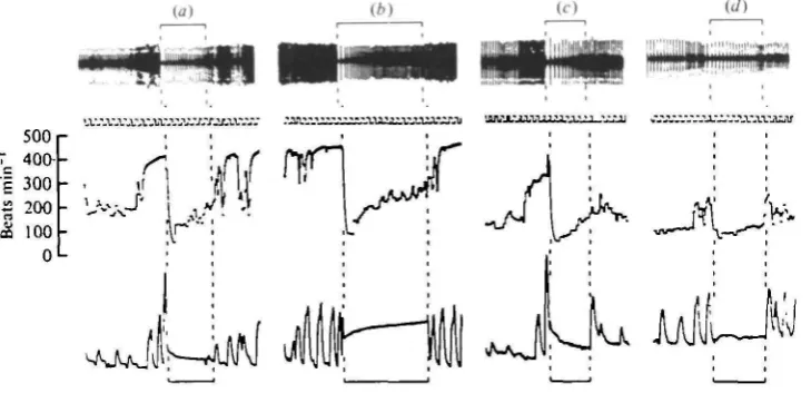
These dives occurred in the absence of any noticeable external stimuli. They often occurred in a series and there could be in excess of 40 dives in fairly quick succession. Typically, the first dive in a series was heralded by increases in both heart rate and respiratory frequency. Immediately upon submersion there was an instantaneous reduction in heart rate to a level below that recorded while the animal was swimming (Fig. 1 a). Heart rate then increased in a hyperbolic fashion during the early stages of the dive and eventually stabilized at a level which was between the values recorded during fast and vigorous swimming activity. The birds tended to breathe out upon submersion and there was often a further expiratory effort upon surfacing before the first large inspiration (Fig. 1 b). An immediate increase in heart rate accompanied the commencement of lung ventilation. Both heart rate and respiratory frequency were relatively high upon surfacing but both then decreased steadily.
Sometimes no further dives occurred after the first one and, on average, it took 14-8 ± 1-7 (6) s for heart rate and respiratory frequency to return to their pre-dive values in tufted ducks and io-8 ± 0-9 (13) s in the pochards. If no further dives occurred before full recovery of these variables, then the next dive that took place was desig-nated the first dive of a series. When isolated dives did occur they often displayed unusual changes in heart rate. On occasions there was no obvious post-dive tachy-cardia (Fig. ic) and more rarely there was no dramatic change in heart rate at all during the period of submersion (Fig, 1 d).
[image:6.451.32.397.412.588.2]The most common occurrence was for another dive to follow the first one before heart rate and respiratory frequency had fully recovered. The average time between first and second dives was 12-6 ± 22 (18) s for tufted ducks and 9-8 ± 1-2 (25) s for
400
300
2
S
20
°
100 •••••
• • • 50
40
30
20
400
300
-0 S i i i i i i i i i i i i i i -0 -10 0 4 0 10
Before After submersion surfacing
Time (s)
I I i I i I i i i i r - 1 0 0 4 0 10
Before After submersion surfacing
Time (s)
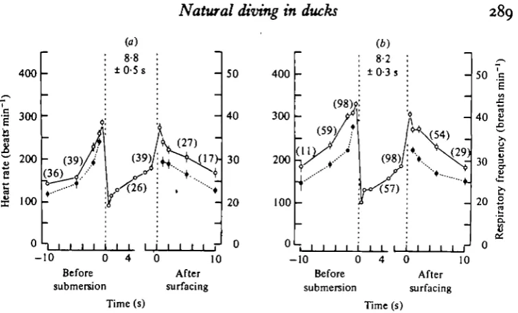
Fig. 3. Graphs showing mean values of heart rate ( O ) and respiratory frequency ( • ) in pochards before, during and after, (a) all recorded first spontaneous dives of a series, and (b) all recorded second and subsequent spontaneous dives of a series. The figure in parentheses above a set of points indicates the number of events contributing to those points and to each successive set of points without a number. Vertical lines associated with each point are ± s.E. of mean. Where vertical lines are absent, the s.E. of mean is within the limits of the symbol. The mean duration of the dives ±S.E. of mean is given between the vertical dotted lines on each graph which delimit the period of the dive.
[image:7.451.38.416.44.272.2]pochards. Thus the anticipatory increases in heart rate and respiratory frequency which preceded the second (or subsequent) dives of a series were on top of already elevated rates (Fig. ib). The average response for pochards can be visualized from Fig. 2 by continuing the 5 s post-dive values of the first dives (Fig. 2 a) with the 5 s pre-dive values for the second and subsequent dives (Fig. zb). Clearly, the shorter the duration between dives the less the reduction in heart rate and respiratory fre-quency, and vice versa. The changes in heart rate during the second and subsequent dives in a series were independent of the position of the dive in the series, so the data for these dives have been grouped together (Fig. 26). In pochards the pattern of changes in heart rate and respiratory frequency in second and subsequent dives were similar to those of the first dive of a series (Fig. 2). This was also the case for the tufted ducks. For both species, however, the heart rates just before diving, immed-iately upon submersion and immedimmed-iately upon surfacing were significantly higher for the second and subsequent dives in a series than they were for the first dives (Table 1). However, the lowest heart rate at the beginning of a dive did not vary significantly with respect to the position of the dive in a series.
Table I. Mean values f S.E. of mean for pochmds and tufted ducks of diere duration and heart rate at spen. tima before, dun'ng and aftcr spatmreoru dives, feeding dives and idiced escape dives (Values arc given for the first dive in a eerie8 (1st) and for second and wbsequent dives combined (and+). The number of events contributing to each mean value is given in parenthesea) Heart rate 5 s before submersion (beats min-l) Highest heart rate immediately before sub- mersion (beats min-') Lowest heart rate immediately after sub- mersion (beats min-') Heart rate I 8 before surfacing at end of dive (beats min-l) Highest heart rate upon surfacing (beats rnin-l) Duration of all dives (a)
Iat 2nd
+
1st and
+
1st 2nd
+
1st 2nd
400
300
200
100
(3) 400
-n i i i i i j i i i i - 1 0 0 4 0 10
Before After submersion surfacing
Time(s)
- 1 0 0 Before submersion
Time (s)
Fig. 3. Graphs showing mean values of heart rate in tufted ducks before, during and after, (a) all recorded first spontaneous dives of a series with a duration between 6 and 8 s, (6) all recorded first spontaneous dives of a series with a duration in excess of ia s. For remainder of caption see Fig. 2.
before surfacing, and immediately upon surfacing, heart rate was significantly higher in the tufted ducks than in the pochards. Having reached its lowest level immediately upon submersion, heart rate increased almost linearly during the first 6-8 s of a dive, but from then on the rate of increase declined steadily. Thus during dives of short duration (< 8 s) heart rate increased continuously during a dive (Fig. 3 a), whereas during dives of longer duration a plateau was reached after the initial rise (Fig. 3^). This occurred in both species.
292 P. J. BUTLER AND A. J. WOAKES
500
E 400
I
to
o
I 300
8 200
100 L
y = a + b log x
where a = 35-6 ± 74-8
b = 314-2 ±75-7
r - 0 - 7
y =
a + bx
where a = 0014 ±00032 b = 0002 ± 000042
12 16 Duration of dive (s)
20 24
Fig. 4. Graphs to show the relationship between the highest heart rate recorded upon sur-facing after the first spontaneous dives ofaseriesand the duration of the dive in pochards ( • ) and tufted ducks ( • ). The line of best fit is drawn through the points. The equation describing each line and the correlation coefficient (r) are given with each line.
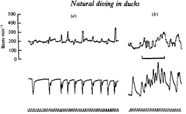
Sometimes a bird would upend itself in the water and ' dabble' at the edge of the pond in a fashion similar to that displayed by mallard ducks. Several 'dabbles' often occurred in a series and there were no consistently large changes in heart rate associated with each period of head submersion, even when the head was under water for 6 s or more (Fig. 5 a). Also, the birds would periodically go through the initial motions of a spontaneous dive to the point of placing the head under water, but then instead of the rest of the body being sumberged, the head would come to the surface. Several of these 'head ducking' manoeuvres would often occur in a series and although heart rate was somewhat irregular during such a series (Fig. 5 b), there were not the large changes in heart rate which accompanied a complete dive.
Feeding dives
spon-500 400
I 300
l
m
100
0
(a) (fc)
[image:11.451.53.365.51.242.2]TTrrrrrrrr
Fig. 5. Traces from 6* tufted duck (0-75 kg) showing changes in heart rate associated with: (a) 'dabbling' under water, (6) 'head ducking'. In (a) the bird upended itself like a mallard duck during the periods of apnoea indicated on the air flow trace. In (6) the bird went through the initial manoeuvres of a dive to the point of ducking its head under water, but instead of submerging itself completely it then removed its head from the water. This procedure was repeated several times during the period indicated by the black bar. Traces in each set are from above downwards: instantaneous heart rate, air flow through the trachea (inspiration-up on trace), time markers).
taneous dives. This was also the case for the tufted ducks as far as the first dive in a series was concerned, but not so for second and subsequent dives. For both species of duck, the heart rate immediately upon submersion was always significantly lower during spontaneous dives than during feeding dives (Table 1). For pochards, heart rate associated with all stages of the second and subsequent feeding dives was signifi-cantly higher than during the first feeding dive of a series. This was not the case for tufted ducks (Table 1). There were no consistent differences in respiratory frequency associated with spontaneous or feeding dives for either species of duck (Table 2).
The average duration of feeding dives was significantly greater than for spon-taneous dives in pochards, but not in the tufted ducks (Table 1). In a number of feeding dives there was a clear indication of heart rate increasing at the end of a dive before lung ventilation commenced (Fig. 6a, b). It is also clear from the respiration traces in Fig. 6 that the birds breathed out several times during a feeding dive. These expirations may have been related to the feeding manoeuvres themselves.
Escape dives
n-
3s
Ls
+I+
g"
8
h h
z2
m-
i??'
-
I-+I -H
500 400 .5 400
E
a 300
2
[image:13.451.41.394.57.278.2]100
Fig. 6. Traces from $ pochard (0-73 kg) showing changes in heart rate and respiratory fre-quency associated with feeding dives, (a) First dive in a series, (6) last two (14th and 15th) dives in the same series. In each case the period of submersion is indicated by dashed, vertical lines joined by horizontal bars. In each set the traces are from above downwards: instantaneous heart rate, air flow through the trachea (inspiration-up on trace), time markers).
the tufteds, heart rate decreased on average by almost 460 beats min"1. Within a
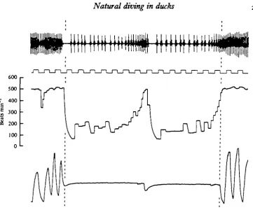
second of submersion, heart rate had increased above the low, initial value, and then rapidly decreased again. This new level was maintained throughout the duration of the dive until just before surfacing (Figs. 7, 8). Thus, there was not the gradual increase in heart rate during the first 6-8 s of a dive as was seen in spontaneous and feeding dives. There was, however, quite often a definite increase in heart rate just before lung ventilation commenced at the end of a dive. This is clearly seen in Fig. 8. During the dive shown, the heart rate began to increase approximately 5-5 s after the
duck had submerged. By 7 s after submersion, heart rate was about 500 beats min"1
and the bird had just begun to breathe out but saw the net above the water and sub-merged again. The heart rate fell immediately upon re-submersion. A similar antici-patory increase in heart rate was apparent just before the bird did eventually surface and ventilate its lungs completely. On average, for the escape dives, heart rate began to increase 0-910-09 (19) s before surfacing in pochards and 0-9210-14(11)8 before surfacing in tufted ducks.
296 P. J. BUTLER AND A. J. WOAKES
-10 0
Before submersion
100
10 After surfacing Time (s)
80
E
£1
60
3
40 o
.5
20
-1 0
Fig. 7. Graph showing mean values of heart rate (O) and respiratory frequency ( • ) before, during and after all recorded induced escape dives of a series for tufted ducks. For remainder of caption see Fig. a.
Laboratory 'dives'
When the ducks were lightly restrained ventral side down in the laboratory,
heart rate, at 115 + 4 (7) beats min"1, was similar in the pochards to those values
recorded in the free range animals when they were asleep on land or drifting on the
water. In the tufteds, resting heart rate in the laboratory was n o ± 1 (3) beats min"1.
The changes in heart rate and respiratory frequency associated with head sub-mersion in the laboratory were completely different from those seen in association with natural dives for both species of birds. There were no significant increases in either of the variables just before head submersion, neither was there an immediate reduction in heart rate upon submersion. Instead, heart rate decreased progressively, during the first 10 s when it reached a level that was maintained for at least the next
10 s. This heart rate was 54 ± 10 (7) beats min-1 for pochards and 22 ± 1 (3) beats
min- 1 for tufteds. These values are significantly lower on average than the mean
lowest rate recorded during any of the natural dives. For ' dives' of 60 8 duration,
heart rate eventually reached a value of 25 ± 2 (3) beats min- 1 in the pochards which is
600
500
7 400
c
I 300
1 200
100
[image:15.451.38.400.51.351.2]0
Fig. 8. Trace from 6* tufted duck (o'7s kg) showing changes in heart rate and respiratory frequency associated with an induced escape dive. T h e period of submersion is between the vertical dashed line. The traces are from above downwards: e.c.g., time marker(s) instantaneous heat rate, air flow through the trachea (inspiration-up on trace).
emersion of the head into air was greater for longer periods of submersion in the pochard (not enough 'dives' were performed for a similar comparison in the tufted ducks), and this post ' dive' tachycardia was significantly greater in the tufted ducks than in the pochards for ' dives' of equal duration.
DISCUSSION
298 P. J. BUTLER AND A. J. WOAKES
transmitter, the areas denuded of feathers and the slight loss of body mass during the first two weeks after the operation, did not appear to affect the cardiac response of the birds to natural diving. Unfortunately it was not possible to do similar checks on the effect of the thermistor probe on respiratory activity, although Butler, West & Jones (1977) found that a similarly placed thermocouple had no noticeable effect on lung ventilation of pigeons in the short term.
The two species of diving duck studied show essentially similar cardiac responses to natural diving and these are substantially different from those seen during head submersion in the laboratory. There are probably psychological and physiological factors involved in these differences. The tachypnoea and tachycardia which precede spontaneous dives may be taken as indications that the birds anticipate their diving behaviour. These anticipatory adjustments may serve to load the animal with oxygen, by increasing the oxygen tension in the air sacs and in the venous blood. The slight exhalation upon diving may serve to reduce the buoyancy of the animal which in terms of its energy budget may more than offset the loss of oxygen in the exhaled air. The instantaneous reduction in heart rate upon submersion, which may precede cessation of ventilation (Butler & Woakes, 1976 A), is more than likely elicited by higher centres of the brain. It is extremely unlikely that the hypothalamic area (area A) which was electrically stimulated by Folkow & Rubinstein (1965) is involved in this response as these authors described a slight initial decrease in heart rate followed by a progressively more intense bradycardia. There was no instantaneous reduction in heart rate associ-ated with ' dabbling' or ' head ducking' manoeuvres, so that it was not likely to be the result of a postural reflex or of sudden changes in pressure in some part of the cardio-vascular system. Also, the absence of a dramatic reduction in heart rate during these two manoeuvres and the fact that heart rate did not decline so rapidly during head submersion in the laboratory as it did during natural dives would suggest that unlike the situation in seals (Daly, Eisner & Angell-James, 1977), stimulation of branches of the trigeminal nerve does not make any significant contribution to the instantaneous bradycardia at the beginning of natural dives. There was no reduction in the initial bradycardia upon submersion during the later spontaneous dives in the series; there was thus no habituation of the initial cardiac response to diving. This argues against the idea that the response to natural dives is similar to the orienting reflex (Goodman & Weinberger, 1970).
increase as the depth of water increases, and as the former is thought to be more or less constant for a given depth (Dewar, 1924). The intensity of the bradycardia at the beginning of the dive and the level of the steady heart rate during the later stages of submersion, both seem to be related to the pre-dive heart rate. The presentation of food caused large increases in heart rate when the birds were on water as it did when they were on land, and a heart rate was consistently higher throughout all stages of the feeding dives compared with the spontaneous dives.
The main physiological difference between natural dives and head submersion in the laboratory, is the fact that the birds were active during the former. The settled heart rate during all dives was not substantially different from that recorded when the birds were swimming relatively quickly on the surface of the water. It has been stated that the bradycardia associated with natural submersion in seals 'probably also reflects the initiation of the redistribution in blood flow' (Jones et al. 1973). Whether or not the obverse is true in ducks, ie. that the absence of a reduction in heart rate below that seen during swimming may be indicative of a lack of redistribution of blood, remains to be seen. For spontaneous dives up to a duration of 25 s at least, the classical idea of selective redistribution of blood accompanying bradycardia and reduction in cardiac output must be viewed with caution. On the other hand, the high values of heart rate and respiratory frequency when the birds were being chased, were no doubt indicative of high values of cardiac output as the birds swam and flapped their wings in efforts to escape. Upon submersion the immediate reduction in heart rate was dramatic, and during these dives this bradycardia was doubtless accompanied by a reduction in cardiac output and may well have reflected a redistri-bution of blood flow, away from the then inactive flight muscles.
Millard, Johansen & Milson (1973) reported that penguins, like seals (Jones et al. 1973), showed anticipatory increases in heart rate before the first ventilatory effort upon emersion. The only time that this was consistently seen in the ducks used in the present observations, was at the end of escape dives, although it was apparent at the end of some feeding dives. Ducks do, then, show all of the cardiac responses to natural submersion that have been described in seals. They anticipate the dive, and the cardiac slowing may precede cessation of lung ventilation, there is an in-stantaneous reduction in heart rate and under certain circumstances there is a clear increase in heart rate before ventilation begins upon surfacing. Heart rate does not, however, stabilize at the low values that have been recorded in free diving seals (Eisner, 1969; Jones et al. 1973) and there may not be in ducks, therefore, the massive reduction in cardiac output that accompanies the bradycardia in seals (Eisner, 1969). Perhaps this is not surprising, as seals stay under water for much longer periods of time than ducks.
300 P. J. BUTLER AND A. J. WOAKES
during the later stages of these dives (cf. Daly et al. 1977). If asphyxic stimulation of the carotid bodies does occur, then central inhibition of the respiratory drive from such stimulation via receptors in the upper respiratory tract (Butler & Jones, 1968; Bamford & Jones, 1974; Blix et al. 1976) may also be important.
This work was supported by the Science Research Council. The authors wish to express their thanks to the Wildfowl Trust, Slimbridge, for supplying the animals.
REFERENCES
ANDERSEN, H. T. (1963). Factors determining the circulatory adjustments to diving. 1. Water immersion. Acta physio!, scand. 58, 173-185.
BAMFORD, O. S. & JONES, D . R. (1974). On the initiation of apnoea and some cardiovascular responses to submergence in ducks. Rapir. Pkytiol. 23, 190-316.
BLIX, A. S., RETTEDAL, A. & STOKKAN, K. A. (1976). On the elicitation of the diving responses in ducks. Acta pkytiol. scand. 98, 478-^+83.
BUTLER, P. J. & JONES, D . R. (1968). Onset of and recovery from diving bradycardia in ducks. J. Pkysiol., Lond. 196, 355-372.
BUTLER, P. J. & JONES, D . R. (1971). The effect of variations in heart rate and regional distribution of blood flow on the normal pressor response to diving in ducks. J. Pkytiol., Lond. 214, 457—479. BUTLER, P. J. & WOAKES, A. J. (1976a). Changes in heart rate and respiratory frequency associated with
natural tubmersion in ducks. J. Pkytiol., Lond. 256, 73-74P.
BUTLER, P. J. & WOAKES, A. J. (19766). Changes in heart rate and respiratory frequency associated with spontaneous submersion of ducks. In Biotelemetry III (ed. T. B. Fryer, H. A. Miller and H. Sandier), pp. 215-318. New York, London: Academic Press.
BUTLER, P. J., WEST, N. H. & JONES, D . R. (1977). Respiratory and cardiovascular responses of the pigeon to sustained level flight in a wind-tunnel. J. exp. Biol. 71, 7-36.
DALY, M. DE B., ELSNER, R. & ANGELL-JAMES, J. E. (1977). Cardiorespiratory control by carotid chemo-receptors during experimental dives in the seal. Am. J. Phyriol. 233, H508-H516.
DEWAR, J. M. (1934). The Bird as a Diver. London: H. F. & G. Witherby.
ELIASSBN, E. (i960). Cardiovascular responses to submersion asphyxia in avion divers. Arbok Univ. Bergen 3, 1-100.
ELSNER, R. W. (1969). Diving mammals. Science Journal 6, 68-74.
FOLKOW, B. & RUBINSTEIN, E. M. (1965). Effects of brain stimulation on 'diving' in ducks. HvaJrad. Skr. 48, 30-41.
GOODMAN, D . A. & WEINBERGER, N. M. (1970). Possible relationships between orienting and diving reflexes. Nature 225, 1153-1154.
HOLLENBERO, N. K. & UVNAS, B. (1963). The role of the cardiovascular response in the resistance to asphysia of avian divers. Acta pkytiol. scand. 58, 150-161.
HOLM, B. & SORENSEN, S. (197a). The role of the carotid body in the diving reflex in the duck. Res. Pkytiol. 15, 303-309.
JOHANSEN, K. (1964). Regional distribution of circulating blood during submersion asphyxia in the duck. Acta pkytiol. scand. 63, 1-9.
JONES, D . R. & PURVES, M. J. (1970). The carotid body in the duck and the consequences of its dener-vation upon the cardiac responses to immersion. J. Pkysiol., Lond. 211, 279-394.
JONES, D . R. & HOLETON, G. F. (1972). Cardiac output in ducks during diving. Comp. Biochem. Pkytiol. 41A, 639-645.
JONES, D. R., FISHER, H. D., MCTAGGART, S. & WEST, N. H. (1973). Heart rate during breath-holding and diving in the unrestrained harbor seal (Phoca vitulina richardt). Can. J. Zool. 51, 671-680. MILLARD, R. W., JOHANSEN, K. & MILSOM, W. K. (1973). Radiotelemetry of cardiovascular responses to
exercise and diving in penguins. Comp. Biochem. Pkytiol. 46A, 227—340.