key themes of celebrating our international research, individual achievement, connecting alumni and leaving a lasting legacy.
University of Warwick institutional repository:http://go.warwick.ac.uk/wrap
A Thesis Submitted for the Degree of PhD at the University of Warwick
http://go.warwick.ac.uk/wrap/3641/
This thesis is made available online and is protected by original copyright. Please scroll down to view the document itself.
Working Memory in N-Back Tasks: ERP Studies
by
Yung-Nien Chen, M.D.
A thesis submitted in partial fulfillment of the requirements for the degree
of Doctor of Philosophy in Psychology
University of Warwick, Department of Psychology
Table of Contents
TABLE OF CONTENTS I
LIST OF ILLUSTRATIONS AND TABLES IX
ACKNOWLEDGEMENT XIV
SUMMARY XV
ABBREVIATIONS XVI
CHAPTER 1. INTRODUCTION 1
1.1. What is referred to as working memory? 2
1.2. WM models 2
1.2.1. Baddeley’s model 2
1.2.2. Cowan’s model 4
1.2.3. Major differences between models 6
1.3. Categories of WM 6
1.3.1. Spatial-verbal dichotomy 7
1.3.3. Attentional and executive functions 11
1.3.3.1. Dual tasks 11
1.3.3.2. Interference exclusion 13
1.3.3.3. Anatomical relationships 15
1.3.3.4. Impairment of attentional and executive functions in diseases 17
1.3.4. Visuospatial processing 20
1.3.4.1. Visuospatial attention and control 20
1.3.4.2. ERP time course effects associated with visuospatial processing 22
1.3.4.3. Spatial-object dichotomy 23
1.3.4.4. Anatomical relationships 24
1.3.5. Verbal processing 27
1.3.5.1. Anatomical relationships 27
1.4. Laboratory tasks and sub-processes of WM 30
1.4.1. Item-recognition tasks and the traditional model: encoding, active maintenance and
recognition 30
1.4.1.1. Experimental evidence of the proposed sub-processes 31
1.4.2. N-back tasks: information manipulation and executive functions 32
1.4.1.1. Task description 32
1.4.2.3. What is lacking in previous studies using the N-back task? 34
1.5. Imaging and electrophysiological methods in working memory research 35
1.5.1. PET 35
1.5.2. fMRI 36
1.5.3. EEG 36
1.5.4. ERPs 37
1.6. The Present Work 39
1.6.1. The aims of this thesis 39
1.6.2. Proposed logical analysis of the N-back task sub-processes 39
1.6.3. Proposed experimental tests of the logical analysis 43
1.6.4. Choice of ERP as methodology 43
1.7. Overview of the following chapters 44
1.7.1. Chapter 2 44
1.7.2. Chapter 3 45
1.7.3. Chapter 4 45
CHAPTER 2. EXPERIMENT 1: IS INFORMATION PROCESSING
2.1. Introduction 48
2.2. Method 50
2.2.1. Participants 50
2.2.2. Stimulus and apparatus 50
2.2.3. Procedure 50
2.2.3.1. Experimental steps 51
2.2.3.2. Experimental groups 53
2.2.4. Electrophysiological recording and data processing 53
2.2.4.1. Acquisition 53
2.2.4.2. Pre-processing 54
2.2.5. Data analysis 55
2.2.5.1. Data trimming 55
2.2.5.2. Behavioral data 55
2.2.5.3. ANOVA of original ERPs 56
2.2.5.4. Replacement and shift effects and t-statistical maps 57
2.2.5.5. ANOVA of replacement and shift 57
2.3. Results 58
2.3.1. Behavioral data 58
2.3.1.2. Error rate 59
2.3.2. Electrophysiological data 60
2.3.2.1. Overall ERPs 60
2.3.2.2. Replacement and shift effects 73
2.3.2.3. Omnibus ANOVA of replacement effects 73
2.3.2.4. Omnibus ANOVA of shift effects 75
2.4. Discussion 78
2.4.1. Overall ERPs 78
2.4.2. Sub-processes of the N-back task 83
CHAPTER 3. EXPERIMENT 2: INTERFERENCE FROM THE IRRELEVANT
DOMAIN IN N-BACK TASKS 86
3.1. Introduction 87
3.2. Methods 89
3.2.1. Participants 89
3.2.2. Stimulus and apparatus 89
3.2.3. Procedure 90
3.2.4. Acquisition 93
3.2.6. Behavioral data analysis 94
3.2.7. Electrophysiological data analysis 94
3.3. Results 95
3.3.1. Behavioral data 95
3.3.2. Electrophysiological data 96
3.3.2.1. The t-statistical maps 96
3.3.2.2. ANOVA for replacement ERPs 101
3.3.2.3. ANOVA for shift ERPs 103
3.4. Discussion 105
3.5. Conclusion 108
CHAPTER 4. EXPERIMENT 3: IS INFORMATION PROCESSING DIFFERENT BETWEEN SPATIAL AND VERBAL STIMULI IN A
DATA-DRIVEN TASK? 109
4.1. Introduction 110
4.2. Method 111
4.2.1. Participants 111
4.2.3. Procedure 112
4.2.4. Electrophysiological recording and data processing 115
4.2.5. Behavioral data 115
4.2.6. ANOVA of general ERPs 116
4.2.7. ANOVA of replacement and shift effects 117
4.3. Results 117
4.3.1. Behavioral data 117
4.3.2. Electrophysiological data 121
4.3.2.1. Omnibus ANOVA for Original ERPs 121
4.3.2.2. Omnibus ANOVA for replacement 123
4.3.2.3. Omnibus ANOVA for shift 126
4.4. Discussion 128
4.4.1. Electrophysiological correlates of verbal and spatial WM 128
4.4.2. Sub-processes of WM 130
CHAPTER 5. GENERAL DISCUSSION 134
5.1. Overview of experimental results 135
5.2.1. Behavioral data 138
5.2.2. General ERP waveforms 139
5.2.3. Difference waveforms 140
5.3. Conclusion remarks: hints on the neurophysiology of WM 142
REFERENCES 146
APPENDIX 169
Appendix 1. Test-words used in the N-back tasks. 169
Appendix 2. Instructions before a block. 170
List of Illustrations and Tables
Figure 1-1. Logical analyses of item-recognition tasks
Figure 1-2. Logical analyses of N-back tasks
Figure 2-1. Analysis of sub-processes involved in 0-back, 1-back, and 2-back tasks.
Figure 2-2. Experimental Trial
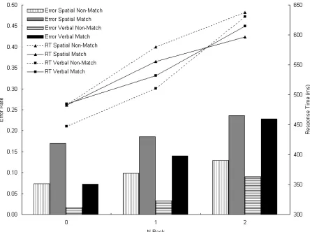
Figure 2-3. Response time (lines) and error rate (bars) in 0-, 1-, and 2-back conditions, separately for spatial and verbal tasks, and separately for match and non-match trials.
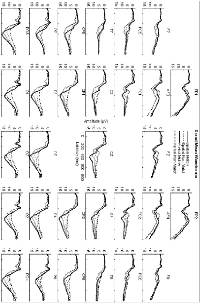
Figure 2-4. Grand mean ERP waveforms, collapsed across the N-back factor, elicited during for spatial (thin lines) and verbal (thick lines) tasks. Solid lines indicate ERPs elicited by matching items are indicated. Dashed lines indicate ERPs elicited by non-matching items.
Figure 2-5. EPC amplitudes elicited during spatial (thin lines) and verbal (thick lines) tasks. Solid lines indicate ERPs elicited by matching items. Dashed lines indicate ERPs elicited by non-matching items.
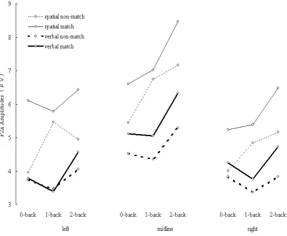
Figure 2-6. P2a amplitudes elicited during spatial (thin lines) and verbal (thick lines) tasks. Solid lines indicate ERPs elicited by matching items. Dashed lines indicate ERPs elicited by non-matching items.
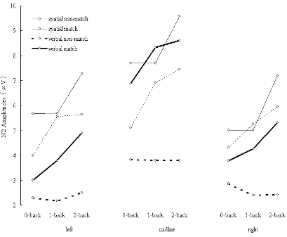
Figure 2-7. N2 amplitudes elicited during spatial (thin lines) and verbal (thick lines) tasks. Solid lines indicate ERPs elicited by matching items. Dashed lines indicate ERPs elicited by non-matching items.
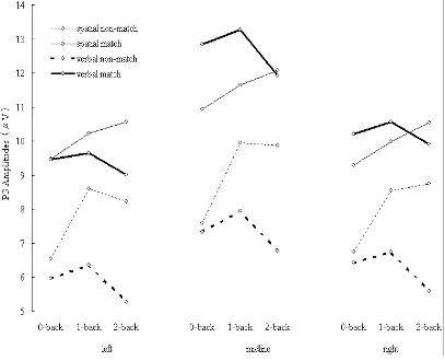
Figure 2-8. P3 amplitudes elicited during spatial (thin lines) and verbal (thick lines) tasks. Solid lines indicate ERPs elicited by matching items. Dashed lines indicate ERPs elicited by non-matching items.
Figure 2-10. Shift effects (2-back minus 1-back difference waveforms), separately for spatial (thin line) and verbal (think line) task instructions, collapsed across stimulus types (match and non-match).
Figure 2-11. Replacement effects (1-back minus 0-back difference) in the three successive latency windows (200-400 ms, 400-600 ms, 600-800 ms), separately for spatial and verbal task instructions, by t-Statistical Maps. White: significant positive; light grey: non-significant positive; dark grey: non-significant negative; black: significant negative.
Figure 2-12. Shift effects (2-back minus 1-back difference) in the three successive latency windows (200-400 ms, 400-600 ms, 600-800 ms), separately for spatial and verbal task instructions, by t-Statistical Maps. White: significant positive; light grey: non-significant positive; dark grey: non-significant negative; black: significant
negative.
Figure 2-13. Replacement amplitudes elicited during spatial (triangle) and verbal (square) tasks. Solid lines indicate ERPs elicited by matching items. Dashed lines indicate ERPs elicited by non-matching items. A: Anterior; C: Central; P:
Posterior; LH: Left Hemisphere; RH: Right Hemisphere.
Figure 2-14. Shift amplitudes elicited during spatial (triangle) and verbal (square) tasks. Dashed lines indicate ERPs elicited by matching items. Solid lines indicate ERPs elicited by non-matching items. A: Anterior; C: Central; P: Posterior; LH: Left Hemisphere; RH: Right Hemisphere.
Figure 3-1. N-back tasks. The upper illustrates logical analyses of n-back tasks. The lower shows trial structure during testing.
Figure 3-2. Response time (lines) and error rate (bars) in 0-, 1-, and 2-back
conditions, separately for ID and IS conditions, for spatial and verbal tasks, and for match and non-match trials.
grey and black labels represent significant positive-going, non-significant positive-going, non-significant negative-going and significant negative-going amplitudes in these electrodes.
Figure 3-4. The t-statistical maps demonstrate effects of spatial shift elicited in IS (Row 1) and ID (Row 2) conditions, and verbal shift elicited in IS (Row 3) and ID (Row 4) conditions, in successive three time windows: 200-400 ms (Column 1), 400-600 ms (Column 2) and 600-800 ms (Column 3). The white, light grey, dark grey and black labels represent significant positive-going, non-significant
positive-going, non-significant negative-going and significant negative-going amplitudes in these electrodes.
Figure 3-5. Replacement (upper) and shift (lower) amplitudes elicited during spatial (triangle) and verbal (square) tasks. Solid lines indicate ERPs elicited under ID condition. Dashed lines indicate ERPs elicited under IS condition. A: Anterior; C: Central; P: Posterior; LH: Left Hemisphere; RH: Right Hemisphere.
Figure 4-1. Analysis of sub-processes involved in 0-back, 1-back, and 2-back tasks.
Figure 4-2. Experimental trial
Figure 4-3. Response time (lines) and error rate (bars) in 0-, 1-, and 2-back conditions, separately for spatial and verbal tasks, and separately for match trials and non-match trials.
Figure 4-4. Grand mean ERP waveforms, collapsed across the N-back factor, elicited during spatial (thin lines) and verbal (thick lines) tasks. Solid lines indicate ERPs elicited by matching items. Dashed lines indicate ERPs elicited by non-matching items.
Figure 4-5. EPC amplitudes elicited during for spatial (thin lines) and verbal (thick lines) tasks. Solid lines indicate ERPs elicited by matching items. Dashed lines indicate ERPs elicited by non-matching items.
Figure 4-7. N2 amplitudes elicited during spatial (thin lines) and verbal (thick lines) tasks. Solid lines indicate ERPs elicited by matching items. Dashed lines indicate ERPs elicited by non-matching items.
Figure 4-8. P3 amplitudes elicited during spatial (thin lines) and verbal (thick lines) tasks. Solid lines indicate ERPs elicited by matching items. Dashed line indicate ERPs elicited by non-matching items.
Figure 4-9. Replacement effects (1-back minus 0-back difference waveforms), separately for spatial (thin line) and verbal (think line) stimuli, collapsed across stimulus types (match and non-match).
Figure 4-10. Shift effects (2-back minus 1-back difference waveforms), separately for spatial (thin line) and verbal (think line) stimuli, collapsed across stimulus types (match and non-match).
Figure 4-11. Replacement effects (1-back minus 0-back difference) in the three successive latency windows (200-400 ms, 400-600 ms, 600-800 ms), separately for spatial and verbal stimuli, by t-statistical maps. White: significant positive; light grey: non-significant positive; dark grey: non-significant negative; black: significant negative.
Figure 4-12. Shift effects (2-back minus 1-back difference) in the three successive latency windows (200-400 ms, 400-600 ms, 600-800 ms), separately for spatial and verbal stimuli, by t-statistical maps. White: significant positive; light grey:
non-significant positive; dark grey: non-significant negative; black: significant negative.
Figure 4-13. Replacement amplitudes elicited during spatial (triangle) and verbal (square) tasks. Solid lines indicate ERPs elicited by matching item. Dashed lines indicate ERPs elicited by non-matching items. A: Anterior; C: Central; P:
Posterior; LH: Left Hemisphere; RH: Right Hemisphere.
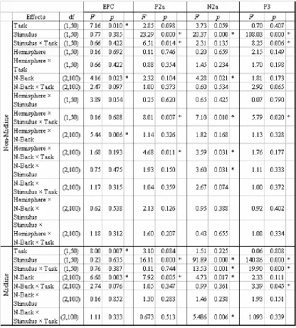
Table 2-1. Omnibus ANOVA of ERP Results (Task = verbal versus spatial task; Stimulus = matching versus non-matching stimulus; Hemisphere = left versus right hemisphere; N-Back = 0-back versus 1-back versus 2-back task;). Significant effects (p < .05) are marked with star.
Table 4-1. Omnibus ANOVA for Original ERPs (Task = verbal versus spatial task; Stimulus = matching versus non-matching stimulus; Hemisphere = left versus right hemisphere; N-Back = 0-back versus 1-back versus 2-back task). Significant effects (p < .05) are marked with star.
Table 5-1. Summary of statistically significant effects in the ANOVA of original ERP waveforms. L/M: Lateral/Medial; L/R: Left/Right; 0/1/2: N-back
Acknowledgement
Summary
Three event-related potential (ERP) experiments investigated the spatial-verbal
dichotomy (emphasized by Baddeley’s model of working memory), selective attention (emphasized by Cowan’s model), and sub-processes in n-back tasks. The studies provide a basis for further clinical research on Alzheimer’s disease.
Experiment 1 studied the spatial-verbal dichotomy using n-back tasks with top-down control. It used identical stimuli in both spatial and verbal tasks, and was designed to eliminate interference from perceptual processes. The spatial and verbal tasks differed only in the instructions given before the tasks. Using a model of the task, sub-processes involved in n-back tasks were delineated and analyzed by
difference waveforms of ERPs. Domain-specific lateralization was found in a shift sub-process but not in a replacement sub-process. Because information from the irrelevant domain could not be totally excluded by top-down control (which
distinguished the spatial and verbal tasks), Experiment 2 recorded information from irrelevant domains. Interactions between irrelevant and relevant domains were found in ERP difference waveforms. Therefore, the results of Experiment 2 suggest that selective attention is unable to exclude interference from the irrelevant domain. Following this conclusion, Experiment 3 adopted a data-driven (bottom-up controlled) methodology, and used different stimuli in spatial and verbal domains in contrast with Experiment 1. In Experiment 3, selective attention was not needed to distinguish spatial tasks from verbal tasks because the spatial and verbal stimuli were different. A different pattern was obtained in Experiment 3; domain-specific lateralization was found only in the replacement sub-process (and not in the shift sub-process).
Abbreviations
AD Alzheimer's disease ANOVA analysis of variance
DLPFC dorsolateral prefrontal cortex EEG electroencephalogram
ERPs event-related potentials
fMRI functional magnetic resonance image MEG magnetoencephalogram
MTL medial temporal lobe
PET positron emission tomography PFC prefrontal cortex
PhD doctor of philosophy RT response time
1.1. What is referred to as working memory?
The term working memory (WM) has been used within cognitive psychology to refer
to the system for temporary maintenance and manipulation of information during a
task (Baddeley, 2002).
1.2. WM models
Two viewpoints on information processing are frequently applied: Baddeley’s and
Cowan’s.
1.2.1. Baddeley’s model
The first one is a computer-like model: domain-specific storage subsystems controlled
by a central processing unit. The multi-component model proposed by Baddeley and
Hitch (1974) is the most influential one for WM, consisting of a visuospatial
sketchpad for storing and manipulating visual and spatial information, a phonological
loop for the corresponding function on phonemic sound information, and a central
executive to control attention and supervise both buffers (Baddeley & Hitch, 1994).
The visuospatial sketchpad was supported by evidence of selective interference with
imagery: the sketchpad can be disrupted by requiring participants to repeatedly
process a specified pattern of locations or keys, a procedure that blocks the use of
visuospatial imagery (Baddeley & Lieberman, 1980). The phonological loop was
are similar in sound is poorer than that of dissimilar items, Conrad & Hull, 1964),
articulatory suppression (with the repetition of an irrelevant sound such as the word
‘the’, the word length effect disappears, Baddeley et al., 1975), irrelevant speech
(articulatory suppression eliminates the effect of phonological similarity when
material is presented visually but not auditorily, Baddeley, Lewis, & Vallar, 1984;
Murray, 1968), and word length effects (the word length effect stems from the greater
fragility of multi-component long words to the processes involved in forgetting, Neath
and Nairne,1995, and Brown and Hulme, 1995). Traces within the phonological
store were assumed to decay over a period of about two seconds (Baddeley & Hitch,
1974). The question of whether short-term forgetting of the phonological loop
represents trace decay or interference remains unresolved (Baddeley, 2002). The
central executive is proposed to be an attentional control system which has access to
long-term memory and is supported by the two buffer systems mentioned above. One
of the functions of the central executive is to integrate information from many
different sources (Baddeley, 1986). The central executive is assumed to play a role
in many cognitive tasks, including those requiring working memory and was
supported by deficits observed in Alzheimer’s disease (AD). AD patients may have
central executive impairment, resulting in the impairment of task performance that
& Spinnler, 1991). There is a very clear tendency in patients for dual task
performance to be impaired while single task performance is maintained. A new
fourth system called the “episodic buffer” is also suggested as a supportive buffer for
the central executive, which acts as the interface between the sub-systems and
long-term memory (Baddeley, 2002). The performance of processing-and-storage
tasks is not clearly distinguishable from storage-oriented tasks in the visuospatial
domain whereas it is distinguishable in the verbal domain (Miyake, Friedman,
Rettinger, Shah, & Hegarty, 2001). This suggests that the visuospatial sketchpad has
a closer relationship with the central executive than the phonological loop.
1.2.2. Cowan’s model
The second viewpoint is that WM is only a temporary emergence of neuronal activity
in the pool (of the whole nervous system). Cowan formulated a model of
information processing integrating memory storage, selective attention, effortful
versus automatic processing, and interactions between these areas (Cowan, 1988).
Cowan's Embedded-Processes Model is a broad-scope information processing
framework originally developed to synthesize a large array of findings on attention
and memory. The mnemonic function preserves information that can be used to do
the necessary work. In this point of view, the memory storage in Cowan’s model is
stating that any processing mechanism that contributes to the desired outcome is said
to participate in the working memory system (e.g., Cowan, 1988). In contrast, some
researchers prefer to define working memory according to the mechanisms
themselves (e.g., Engle, Kane & Tuholski, 1999). Though Cowan's framework has
much in common with those of other researchers, a functional definition of working
memory seems more likely to encourage a consideration of diverse relevant
mechanisms. Some theories of working memory equate it to focus of attention and
awareness (Baddeley, 1993) and some equate it to the sum of activated information
(Atkinson and Shiffrin, 1971).
There are two phases of sensory storage in Cowan’s model (Cowan, 1988). The
first phase extends sensation for several hundred milliseconds, whereas the second
phase is a vivid recollection of sensation. A distinctive aspect of the model is that
attentional focus is determined by automatic attending (Shiffrin and Schneider, 1977),
the automatically preferred processing of cues that have been well-learned, without
the need for an attentional filter. In other words, this model emphasizes automatic
rather than effortful processing in the first phase. Interference between tasks that
occurred in the retrieval of a verbal load during maintenance of a visual array
suggested that verbal and visual loads are stored together in the same buffer (Morey &
during maintaining verbal loads, especially when these items were read aloud.
According to this view, all input information is stored in domain-general neural
networks, but they are retrieved and revived only when attention applies. Whether a
stimulus is spatial or verbal is not determined by the nature of information while
storing the data, but by attention while reviving them. For example, the location of a
verbal code would be retrieved if participants were asked about spatial attributes.
1.2.3. Major differences between models
The major differences between these two models are: first, buffers in Baddeley’s
model are domain-specific whereas the buffer in Cowan’s model is domain-general;
second, an independent central executive controls both buffers in Baddeley’s model
whereas the attentional focus in Cowan’s model is WM per se and refers to only a small part of the so-called buffer. Although different in buffers, both models
separate executive functions from storage and suggest that the latter is manipulated by
the former.
1.3. Categories of WM
Working memory can be viewed in multiple aspects: by models or by tasks. In both
independent unit. Separate spatial and verbal buffers are suggested in Baddeley’s
model but a domain-general buffer was suggested in Cowan’s model. Because the
kinds of buffers are different among models, it is possible that different models have
different information processing. Furthermore, different tasks also possibly have
different information processing. Because WM is defined as the system for
temporary maintenance and manipulation, namely information processing during a
task, it is possible that different models and tasks define different kinds of WM.
1.3.1. Spatial-verbal dichotomy
The spatial-verbal dichotomy is emphasized in Baddeley’s model. Early lesion
studies suggested that the right hemisphere is related to spatial information processing
(McFie, Piercy, & Zangwill, 1950) and the left hemisphere is related to verbal
information processing (Alekoumbides, 1978). Thus, hemispheric specialization in
information processing is domain-specific. Given the bilateral distribution of several sensory-specific brain areas, this generalized conclusion cannot be applied to
more specific regions. Hemispheric specialization in the PFC is considered as
domain-specific between spatial and verbal materials, where verbal materials
activated the left prefrontal cortex (PFC) and visuospatial materials activated the right
Smith & Jonides, 1999) or process-specific between maintenance and manipulation (Owen et al., 1998; Petrides, 1994). In addition to visual materials, an ERP study during auditory N-back tasks (Anourova et al., 1999) showed that there was also a load-dependent segregation between spatial and non-spatial information processing in
auditory working memory.
In addition to topographical differences, spatial-verbal differences in the time
course of processing were also studied. An ERP experiment requiring participants to
maintain memoranda for arithmetical tasks and recording during a task requiring rapid
storage, modification and retrieval of multiple stimuli showed that synchronous peaks
were distributed in occipital, parietal and prefrontal sites from 130 ms after stimulus
onset and continuing after 500 ms (Halgren, Boujon, Clarke, Wang, & Chauvel,
2002). Activity reflecting visual processing occurred in the visual association cortex
from 90 to 130 ms, and projected to fronto-parietal areas from 130 to 280 ms, then the
activity reflecting visual processing occurred from 300 to 400 ms back to the visual
scratchpad in the dorsolateral occipital cortex. Following that, there was a second
reversal from 420-600 ms back to frontal-parietal sites for data renewal. Lateralized
perisylvian oscillations suggested an articulatory loop. A fronto-centro-parietal
'central executive', an occipital visual scratch pad, a perisylvian articulatory loop and
mental operation. This experiment showed interactions among the visual scratch pad,
articulatory loops and the central executive.
The spatial-verbal dichotomy is not clear-cut in some respects. A
magnetoencephalographic (MEG) study showed that the maintenance of words in
working memory activated superior frontal gyri, DLPFC, and superior and inferior
parietal lobes, which are traditionally associated with visuospatial working memory
(Campo, Maestu, Ortiz et al., 2005). Thus, the words were processed as if they were visuospatial information.
To sum up, the spatial-verbal dichotomy has been studied in topography and time
course with different WM tasks, showing left-right hemispheric differences in the
frontal lobe and the whole brain, and different time course of activity in the areas
associated with spatial or verbal processing.
1.3.2. Storage-executive dichotomy
Both Baddeley and Cowan’s models comprise of two different components:
information storage and the executive that operates the storage. Cohen et al. (1997) has studied this dichotomy with fMRI. N-back tasks were used with time-sustaining
memory loads to test active maintenance of memory and time-changing memory loads
to test manipulation. Traditional views suggested that manipulation would activate
regions. Cohen et al.’s study reported that not only posterior regions but also PFC
was involved in active maintenance. In addition to this frontal-posterior difference,
another fMRI study suggested that evidence of both manipulation and maintenance
activity existed in PFC, but DLPFC activity was greater in manipulation (D'Esposito,
Postle, Ballard, & Lease, 1999). In contrast to N-back tasks, Sternberg tasks
(item-recognition tasks) (Sternberg, 1966) separate retention and recognition, which
enables us to investigate maintenance between these two steps. An MEG study
using Sternberg tasks with visually presented digits (Jensen & Tesche, 2002)
suggested theta oscillations generated in the frontal areas during tasks requiring
memory maintenance. An fMRI study during memory tasks with number-letter
distracters (Sakai & Passingham, 2004) showed that both PFC and medial temporal
lobes (MTL) were involved in memory retrieval. PFC was involved in interference
resolution and MTL in rehearsal or reactivating stored information. Transcranial
magnetic stimulation (TMS) and lesion studies (Pierrot-Deseilligny, Muri, Nyffeler, &
Milea, 2005) showed that the DLPFC controlled short-term spatial working memory,
whereas medium-term spatial memory (after 25 s) might be controlled by the MTL.
An fMRI study using verbal working memory tasks reported that both maintenance
and manipulation were affected in schizophrenia (Tan, Choo, Fones, & Chee, 2005).
was more affected than maintenance, and DLPFC activation was relatively reduced
but VLPFC activation relatively increased, in comparison to normal persons. The
results suggest that DLPFC is related to manipulation and VLPFC is related to
maintenance.
1.3.3. Attentional and executive functions
Although working memory is defined as the temporary buffer in tasks, the majority of
WM studies are not related to the stored materials, but to process-specific functions
(Kessels, Postma, Wijnalda, & De Haan, 2000). Thus, what they studied was
theoretically linked to the central executive in Baddeley’s model, whose function is to
control attention and supervise both buffers (Baddeley & Hitch, 1994). Attentional
control is part of WM (the function of the central executive) in Baddeley’s model
whereas attentional focus is in actually WM per se in Cowan’s model. Whether something is in attentional focus is usually indicated by the variation of efficiency in
information processing (e.g., response time or accuracy). In addition to direct task
performance, researchers also use interference to show the effects of attention.
Precisely speaking, attention is regarded as the ability to resolve interference.
An age-comparison study suggested that dual tasks are specific to testing attention
and executive functions (Holtzer, Stern, & Rakitin, 2005). In single tasks, memory
and motor speed were strongest predictors of age, whereas in dual tasks, the attention
and executive factors were the most important predictors of age. The ability to
ignore irrelevant information is an important difference between working memory and
long-term memory (Oberauer, 2001). Patients with frontal lobe lesions and
dys-executive syndrome showed impairment for dual-task coordination, but no
impairment in card sorting (a single visuospatial task) and verbal fluency (a single
verbal task) (Baddeley, Della Sala, Papagno, & Spinnler, 1997).
Topographical and temporal pattern variations were shown during dual tasks. A
functional magnetic resonance image (fMRI) study on dual task performance showed
activation of the prefrontal cortex (PFC) when both tasks were performed together,
but inactivation when they were performed separately (D'Esposito et al., 1995). Activation gain with increasing task loading was also smaller in patients. Another
study demonstrated that thalamic and medial prefrontal cortical regions were activated
in a decision-making task for ambiguous categorization, which reflected the function
of the central executive (Scott, Holmes, Friston, & Wise, 2000). The more
ambiguous difference caused the less activation. In contrast, tasks detecting auditory
central executive participation (Kaiser, Walker, Leiberg, & Lutzenberger, 2005).
1.3.3.2. Interference exclusion
The ability of interference exclusion was shown in a Sternberg’s task where a cue was
given after a memory set to indicate whether the set was relevant or irrelevant.
Increasing the set size usually causes longer response time in the Sternberg’s task.
Setsize effects to negative probes (i.e., probes were from the irrelevant memory set)
last for only one second after the cue. In contrast, setsize effects to intrusion probes
(i.e., probes that were not from the irrelevant memory set) last for at least five seconds
after the cue. These results suggested that irrelevant information is excluded in
working memory after one second. Studies on exclusion also suggested that
attentional focus has limited capacity and is liable to interference. A study using
N-back tasks suggested that only one item could fall within attentional focus at a time
(McElree, 2001). The inability to perfectly maintain a target in focus was evidenced
by decrease of accuracy. Other information outside attentional focus should be
retrieved though a slow search process, which was evidenced by the difference in accuracy between inclusion and exclusion tasks. A study on twin pairs showed that
memory search rate is heritable (Stins et al., 2005).
Neurophysiological studies have also used interference to study selective
memory tasks revealed differences in attention allocation between distracter and
memorized items (Wolach & Pratt, 2001). The N1 (about 200 ms) and P2 (about
200 ms) components differed between distracter items and memorized items,
indicating different attention allocation. The P2 and N2 (about 300 ms) components
indicated differences between probes. P2 was enhanced in response to target stimuli
(“go” response), whereas N2 was enhanced in response to non-target stimuli (“no-go”
response). The P3 component indicated different speeds of scanning and
comparison. P3a amplitude increased with increasing memorized set size, whereas
amplitudes in late P3 components (P3b and P3c) increased with faster and more
accurate response. Another ERP study revealed that distraction elicited a negativity
followed by P3a (350 ms post-stimulus) and then a re-orienting negativity (500 ms
post-stimulus) (Berti & Schroeger, 2001). This result suggested that irrelevant
information causes an attention shift.
Further studies focused on spatial attention research. Selective attention that
shifts on a trial-by-trial basis is called transient selective attention (where participants are informed of the target by a precue at the beginning of every trial), whereas
selective attention that is focused on the same location during the entire experimental
both transient and sustained selective attention, but in the transient condition these
components had shorter latencies and larger amplitudes than in the sustained
condition (Eimer, 1997). P3 target effects in non-spatial visual attributes only
appeared in sustained selective attention conditions but not in transient selective
attention conditions.
1.3.3.3. Anatomical relationships
Frontal areas were suggested to be involved in attentional and executive functions by
neurophysiological studies. Single cell recordings in monkeys reported that the
lateral PFC provided top-down control during visual working memory tasks (Kessler
& Kiefer, 2005). The PFC counteracted interference, whereas the middle temporal
lobe was involved in retrieval. A magnetic resonance spectrometry (MRS) study
showed N-acetylaspartate in the dorsolateral PFC (DLPFC) correlated with activation
of the working memory network including the DLPFC, temporal and inferior parietal
cortices (Bertolino et al., 2000). An fMRI study revealed that anterior PFC was more active during the recognition phase in a working memory task, and more active
in response to non-target probes than to target probes (Leung, Gore, &
Goldman-Rakic, 2005). These findings support the conclusion that the anterior PFC
stimulus distinction. A lesion study of the rat striatum (Bailey & Mair, 2004)
reported a double dissociation between visuospatial response time and radial maze,
delayed non-matching tasks. Dorsal prefrontal lesions caused attentional impairment
that had not been noted in striatal or thalamic lesions and suggested contributions of
PFC to attention. The dissociation between lateral PFC activity and basic memory
demand suggests that the function of the lateral PFC is to reorganize information and
thus reduce task difficulty (Bor, Cumming, Scott, & Owen, 2004). Mathematically
structured sequences encourage chunking and elicit greater lateral PFC activation.
An fMRI study using N-back tasks with letters and fractal figures showed that
maintenance (1-back minus 0-back) activated inferior parietal and DLPFC, with
activation in right ventrolateral PFC (VLPFC) in letter tasks and left lingual gyrus in
fractal tasks (Ragland et al., 2002). Maintenance plus manipulation (2-back minus 0-back) activated inferior parietal, Broca’s area, insula, DLPFC and ventral PFC, with
greater right DLPFC activation for letter tasks. Manipulation-only (2-back minus
1-back) produced equivalent DLPFC and anterior cingulated activation in both tasks.
Nevertheless, a study on patients with partial frontal lesions suggested that executive
processes are not exclusively sustained in the frontal cortex because no difference was
seen between normal subjects and patients during dual tasks (Andres & Van der
attend to location) is mediated by a left-hemisphere dominant parietal-frontal system,
whereas temporal orienting (instruction to attend to time interval) is associated with
sensorimotor areas (Nobre, 2001). An fMRI study on multiple sclerosis patients
with attention and working memory disorder (Cader, Cifelli, Abu-Omar, Palace, &
Matthews, 2006) reported that patients showed relatively reduced activation in the
superior frontal and anterior cingulate gyri than normal persons. A
magnetoencephalogram (MEG) study showed that early spikes or slow waves indicate
attention or task relevance (Aine, Stephen, Christner, Hudson, & Best, 2003). Early
spike-like activity (200 ms post-stimulus) was evoked by a visual working memory
task. In an auditory task, this spike was absent but replaced by slow waves. These
auditory-presented words evoked activity in occipital cortex even though visual
stimuli were not present. Prefrontal activity by the working memory task, which
was along the superior frontal sulcus, was active later than the earliest effect of
attention modulation in visual cortex. This suggests that visual input is
auto-focused.
1.3.3.4. Impairment of attentional and executive functions in diseases
Impairment in active processes of working memory was suggested as a feature of
domain-specific processes seemed not to be affected. Patients in early dementia
have an obvious impairment in the central executive system, which was implicated by
loss of synchrony between brain regions, but the phonological loop system,
represented by Broca’s areas, was shown by positron emission tomography (PET) as
intact at the same time (Morris, 1994). Patients with very mild AD had difficulty in
learning and maintaining words in the middle part of the word list (interfered by both
former and latter words) rather than the words in the beginning of the list (Hashimoto
et al., 2004). This fact implied a functional deficit of the central executive system. Patients with AD suffered from prominent impairments in the shifting and division of
attention (Parasuraman & Haxby, 1993). The first cognitive indicator of neocortical
dysfunction in early AD is attentional dysfunction. Disconnection between frontal
and posterior parietal areas might be the cause of attentional dysfunction in AD
(Clark, Iversen, & Goodwin, 2001). By using tasks selected primarily for the
detection of localized neural disruption within PFC, deficits in sustained attention and
verbal learning were best indicators of manic performance rather than deficits on any
of the tests of executive functioning. In patients with frontotemporal dementia, the
activation gain with increasing memory loads was even more decreased than those
Parkinson’s disease had insufficient mental resources for the central executive to use
and caused poor performance in a dual-task paradigm (Fournet, Moreaud, Roulin,
Naegele, & Pellat, 2000). The assessment for the central executive, for example, the
visual memory span backwards, was able to predict dementia severity such as clinical
dementia rating (CDR) (Cherry, Buckwalter, & Henderson, 1996). Patients of
multiple sclerosis with a working memory problem usually reflected an impaired
central executive system (D'Esposito et al., 1996). A study on multiple sclerosis patients using an auditory N-back task and a task with a significant central executive
component revealed that the primary working memory impairment in multiple
sclerosis patients was within the central executive rather than the phonological loop
(Lengenfelder, Chiaravalloti, Ricker, & DeLuca, 2003). Greater cautiousness and
increased mental effort were found in closed head injury patients recovered from
posttraumatic amnesia (Veltman, Brouwer, van Zomeren, & van Wolffelaar, 1996).
Central executive resources reduced in schizophrenia (Granholm, Morris, Sarkin,
Asarnow, & Jeste, 1997). Schizophrenia patients showed impairment on both the
forward digit span task, a measure of general attention, and the backward digit span
task, a measure of verbal working memory (Conklin, Curtis, Katsanis, & Iacono,
2000). Their non-psychotic relatives showed only impairment on the backward digit
DLPFC dysfunction (Perlstein, Carter, Noll, & Cohen, 2001). The pattern of
performance was not associated with storage but was related to executive functions.
An fMRI study on schizophrenia patients showed that the DLPFC activation
decreased and VLPFC activation increased in the manipulation plus maintenance
verbal task (Tan et al., 2005). Manipulation was more affected in schizophrenia patients. In clinically stable patients, left DLPFC and right cerebellum were still
under-activated and left cerebellum, medial frontal, anterior cingulated and left
parietal cortices were still over-activated (Mendrek et al., 2005). Schizophrenia patients who performed an N-back task used greater prefrontal resources but achieved
lower accuracy (Callicott et al., 2003). Prefrontal-parietal functional disconnection, prefrontal dissociation and abnormal prefrontal-parietal interaction were found during
working memory processing in schizophrenia patients (Kim et al., 2003). A PET study found that schizophrenia patients’ performance in working memory tasks was
vulnerable to high memory load, along with a reduction of blood flow in right DLPFC
(Carter et al., 1998).
1.3.4. Visuospatial processing
1.3.4.1. Visuospatial attention and control
systems exist in the brain. A case report of a patient who experienced two
successive strokes in the right hemisphere showed that stimulus-centered left neglect
happened after the first stroke but body-centered left neglect occurred after the second
stroke (Ota et al., 2003). A study on visual neglect patients found that frequent re-fixating of targets by patients might be due to a lateral bias combined with
impairment of spatial working memory for the fixation point (Husain et al., 2001). A phenomenon termed the attentional blink, which refers to the detection or
discrimination of the second of two successive targets in a rapid serial visual
presentation task was often temporarily impaired (Olivers, 2004). It was suggested
to be due to spatial compression, a systematic localization bias toward the fovea.
During spatial rehearsal, attention was oriented toward the target locations, which was
evidenced by increases in visual processing efficiency for these locations (Awh et al., 1998). The increases were not found in a non-spatial memory task using identical
stimuli. When participants’ attention to memorized locations was blocked, spatial
working memory was impaired. ERP modulations during spatial attention start
about 80 ms after stimulus onset whereas non-spatial visual attention starts about
100-150 ms post onset (Hillyard, 1998). A sequence-learning experiment that
investigated interference between relevant objective information and irrelevant
Soetens, 2006). The participant has to fight automatically captured attention and
leads to longer response time. Threat-evoked anxiety and spatial working memory
shared a common visuospatial attention mechanism (Lavric, Rippon, & Gray, 2003).
A threat of shock was present or absent while performing spatial or verbal N-back
tasks. Anxiety was measured by heart rate. The results revealed that anxiety
blocked performance of spatial tasks but had no influence on verbal tasks.
1.3.4.2. ERP time course effects associated with visuospatial processing
An EEG study evaluating the peak latency of the posterior contralateral negativity
suggested that the time spent in the brain was proportional to the response time in
attentional cueing tasks and stimulus localization tasks, but the effect was weaker in
visual search (Wolber & Wascher, 2005). A study that applied transcranial magnetic
stimulation over the right posterior parietal cortex at different latencies during a
visuospatial task suggested that the effective interference occurred at an early stage of
50 ms post-stimulus, which provided the accurate time course for visuospatial
processing that the right posterior parietal cortex contributes (Pourtois, Vandermeeren,
Olivier, & de Gelder, 2001).
TMS on the bilateral middle temporal area increased reaction time in the
the visuospatial task (Oliveri et al., 2001). The interference was most evident at the latency of 300 ms in both middle temporal and parietal areas. TMS on the superior
frontal gyrus increased response time in visuospatial working memory task, whereas
those on the DLPFC increased response time and error rate in both tasks. The
interference was most evident at the latency of 600 ms rather than 300 ms in both
superior frontal gyrus and DLPFC areas. These findings suggested that there are
separate buffers for object and spatial working memory in the posterior, and DLPFC
for the executive functions regardless of the stimulus types.
1.3.4.3. Spatial-object dichotomy
The disruption of a visual working memory task from visual interference was stronger
than the one from spatial interference, whereas the disruption of a spatial working
memory task from spatial interference was also stronger than the one from visual
interference (Klauer & Zhao, 2004). This phenomenon suggested that visuospatial
working memory should be divided into separate visual and spatial components.
The fact that spatial working memory could be interfered with in the encoding stage
by irrelevant location information also suggested dissociation of spatial and object
memory (Hale, Myerson, Rhee, Weiss, & Abrams, 1996). A study of the
memory tasks reported that in the initial learning tasks Chinese participants tended to
activate the dorsal stream for spatial feature analysis, whereas Caucasians tended to
recruit the ventral stream for object identification (Gron, Schul, Bretschneider,
Wunderlich, & Riepe, 2003).
1.3.4.4. Anatomical relationships
The right hemisphere is associated with global-level processing and spatial coordinate
judgments, whereas the left hemisphere is associated with intact local and categorical
judgments (Schatz, Craft, Koby, & DeBaun, 2004). A study on two callosotomy
patients revealed right-hemisphere superiority for spatial judgements and
left-hemisphere superiority for identity judgments (Corballis, Funnell, & Gazzaniga,
1999). Persons with atypical right hemispheric dominance for language have more
bilateral activation during spatial judgment than typical persons (Jansen, Floel,
Menke, Kanowski, & Knecht, 2005).
A study on monkeys with lesions of areas 9 and 46 in DLPFC reported that
egocentric spatial memory which refers to the map from their own viewpoint was
impaired but allocentric spatial memory which refers to memory of a 3D
environmental map was intact (Ma, Tian, & Wilson, 2003). Prefrontal neurons
related to stimulus identity were different by function and region from neurons related
patients with intracranial tumour after resection also revealed that object memory and
positional memory were separate systems (Kessels, Postma, Kappelle, & de Haan,
2000). Spatial memory problems existed in patients with lesions in either posterior
parietal lobe or in the right hemisphere. An fMRI study for a facial working
memory task presented that three occipitotemporal areas responded transiently to
stimuli indicated perceptual processing (Courtney, Ungerleider, Keil, & Haxby, 1997),
whereas sustained response in three prefrontal areas suggested working memory.
Different degrees of selectivity in visual areas and different strengths of sustained
activity in prefrontal areas revealed a functional specialization from occipital to
prefrontal areas. Another fMRI study on children using a spatial working memory
task (Nelson et al., 2000) reported that subtraction of the activation of the motor condition from the memory condition revealed activity in the dorsal PFC and in the
posterior parietal and anterior cingulate cortex. A study using repetitive TMS to
block the DLPFC (Robertson, Tormos, Maeda, & Pascual-Leone, 2001) revealed that
learning of position were also blocked but object learning was preserved. This effect
could not be found in the similar TMS treatment on parietal lobes. These results
suggested that DLPFC plays an important role in spatial working memory.
The right posterior parietal cortex (PPC) was implicated in visuospatial
study on right hemisphere stroke patients with and without hemineglect (Malhotra et al., 2005) showed that spatial working memory capacity was correlated with severity of left neglect, and the anatomy findings of lesion in the parietal white matter and
insula. In a mental rotation task, female participants presented strong
right-lateralized ERP bias when making non-dominant hand responses (Johnson,
McKenzie, & Hamm, 2002). Male participants showed a right parietal bias
regardless of response hand. The results suggested the importance of considering the
factors of sex and handedness in a spatial manipulation task.
The anterior thalamic nuclei and the hippocampus were critical areas for spatial
memory and work dependently during the performance of certain spatial learning
tasks (Henry, Petrides, St-Laurent, & Sziklas, 2004). An animal study showed that
mice with hippocampal rather than parietal cortex lesion had deficits in measuring
egocentric distance and place map, which implies that the hippocampus appears to be
involved in working memory for egocentric distance and spatial location information,
whereas the parietal cortex was not (Long & Kesner, 1998). A study surveying
children with chromosome 22q11.2 deletion syndrome suggested that the significant
reduction of thalamus, including the pulvinar nucleus, causes visuospatial deficits in
this group (Bish, Nguyen, Ding, Ferrante, & Simon, 2004). An MEG study using a
the MTL showed sustained activation from 200 to 800 ms post-stimulus in a spatial
working memory task, whereas the activation sustained from 200 to 400 ms
post-stimulus in perceptual tasks (Campo, Maestu, Capilla et al., 2005). In the period 200 to 400 ms post-stimulus, both tasks presented the same activation. This
result suggests that pure encoding starts only after 400 ms post-stimulus. A study on
temporal lobectomy patients reported that the right anterior temporal lobe stores
long-term allocentric spatial memory for the reference of spatial working memory
(Feigenbaum & Morris, 2004).
1.3.5. Verbal processing
1.3.5.1. Anatomical relationships
Verbal working memory is generally thought to be housed in a left-dominant neural
network, including parietal, temporal and PFC. An evoked-potential study on aphasic
patients reported that in normal persons and non-aphasic brain-injured patients the left
hemisphere was activated during a verbal memory task, whereas in recovered
aphasics the right hemisphere was activated (Papanicolaou, Levin, & Eisenberg,
1984). Left parietal and right frontal positivities are usually observed by ERP
studies in verbal recognition tasks (Graham & Cabeza, 2001). For non-verbal
effect was noted for neutral faces. The parietal positivities could not be seen in
non-verbal stimuli, implying that the left lateralization of parietal activities reflected
verbal processing.
A study using repetitive TMS during and after the performance of a verbal
working memory task suggested a symmetrical, bilateral parietofrontal verbal
working memory network (Mottaghy, Doring, Muller-Gartner, Topper, & Krause,
2002). The result was explained as a parietofrontal central executive network during
the processing of semantic and objective features. Verbal working memory
performance activated Broca's area, the left premotor cortex, the cortex along the left
intraparietal sulcus and the right cerebellum (Gruber, 2001). After silent articulatory
suppression that blocked the participant’s rehearsal, no significant memory-related
activation was found in these areas. However, non-articulatory maintenance
occurred instead in anterior prefrontal and inferior parietal lobes. An fMRI study
suggested that rehearsal was optional but encoding was an obligatory component of
the phonological loop, which is located in the left inferior frontal gyrus, right lateral
cerebellum and medial frontal gyrus (Li et al., 2004). Another fMRI study using a sentence-pair matching task revealed that left dorsal frontal and left inferior parietal
regions were more activated with increasing memory load. The left parietal lobe
in mismatches (D'Arcy, Ryner, Richter, Service, & Connolly, 2004).
1.3.5.2. Word and sentence processing
An fMRI study on verbal working memory revealed that supplementary motor,
premotor and inferior frontal areas are associated with maintenance, and left inferior
frontal and supplementary motor regions are associated with articulatory rehearsal
(Chein & Fiez, 2001). Another fMRI study on Chinese reading in dyslexia patients
revealed that both the processes from orthography to syllable and from orthography to
semantics involve left middle frontal gyrus (Siok, Perfetti, Jin, & Tan, 2004). An
ERP study also on Chinese reading found that N2 on the right anterolateral scalp in
the time window 182-240 ms post-stimulus was significantly larger for target stimuli
in the visual word recognition task, whereas the N2 on the left anterolateral scalp in
the time window 262-350 ms post-stimulus was larger for non-target stimuli (Wang,
Tang, Kong, Zhuang, & Li, 1998).
Sentence comprehension involves the posterior middle and superior temporal
gyri which process sentence structure, the anterior temporal gyrus which processes
sentence context, and left inferior frontal cortex which supports verbal working
memory task is related to different working memory modules (Roberts & Gibson,
2002). A PET study showed that sentence listening activated the bilateral superior
and middle temporal gyri but left side activation was stronger. Sentence generation
involved the left middle and inferior frontal gyri, and left inferior temporal lobe
(R.-A. Muller et al., 1997). An fMRI study on schizophrenia patients suggested that activation in the left inferior frontal gyrus is reduced in patients with verbal working
memory deficits (Stevens, Goldman-Rakic, Gore, Fulbright, & Wexler, 1998).
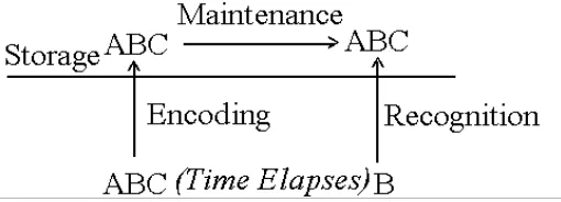
[image:49.595.114.369.441.533.2]1.4. Laboratory tasks and sub-processes of WM
Figure 1-1 Logical analyses of item-recognition tasks
1.4.1. Item-recognition tasks and the traditional model: encoding, active maintenance and recognition
Sternberg, 1966) comprise of clear-cut stages of encoding, maintenance and
recognition (See Figure 1-1) and reflect the traditional model of
encoding-storage-retrieval processes. Encoding is a conscious process which
transfers and screens physical information into relevant neural signals. Active
maintenance is conscious processes keeping neural signals alive. Recognition is the
stage that compares the retrieved memory with the target and generates task
performance indices, namely response time and error. However, not every WM task
has clear steps of encoding, active maintenance and recognition, for example, N-back
tasks, which mix these steps together.
1.4.1.1. Experimental evidence of the proposed sub-processes
An ERP study of an item-recognition task (Ruchkin et al., 1992) required participants to memorize items in the beginning and then recognize them later. It showed that
domain-specific differences occurred in early ERPs in the posterior N220 component,
and a phonological loading effect was found at the P300 component by increasing
amplitudes with increasing loads, but visuospatial loading effects were not found at
the same latency. The encoding period for visuospatial stimuli started by an abrupt
maintenance rehearsal, with a significant shift in lateralization from right dominant to
the midline in the retention interval. The topographic transition at 600, 1000 and
3000 ms post-stimulus marked the more complex encoding activity of the
phonological task.
1.4.2. N-back tasks: information manipulation and executive functions
Although the traditional encoding-storage-retrieval model is broadly assumed in
memory research, the stepwise computational model is criticized because the
components are chosen arbitrarily and represents merely a computational account that
may not reflect actual human memory (Malafouris, 2005; O'Reilly, Braver, & Cohen,
1997; Riegler, 2005). The N-back tasks provide another viewpoint for working
memory, which has been commonly used in electrophysiological and imaging studies
on WM (Gevins & Cutillo, 1993; Jansma, Ramsey, Coppola, & Kahn, 2000; Smith &
Jonides, 1997).
1.4.1.1. Task description
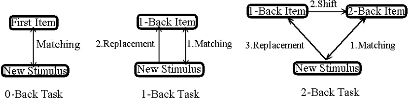
In the N-back task, the participant is shown a series of items (e.g., letters, words or
location markers) and is asked to decide, upon presentation of each item, whether a
presentations back. Figure 1 displays a schematic diagram of the 0-, 1- and 2-back
tasks. If n = 0, each new item is matched against the very first item in the series. If
n = 1, each new item is matched against the immediately preceding item, and if n = 2,
the new item is matched against the item presented just before the preceding item.
Researchers currently prefer the N-back task in studies of WM because it taps
into processes involving manipulation as well as maintenance of information in WM.
(e.g., Meegan, Purc-Stephenson, Honsberger, & Topan, 2004; Ragland et al., 2002). Impairment in executive function of WM is suggested as a feature of early
Alzheimer’s dementia (Hashimoto et al., 2004; Morris, 1994; Vecchi & Cornoldi, 1999; Vecchi, Saveriano, & Paciaroni, 1998), and, in these patients, continuous
deterioration of WM performance is noted in dual rather than single task settings
(Baddeley, Bressi, Della Sala, Logie, & Spinnler, 1991). Thus, a clear delineation of
WM processes is important from both theoretical and clinical points of view.
1.4.2.2. Experimental evidence from N-back tasks
Intuitively, matching and other WM manipulations in N-back tasks are simultaneous.
An event-related potential (ERP) study found that P300 latency was constant with
increasing N but P300 amplitudes increased with increasing N (Watter, Geffen, &
better) whereas P300 amplitude reflects attention and memory loading (the larger the
harder). As a result, the N-back task is considered a dual task with parametrically
increasing attentional and memory loading along with constant loading of an
implanted matching subtask. Because matching load is purportedly constant,
matching effects can be potentially eliminated by comparing different N-back tasks,
and memory effects clarified.
1.4.2.3. What is lacking in previous studies using the N-back task?
Although N-back tasks are broadly used in WM studies, its sub-processes have not
been sufficiently elucidated. It has been suggested, however, that the literature in
fact lacks a thorough task analysis of the N-back paradigm, and that this leaves room
for seemingly reasonable assumptions that may prove to be unsustainable under closer
inspection. For example, task analysis (Meegan, Purc-Stephenson, Honsberger, &
Topan, 2004) has cast doubt on the commonly held assumption that spatial and verbal
N-back tasks actually tap into spatial and verbal WM processes, respectively. Based
on behavioral studies using letter and position N-back tasks, these authors concluded
that irrespective of the actual stimulus material or task demands, N-back task
performance always involves both spatial and verbal processing (See also Meegan &
Jonides, 1997) showing that in an N-back task where letters were presented at
different positions, activity in both cerebral hemispheres was obtained both under
verbal task instructions (matching letter identity) and under spatial task instructions
(matching letter position). However, because activity was lateralized slightly to the
left under verbal instructions, and slightly to the right – at least in some areas – under
spatial instructions, these authors concluded that verbal and spatial WM are in fact
mediated by different neural substrates. Because temporal resolution was too low to
analyze sub-processes in previous imaging studies (Ragland et al., 2002; Smith & Jonides, 1997), earlier studies revealed only summation of overlapped sub-processes
during a particular long period.
1.5. Imaging and electrophysiological methods in working memory
research
Modern technology enables us to show brain activity directly. Here I compare PET,
fMRI, EEG and ERPs and elaborate on ERPs.
1.5.1. PET
This method applies radioactive isotopes (usually Xe133) to label blood molecules.
detected. PET has excellent topographical resolution of a few millimetres (Aguirre,
2003). However, it has very poor temporal resolution limited by the half-life of the
radioisotope used. It limits the ability of the method to dynamically track changes in
neural activity related to cognitive processes.
1.5.2. fMRI
Functional MRI is based on the increase in blood flow to the local vasculature that
accompanies neural activity in the brain. This results in a corresponding local
reduction in deoxyhemoglobin because the increase in blood flow occurs without an
increase of similar magnitude in oxygen extraction (Fox and Raichle, 1985). fMRI has
good spatial and temporal resolution. Because it can provide real-time information,
an event-related fMRI is often applied in cognitive psychology experiments. A
problem is that fMRI is expensive. Furthermore, although the temporal resolution of
fMRI is higher than PET, it is not high enough for psychological responses that occur
at the level of a few milliseconds. Also, participants need to keep steady during
testing to avoid motion artifacts.
1.5.3. EEG
Electrophysiological tools provide excellent temporal resolution at the level of
electrical potentials at the scalp, and is suggested to reflect the activity of the cortex
below the electrodes. The analysis of EEG is usually frequency and amplitude-based.
Electrical pulses from muscles, (for example, eye-blinking) usually cause artifacts.
Magnetoencephalogram (MEG) records magnetic field instead of voltages and
provides higher spatial resolution than EEG. ERPs are EEG per se with further
processing.
1.5.4. ERPs
ERPs are superimposed time-locked EEG epochs. Therefore, peaks, wave troughs
and noises are neutralized in ERPs. ERPs provide more detailed temporal
information of the underlying processes involving in a task. It actually offers the
best temporal resolution of all imaging techniques. The main limitation is that it
needs multiple presentations of stimuli. Furthermore, disadvantages in EEG, for
example, vulnerability to muscle artifacts and poor spatial resolution, also exist in
ERPs.
The traditional task inducing ERP components is the oddball paradigm. The
most discussed ERP components are P3 (P300), whereas N1, P2 and N2 have also
been discussed for more than 20 years (Knight, Hillyard, Woods, & Neville, 1981;