Original Article
Analysis of different methods of extracting DNA and
RNA from paraffin-embedded tissues
Qingqing Yi1*, Guowei Jiang2*, Junfeng Shi4, Dongyu Liang1, Shuang Sha4, Rong Yang3,Qing Chang1
1Clinical Research Center, Departments of 2Pharmacy, 3Pathology, Jiading District Central Hospital Affiliated
Shanghai University of Medicine & Health Sciences, No. 1 Chengbei Road, Jiading District, Shanghai 201800, China; 4Shanghai Key Laboratory for Molecular Imaging, Shanghai University of Medicine and Health Sciences,
279 Zhou Zhu Highway, Shanghai 201318, China. *Equal contributors.
Received January 7, 2019; Accepted February 12, 2019; Epub May 15, 2019; Published May 30, 2019
Abstract:The aim of this research study was to identify the optimal method of extracting DNA and RNA from for-malin-fixed, paraffin-embedded (FFPE) tissues, and to improve amplification of gene fragments with the method. Twenty FFPE specimens from 2018 were randomly selected. Total DNA was extracted using a kit method and phe-nol/chloroform method, and total RNA was extracted using a kit and Trizol method. DNA and RNA concentrations and OD260/OD280 ratio were determined using a NanoDrop-2000 spectrophotometer. The human β-globin gene was amplified using a LightCycler 480 Real-Time PCR System. Gel electrophoresis was performed using 1% agarose for PCRamplification products. One-way ANOVA was used to assess the DNA and RNA quality extracted by different techniques. The experimental results showed that the concentration and purity of DNA and RNA and the success rate of fluorescence quantitative PCR amplification were all higher when extraction was performed using the kit. This shows that the quality of DNA and RNA extracted from the FFPE tissue samples using the kit method is reliable and the method can be used for clinical amplification of some gene fragments.
Keywords: FFPE tissues, DNA and RNA extraction, internal reference gene amplification, comparison of extraction methods
Introduction
Formalin-fixed, paraffin-embedded (FFPE) tis-sues are the most widely used and easily ob- tained specimens in clinicopathological diagno-sis [1]. Extraction of DNA and RNA from the samples not only solved the problem of the small number of cases, but it also effectively combined the results of gene detection with retrospective analysis of tumors [2]. However, formaldehyde immobilization and paraffin em- bedding can easily cause DNA and RNA to degrade and encourage cross-linking of his-tones. Thus, it is difficult to amplify PCR prod-ucts, which limits the research work. Studies have shown [3] that the purity, concentration, and PCR amplification rate of DNA and RNA obtained by different extraction methods are quite different. In this paper, two DNA extrac-tion methods (kit method and phenol/chloro-form method) and two RNA extraction methods (kit method and Trizol method) were compared
in order to select the most suitable methods of DNA and RNA extraction.
Materials and methods
Reagents
Faure Marin, ethanol, xylene, phenol, chlorofo- rm and isoamyl alcohol were purchased from the National Pharmaceutical Corporation (Sh- anghai, China). DNA/RNA extract kits were got-ten from Qiagen (Germany). The primers were synthesized by Sangon Biotech Co., Ltd (Sha- nghai, China). 5× All-In-One MasterMix are pur-chased from abm (Zhenjiang, China). Premix Ex TaqTM II were bought from TaKaRa (Japan).
Tissue specimens
Jiading District Central Hospital Affiliated Sh- anghai University of Medicine & Health Scie- nces. Paraffin blocks were selected to carry out complete serial sections, according to the ar- chived HE staining sections. All of the speci-mens were fixed using 10% neutral formalin solution and embedded in conventional paraf-fin. The selected specimens were preserved completely, and these were observed under a microscope with no tissue autolysis, necrosis, or massive hemorrhage. All of the articles were sterilized and 4-6 sections of FFPE 20-μm thick were placed in a 1.5-mL EP pipe for backup.
DNA extraction
Phenol/chloroform extraction:① Dewaxing: 1 mL of dimethylbenzene was added to FFPE tis-sues in the 1.5-mL EP pipe and thoroughly shaken using 3000 rpm centrifugation for 2 minutes. Then, the supernatant was discarded and 1 mL of absolute ethanol was put in to mix, with 3000 rpm centrifugation for 2 minutes. Subsequently, the supernatant was discarded and the specimen was repeatedly dewaxed. Finally, 100%, 95%, and 75% gradient ethanol was affiliated in turn to wash the residual tolu-ene. ② Digestion: The dewaxed tissues were
incubated in a 37°C incubator for 15-30 min-utes, and then 200 μL STE solution (100 mmol/L Tris-HCl, pH 8.0, 20 mmol/L EDTA, 0.8% (W/V) SDS), was added. The mixture was heated in water bath at 90°C for 10 minutes, then 20 g/L protease K was affiliated and incu-bated in water bath at 55°C for 3 hours until the floc disappeared. After that, the mixture underwent 3000 rpm centrifuge for 1 minute and then the supernatant was obtained. ③
Purification: DNA was extracted using saturat-ed phenol, and phenol/chloroform/isoamyl al- cohol (25:24:1), successively. Then, 2 times vol-ume of anhydrous ethanol and 1 times volvol-ume of 3% sodium acetate was added differentially. Finally, the purified product was stored in -20°C refrigerator for the night, and DNA was precipi-tated. ④ Dissolution: 10,000 rpm centrifuga-tion for 8 minutes, then DNA was dissolved with TE buffer (100 μL pH 8.0), and reserved at -20°C.
Kit method: DNA was extracted and purified according to the instructions of QIAGEN All Prep® DNA/RNA FFPE Kit. The main steps were as follows: ① Xylene dewaxing. ② Cell lysis + protease digestion. ③ Adsorption column for DNA. ④ TE buffer for DNA dissolution.
RNA extraction
Trizol method:① 10 μL Proteinase K was added
to the EP tube containing tissue slices for diges-tion. ② 1 ml Trizol was used according per
50-100 mg of tissue samples and transferred into the centrifugal tube. ③ Centrifugation for 5 minutes at 12,000 rpm to discard the precipita-tion, then 200 ml chloroform was added accordingly to 1 mL Trizol, shaken and mixed for 15 seconds, and stored at room tempera-ture for 10 minutes. ④ Centrifugation for 15
minutes at 12,000 rpm, then the upper water phase was absorbed. Then, 0.6 mL isoamyl alcohol was added accordingly to 1 mL Trizol, mixed, and stored for 5-10 minutes at room temperature. ⑤ Centrifugation at 4°C for 10
minutes at 12,000 rpm and supernatant was discarded, then 1 mL 75% ethanol was mixed accordingly to 1 mL Trizol, oscillated gently, and suspended. ⑥ RNA was dried at room
tempera-ture, dissolved with 50 μL RNA-free ddH2O, and reserved at -20°C.
Kit method: RNA was extracted and purified according to the instructions of QIAGEN All Prep® DNA/RNA FFPE Kit. The main steps were as follows: ① Xylene dewaxing. ② Cell lysis +
protease digestion. ③ Adsorption column for RNA; ④ RNA-free ddH2O for DNA dissolution.
DNA and RNA quality control
Concentration and purity of DNA and RNA: The yield and quality (OD260/OD280) of the DNA and RNA products were measured by a spectropho-tometer (NanoDrop-2000, Thermo Scientific). When OD260/OD280 is 1.8±0.1, the total DNA extracted is qualified, while OD260/OD280 is 2.0±0.1, the total RNA extracted is qualified. DNA and RNA electrophoresis: electrophoresis on 1% agarose gels stained with ethidium bro-mide. Degradation of DNA and RNA, and ampli-fication of internal reference genes were ob- served using BTS-20.M automatic digital imag-ing system and LUV-260D ultraviolet projector (Tanon 1600, Tanon).
Amplification of internal reference gene
Data analysis
Data are represented as the mean ± stan-dard error of the mean. Statistical signifi-cance was analyzed by unpaired Student’s
[image:3.612.88.303.147.394.2]t test or oneway analysis of variance, using GraphPad v5.0 software (GraphPad Sof- tware, Inc., La Jolla, CA, US). P<0.05 was
Table 1. Primer sequences and fragments of amplified products of β-globin gene
Primers Sequences Products Forward Primer 5’-GAAGAGCCAAGGACAGGTAC-3’ 268 bp Reverse Primer 5’-CAACTTCATCCACGTTCACC-3’
Fluorescence quantitative PCR amplification was performed using TaKaRa SYBR® Premix Ex TaqTM II (Tli RNaseH Plus). The cycling protocol was as follows: denaturation at 95°C for 30 seconds, 30 cycles of amplification (5 seconds at 95°C, and 20 seconds at 60°C). After PCR, the amplification of internal reference genes was analyzed by curve and Cp value of PCR amplification. Meanwhile, the amplified prod-ucts of PCR were electrophoresed on 1% aga-rose gel containing ethidium bromide. A 100 bp DNA ladder marker was used as the standard reference (Marker). Pictures were taken after electrophoresis to observe whether the size of the electrophoretic band met the experimen- tal requirements. Primer sequences and frag-ments of amplified products of β-globin gene were seen in Table 1.
considered to indicate a statistically significant difference.
Results
OD260/OD280 ratio of specimens in each group
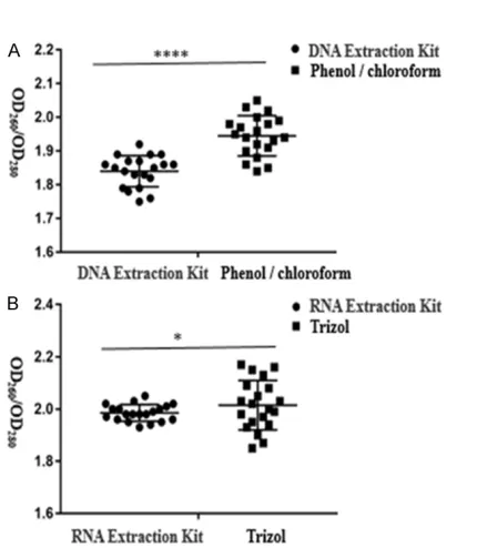
The OD260/OD280 ratio of DNA extracted by the two methods is shown in Figure 1A: OD260/ OD280 of DNA extracted by kit was 1.84±0.01; while OD260/OD280 of DNA extracted by phenol/ chloroform was 1.945±0.01. The OD260/OD280 ratio of RNA extracted by the two methods is shown in Figure 1B: OD260/OD280 of RNA extract-ed by kit was between 1.9 and 2.1; while OD260/ OD280 of RNA extracted using the trizol method differed significantly between 1.85 and 2.20. These results show that the purity of DNA and RNA extracted using the kit method was great-er than by the othgreat-er method, and the diffgreat-erence was statistically significant.
Comparison of DNA and RNA concentration in each group
The concentration of DNA extracted by the two methods is shown in Figure 2A. The concent- ration of DNA extracted using the kit was 244.7±10.89 ng/μL, while the concentration of DNA extracted using phenol/chloroform was 195.3±9.11 ng/μL. The concentration of RNA extracted by the two methods is shown in Fig- ure 2B. The concentration of RNA extracted by kit was 291.7±16.81 ng/μL, and the concentra-tion of RNA extracted using the Trizol method was 212±16.21 ng/μL. The results show that the concentration of DNA and RNA extracted by the kit method was higher, and the difference was statistically significant.
Electrophoresis of DNA and RNA with different extraction methods
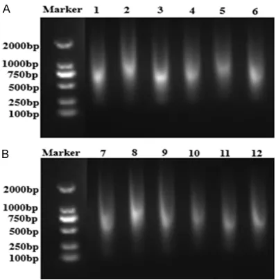
The molecular weight of DNA and RNA obtained using the kit extraction method and the tradi-tional extraction method were not found to be significantly different upon electrophoresis. Bo- th of them were 2000-100 bp diffusion bands. Figure 1. Purity of DNA and RNA extracted by
As shown, the extracted DNA and RNA were all degraded seriously (Figure 3A, 3B).
Amplification results of human β-globin gene
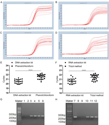
Human β-globin gene in DNA and RNA extract-ed using different methods was amplifiextract-ed by fluorescence quantitative PCR, and the kinetic curves (Figure 4A-D) and CP values (Figure 4E,
4F) were analyzed. The CP value of amplifica-tion products from DNA extracted using the kit and phenol/chloroform was partly 20.85±0.16 and 21.71±0.15 and the CP value of amplifica-tion products from RNA extracted by kit and Trizol method was 20.96±0.09 and 21.55±0.12,
respectively. The results show that the Cp value of DNA and RNA extracted with kit was smaller and the success rate of amplification was hi- gher.
PCR products of human β-globin gene were detected using 1% agarose gel. Clear internal reference bands of DNA and RNA extracted by the kit were found at 268 bp (Figure 4G, 4H). The internal reference bands of DNA and RNA extracted using traditional phenol/chloroform extraction and Trizol extraction were fuzzy or invisible (Figure 4G, 4H). The results show that the success rate of amplification of DNA and RNA extracted by kit was higher.
Discussion
The large number of FFPE tissues collected by hospital Pathology Departments make up an important source of research materials. The most difficult problem of DNA and RNA extrac-tion from FFPE tissues is the degradaextrac-tion of DNA and RNA and protein cross-linking [4-6]. Most scholars believe that the cause of DNA and RNA degradation is that formalin is easily oxidized into formic acid in the air [7, 8], and formic acid has a strong degradative effect on DNA and RNA. Moreover, tissue immobilization causes extensive cross-linking of nucleic acids and histones, forming a strong complex, which can easily cause DNA and RNA breakage. Through experiments and literature review, the following conclusions have been made. ①
[image:4.612.82.524.72.164.2]Selection: It is advisable to avoid performing examination of parts of the sample containing hemorrhage, necrosis, or autolysis, and select densely packed areas. ② The thickness of slic-es: The tissues fixed by formalin and embedded in paraffin become tough and difficult to homog-enize. In the past, FFPE tissues were cut into Figure 2. The concentration of DNA and RNA extracted by different methods. A. The concentration of DNA extracted by kit and phenol/chloroform was compared. B. The concentration of RNA extracted by kit and Trizol method was compared. **P<0.01.
[image:4.612.90.289.225.427.2]3-5-μm slices. However, some studies have suggested that [9] DNA and RNA are easily switched off if the slices of FFPE tissues are so thin. It also affects the acquisition of
[image:5.612.93.522.71.560.2]RNA were all better. ③Other factors: Repea- tability of PCR amplification and complex oper-ation conditions are difficult to control using traditional organic solvents for extraction of DNA and RNA. Furthermore, the extracted DNA and RNA have poor practicability. In contrast, DNA and RNA extracted by the kit method are more conducive to PCR amplification [10]. In this experiment, DNA and RNA were extracted by a Qiagen All Prep® DNA/RNA FFPE Kit. This kit can extract DNA and RNA simultaneously. The purity of DNA and RNA basically met the experimental requirements. Over 99% of the samples were able to amplify the internal refer-ence bands.
In summary, the broken fragments of nucleic acids extracted from FFPE are arbitrary, which depends on many factors, such as the time intervals from sampling to fixing in the agent, the time of preservation of wax blocks, the ty- pes of fixer, the fixed time and the fixed tem-perature. These factors are influential in molec-ular biology research of FFPE specimens. DNA was extracted using the kit method and phenol/chloroform method and RNA was ext- racted using the kit method and Trizol method, both from 20 FFPE tissues in this research. The results show that the purity and concentration of DNA and RNA extracted using the kit method are higher than those of another traditional extraction method. The 1% agarose gel electro-phoresis showed that the DNA and RNA extract-ed using different methods all had different degrees of degradation. Fluorescence quanti-tative PCR was used to amplify human β-globin gene in the extracted DNA and RNA. It was found that the Cp values of DNA and RNA extracted by kit method were smaller and the amplified bands were clearer. In summary, the extraction of DNA and RNA by kit is more suc-cessful than traditional extraction methods.
Acknowledgements
We would like to thank LetPub (www.letpub. com) for providing linguistic assistance during the preparation of this manuscript. This study was supported by institutional funding (SFP-18-20-16-007, seed fund of Shanghai University of Medicine & Health Sciences), the National Natural Science Foundation of China (Grant Numbers: 81670968, 81702284), the Natural
Science Foundation of Shanghai, China (Grant Numbers: 16ZR1430200).
Disclosure of conflict of interest
None.
Address correspondence to: Qing Chang, Clinical Research Center, Jiading District Central Hospital Affiliated Shanghai University of Medicine & Health Sciences, No. 1 Chengbei Road, Jiading District, Sh- anghai 201800, China. Tel: 021-67073029; E-mail: [email protected]
References
[1] Van Wesenbeeck L, Janssens L, Meeuws H, La- gatie O, Stuyver L. Droplet digital PCR is an ac-curate method to assess methylation status on FFPE samples. Epigentics 2018; 13: 207-213. [2] Lee JW, Shin JY, Seo JS. Identification of novel
mutations in FFPE lung adenocarcinomas us-ing DEPArray sortus-ing technology and next-gen-eration sequencing. J Appl Genet 2018; 59: 269-277.
[3] Zlatko J, Renata JA, Aleksandar S, et al. Com- parative study of two dna extraction methods in different tissues and conditions of degrada-tion. Forensic Science International: Genetics Supplement Series 2015; 5: e403-e404.
[4] Choudhary A, Mambo E, Sanford T, Boedig- heimer M, Twomey B, Califano J, Hadd A, Oliner KS, Beaudenon S, Latham GJ, Adai AT. Eva- luation of an integrated clinical workflow for targeted next-generation sequencing of low-quality tumor DNA using a 51-gene enrichment
panel. BMC Med Genet 2014; 7: 62.
[5] Malentacchi F, Pazzagli M, Simi L, Orlando C, Wyrich R, Hartmann CC, Verderio P, Pizzamiglio S, Ciniselli CM, Tichopad A, Kubista M, Gelmini S. SPIDIA-DNA: an external quality assessment for the pre-analytical phase of blood samples used for DNA-based analyses. Clin Chim Acta 2013; 424: 274-286.
[6] Zhang H, Korenková V, Sjöback R, Švec D, Bj- örkman J, Kruhøffer M, Verderio P, Pizzamiglio S, Ciniselli CM, Wyrich R, Oelmueller U, Kubista M, Lindahl T, Lönneborg A, Rian E. Biomarkers for monitoring pre-analytical quality variation of mRNA in blood samples. PLoS One 2014; 9: e111644.
[7] Luciano DJ, Vasilyev N, Richards J, Serganov A, Belasco JG. Importance of a diphosphorylated intermediate for RppH-dependent RNA degra-dation. RNA Biol 2018; 15: 703-706.
[10] Ghatak S, Lallawmzuali D, Lalmawia, Sapkota R, Zothanpuia, Pautu JL, Muthukumaran RB, Senthil Kumar N. Mitochondrial D-loop and cy-tochrome oxidase C subunit I polymorphisms among the breast cancer patients of Mizoram, Northeast India. Curr Genet 2014; 60: 201-212.
ensure accurate DNA repair. J Biol Chem 2017; 292: 10779-10790.