© 2018 IJSRST | Volume 4 | Issue 2 | Print ISSN: 2395-6011 | Online ISSN: 2395-602X Themed Section: Science and Technology
Identification and Characterization of Gut Microflora of Local Earthworms – A
Controlled Laboratory Study
Sethulakshmi K .C*1,Lakshmi R.2, A. P. Thomas3
*1School of Environmental Sciences, Mahatma Gandhi University,Priyadarshini Hills PO, Kottayam, Kerala, India
2Kerala Veterinary and Animal Sciences University, College of Veterinary and Animal Sciences, Pookode, Wayanad. Lakkidi, Kerala, India
3Advanced Center of Environmental Studies and Sustainable Development, School of Environmental Sciences, Mahatma Gandhi University, Priyadarshini Hills, Kottayam, Kerala, India
ABSTRACT
A study was carried out on the Isolation, identification and characterization of gut microflora of local earthworms (Pontoscolex corethrurus, Megascolex konkanensis, Drawida ghatensis). The present research was carried out to identify the microbial analysis of earthworm gut and Isolation, identification and characterization of gut microflora of local earthworms. A bacterium was observed throughout the gut. The fungal and actinomycetes were isolated from the gut. Isolated microflora were identified as Streptococcus sp, Bacillus subtilus, Bacillus cereus, Pediococcus acidilactici, Staphylococcus aureus, Bacillus acidicola, Salmonella sps.,
Pseudomonas sp, Strepto coccus sp, Bacillus sps, Candida spp. and Aspergillus spp was observed.
Keywords : Bacteria, Fungi, Actinomycetes, Gut Contents.
I.
INTRODUCTION
Earthworms are natural invertebrates of agro ecosystem belonging to the Phylum Annelida, Class – Chaetopoda and Order Oligochaeta and dominant in the temperate and tropical soils. Earthworms, as ecosystem engineers, play an important role in many soil ecosystems and are one of the numerous ranges of burrowing organisms, which improve soils (Lavelle 1997). Due to their relatively large size and characteristic feeding behaviour, certain species have significant impacts on soil structure, soil fertility, plant growth and crop yields. The activities of earthworms in soils have been shown to have profound impart on the soil ecosystem functioning as well as on the types and numbers of micro-flora and micro-fauna (Pederson et al. 1993). Earthworms ingest soil microorganisms along with organic residues from the soil and during passage through the earthworms intestinal tract, their population may increase. .
Earthworm activity changes the microbial community structure of soil. It has been proposed that earthworms have a mutualistic relationship with microorganisms (Barois and Lavelle 1986; Lavelle et al., 1995) and may contribute to the maintenance of soil fertility and soil microbial diversity.
Objectives:
To analyse the gut microflora of local earthworms
(Pontoscolex corethrurus, Megascolex konkanensis,
Drawida ghatensis).To identify the morphological and
biochemical characterization of the earthworm gut microflora.
II.
MATERIALS AND METHODS
The study mainly focused on three species of earthworms both exotic and native such as
konkanensis (Fedarb,1898), and Drawida ghatensis
(Michaelsen,1910).
1. Pontoscolex corethrurus (Muller,1856)
Figure 1. Pontoscolex corethrurus
Phylum : Annelida Class : Clitellata Order : Haplotaxida Suborder : Lumbricina Family : Glossoscolecidae Genus : Pontoscolex
Pontoscolex is an exotic species. Its invasion occurred probably in the past five decades and is now causing serious problems to soil ecosystems. Length 92-128mm, clitellum width 3.5-4.3mm, segments number 167-220.
2. Megascolex konkanensis – (Fedarb,1898)
Figure 2. Megascolex konkanensis
Phylum : Annelida Class : Clitellata Order : Haplotaxida Suborder : Lumbricina Family : Megascolicidae
Genus : Megascolex
Length 1 meter, the maximum diameter (at 5th segment) 8 mm, minimum (towards the tail) 4 mm. Segments: 370. Colour gray, darker on the back. Setal ring of each segment protrudes in a special way especially in the mid and hind body.
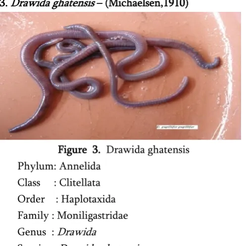
3. Drawida ghatensis – (Michaelsen,1910)
Figure 3. Drawida ghatensis Phylum: Annelida
Class : Clitellata Order : Haplotaxida Family : Moniligastridae Genus : Drawida
Species : Drawida ghatensis
It is a type of native species .The body colour is grey brown with bluish green pigmentataion, length 80-195mm. Diameter 2-7mm, number of segments 145-186, clitellum 10-13 interrepted between lines of seate, prostomoium become prolobous or zygolobous, male pore is in the midbody.
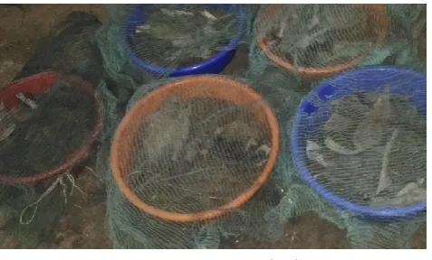
Preparation of Culture bed
Figure 4. Vermibed
Dissection of the earthworm
Each specimen to be dissected was washed in sterile distilled water, placed across the second, third and fourth fingers of the left hand (gloved) with the anterior end pointing forward. The fine edge of a flamed pair of dissecting scissors was inserted into the ventral surface at the region of the clitella and with the body wall slightly raised up with the scissors; an incision was made longitudinally along the earthworm. Sterile dissecting pins were used to hold the earthworm down on a board, stretching out the body wall to expose the internal structures. The gut was then freed from surrounding blood vessels and nephridia with a flamed forceps and separated into three sections: foregut, mid gut and hindgut. The gut sections were washed in sterile distilled water to free their contents before being suspended in other bottles containing clean sterile distilled water. The gut sections were washed with sterile distilled water to free their contents before being suspended in other bottles containing clean sterile water which were used later for further microbiological studies (Mahalingam and Thilagavathy, 2008).
Isolation of earthworm gut microflora A) Preparation of samples dilution
One gram of the content from the three gut regions of the earthworm, suspended in sterile distilled water. Subsamples (1ml each) were taken from each of the above preparations and homogenized in 9ml sterile distilled water blanks to make 10 fold serial dilutions upto 10-5. Then, the resultant suspension was allowed
to settle for almost 30 min. The clear culture supernatant was plated for the determination of microbial population.
B) Medium used for Culturing Microflora
Muller Hinton agar for bacterial culture incubated at 37ºC for 24 hrs, Kustors agar for Actinomycetes culture incubated 35 ºC for 3 to 7 days and Fungus were prepared on the Sabouraud’s Dextrose Agar incubated at room temperature (28ºC) for 3 days.
Identification and characterization of Bacteria, Actinomycetes and Fungal Isolates
Biochemical characterization and identification of various bacterial isolate were carried out according to the methods of Cappuccino and Sherman (1999). Identification Tests for the characterization of bacteria are Gram’s Staining, Spore Staining, Indole Production Test, Methyl Red Test, Citrate Utilization Test, Triple Sugar Iron Test, Reduction Test, Catalase test and Oxidase test. The morphological method consists of macroscopic and microscopic characterization.
Macroscopically the Actinomycetes isolates were differentiated by their colony characters, e.g. size, shape, color, consistency etc. For the microscopy, the isolates were grown by cover slip culture method (Kawato & Sinobu 1979). The observed morphology of the isolates was compared with the Actinomycetes morphology provided in Bergey’s Manual for the presumptive identification of the isolates. Various biochemical tests performed were catalase, oxidase, citrate utilization, nitrate reduction, starch hydrolysis, starch hydrolysis, gelatin hydrolysis, NaCl resistance and temperature tolerance.
production before being identified according to Kreger van Rig (1984) and Barnett et al.(1990). They were then identified by their micro-morphology as well as the color and micro-morphology of their
sporulating structures and conidia according to Barnett and Hunter (1972) and Onions et al.(1981).
III.
RESULTS AND DISCUSSIONS
Table 1. Characterization of bacteria isolated from gut tissues of earthworm CONTENTS P.corethrurus M.konkanensis D. ghatensis
Gram’s Staining Both gram +ve and –ve presesnt
Gram +ve and –ve bacteria is present
Both gram +ve and – ve presesnt
Spore Staining Present Present Present
Indole Production Test - - -
Methyl Red Test - + -
Citrate Utilization Test - Blue colour formed (+) - Triple Sugar Iron Test Acid slants Alkaline and acid slants Acid slants
Reduction Test + + -
Catalase test -ve bacteria Both +ve and –ve bacteria
-
Oxidase test - +ve -
The Symbols -, +,Indicates No Growth, Mild Growth, Good Growth Respectively The morphological and cultural characteristics of the
most active isolate of bacteria from earthworm gut were studied. The growth, characteristics were observed. The observation revealed that the morphological characterization shows both gram negative and gram positive bacteria and spores are
present in all bacteria. Biochemical tests were conducted for indole production test,methyl red test, citrate utilization test, triple sugar iron test, reduction test and catalase test. All the isolates showed positive results in triple sugar iron test.
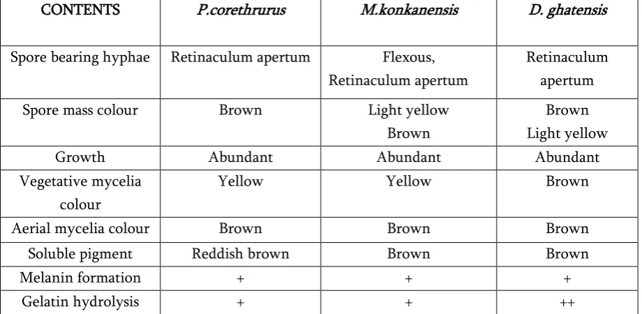
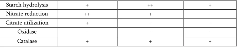
Table 2. Characterization of Actinomycetes isolated from gut tissues of earthworm CONTENTS P.corethrurus M.konkanensis D. ghatensis
Spore bearing hyphae Retinaculum apertum Flexous, Retinaculum apertum
Retinaculum apertum
Spore mass colour Brown Light yellow Brown
Brown Light yellow
Growth Abundant Abundant Abundant
Vegetative mycelia colour
Yellow Yellow Brown
Aerial mycelia colour Brown Brown Brown
Soluble pigment Reddish brown Brown Brown
Melanin formation + + +
Starch hydrolysis + ++ +
Nitrate reduction ++ + -
Citrate utilization + - -
Oxidase - - -
Catalase + + +
The Symbols -, +, ++ Indicates No Growth, Mild Growth, Good Growth Respectively
The morphological and cultural characteristics of the most active isolate of actinomycetes from earthworm gut actinomycetes were studied. The growth characteristics, presence of mycelium and soluble pigments were observed. The morphological characters of the active isolates were also studied microscopically under oil-immersion (100x) after gram-staining. The observations revealed that all the
isolates are gram positive. Biochemical tests were conducted for melanin formation, nitrate reduction, gelatin hydrolysis, starch hydrolysis, citrate utilization, oxidase and catalase. All the isolates showed positive results in melanin formation, gelatin hydrolysis and starch hydrolysis.
Table 3. Characterization of Fungus isolated from gut tissues of earthworms CONTENTS P.corethrurus M.konkanensis D. ghatensis
Pseudomycelium formation Elongated Elongated Elongated
Vegetative mycelia Brown Yellow Brown
Urease production + - +
Starch hydrolysis + - +
Cellulose hydrolysis + + +
Lignin Degrading + + +
Phosphate Solubilizing ++ + +
The Symbols -, +, ++ Indicates No Growth, Mild Growth, Good Growth Respectively
Among the isolates of fungus the observed morphology of the isolates was compared with the morphology and identification of the isolates. Various biochemical tests performed were pseudo mycelium formation, urease production, starch hydrolysis, cellulose hydrolysis, lignin and phosphate solubilizing. The isolated fungal colonies over the surface of media
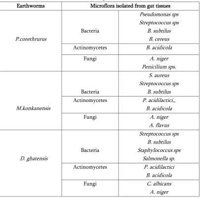
Table 4. Microflora isolated from gut tissues of local earthworms Earthworms Microflora isolated from gut tissues
P.corethrurus
Bacteria
Pseudomonas sps Streptococcus sps
B. subtilus B. cereus
Actinomycetes B. acidicola
Fungi A. niger
Penicilium sps.
M.konkanensis
Bacteria
S. aureus Streptococcus sps
B. subtilus
Actinomycetes P. acidilactici,, B. acidicola
Fungi A. niger
A. flavus
D. ghatensis
Bacteria
Streptococcus sps B. subtilus Staphylococcus sps
Salmonella sp.
Actinomycetes P. acidilactici B. acidicola
Fungi C. albicans
A. niger
Microflora isolated from the gut tissues of local earthworms were given in the table:4. The earthworms selected for this study was P.corethrurus,
M.konkanensis, and D. ghatensis. The bacterial flora isolated from the gut of the three local earthworms include Pseudomonas sps, Streptococcus sps, B. subtilus, B. cereus, S. aureus, Staphylococcus sps and Salmonella sps.The actinomycetes flora isolated from the gut of the three local earthworms include P. acidilactici and B. acidicola the fungal isolates flora isolated from the gut of earthworm include A. niger Penicilium sps, A. flavus and C. albicans.
IV.
SUMMERY AND CONCLUSIONS
The earthworms and microorganisms have the perfect relationship to each other. Similarly, says that, earthworm gut which is described as little bacterial factory. The isolation identification and characterisation gut microflora of local earthworms were studied. Morphological and cultural characteristics of the most active isolate of bacteria, actinomycetes and fungus from earthworm gut microflora were studied.
negative and gram positive bacteria and spores are present in all bacteria. Biochemical tests were conducted for indole production test, methyl red test, citrate utilization test, triple sugar iron test, reduction test and catalase test. All the isolates showed positive results in triple sugar iron test.
The growth characteristics, presence of mycelium and soluble pigments were observed. Biochemical tests were conducted for actinomycetes melanin formation, nitrate reduction, gelatin hydrolysis, starch hydrolysis, citrate utilization, oxidase and catalase. All the isolates showed positive results in fungal isolates melanin formation, gelatin hydrolysis and starch hydrolysis.Various biochemical tests performed were pseudo mycelium formation, urease production, starch hydrolysis, cellulose hydrolysis, lignin and phosphate solubilizing. The selected biochemical test shown very good growth of the some fungal strains and shown average in cellulose hydrolysis, lignin degrading and phosphate solubilizing.
Microflora isolated from the gut tissues of local earthworms were given in the table:4. The earthworms selected for this study was P.corethrurus,
M.konkanensis, and D. ghatensis. The microflora
isolated from the gut of the three local earthworms include Pseudomonas sps, Streptococcus sps, B. subtilus, B. cereus, S. aureus, Staphylococcus sps , Salmonella sps ,P. acidilactici , B. acidicola A. niger Penicilium sps, A. flavus and C. albicans.
V.
ACKNOWLEDGEMENT
The author, Sethulakshmi, is thankful to Dr.A.P.Thomas, Director, ACESSD, M.G.University, Kottayam providing laboratory facility to carry out work and manuscript preparation.
Funding: MG University, Kottayam.
Abbreviations
P.corethrurus - Pontoscolex corethrurus,
M.konkanensis - Megascolex konkenensis , D.ghatensis - Drawida ghatensis B.subtilus - Bacillus subtilus B.cereus - Bacillus cereus
P. acidilactici -Pediococcus acidilactici S. aureus - Staphylococcus aureus B. acidicola -Bacillus acidicola
Ethical Approval: This article does not contain any studies with human participants or animals performed by any of the authors.
VI.
REFERENCES
[1]. Barnett, H.L. and Hunter, B.B. (1972). Illustrated Genera of Imperfect Fungi. 3rd Edition, Burgess Publishing Co., Minneapolis, 241 .
[2]. Barnett,J.A., Payne, R.W. and Yarrow, D. (1990). Yeasts: characterisation and identification, Cambridge University Press, Cambridge.
[3]. Barois, I. and Lavelle,P. (1986). Changes in respiration rate and some physicochemical properties of a tropical soil during transit through Pontoscolex corethrurus
(Glossoscolecidæ, Oligochæta). Soil Biology and Biochemistry 18 (5): 539-541.
[4]. Cappuccino, G.j. and Sherman,N.(1999). Microbiology: A Laboratory Manual.
[5]. Kreger-van Rij, N.J.W. (1984). The Yeasts: a taxonomic study. :1-1082
[6]. Lavelle, P. (1997). Faunal activities and soil processes: Adaptive strategies that determine ecosystem function. Advanced Ecological Research 24: 93-132.
[7]. Lavelle, P., Lattaud,C., Trigo,D. and Barois,I. (1995). Mutualism and biodiversity in soils. Plant and Soil 170: 23-33.
[9]. Perdesen ,J. C. and Hendriksen, N. B.(1993). Effects of passage through the intestinal tract of detritivore earthworms (Lumbricus spp) on the number of selected gram-negative and total bacteria. Biol. Fertil. Soils. 16: 227-232.
[10]. Ismail, S.A. (1997) Vermicology: The biology of Earthworms. Orient Longman Limited, Chennai, 92.
[11]. Kawato,M. and Shinobu.,R.(1979). A simple