by Philippe André Lacoux
Thesis presented for the degree of Doctor
of Medicine of the University of London
All rights reserved
INFORMATION TO ALL USERS
The quality of this reproduction is dependent upon the quality of the copy submitted.
In the unlikely event that the author did not send a complete manuscript and there are missing pages, these will be noted. Also, if material had to be removed,
a note will indicate the deletion.
uest.
ProQuest 10046079
Published by ProQuest LLC(2016). Copyright of the Dissertation is held by the Author.
All rights reserved.
This work is protected against unauthorized copying under Title 17, United States Code. Microform Edition © ProQuest LLC.
ProQuest LLC
789 East Eisenhower Parkway P.O. Box 1346
I hereby declare that this thesis has been composed entirely by myself and that all the work described
herein was carried out by myself alone, except as
otherwise stated in the acknowledgements.
LIST OF FIGURES 8
ACKNOWLEDGEMENTS 9
SUMMARY 11
1. INTRODUCTION 13
1.1. The genus Enterococcus in historical perspective 13
1.2. Present day identification of species 18
1.3. The clinical importance of the enterococci 22
1.3.1. Endocarditis 23
1.3.2. Bacteraemia 24
1.3.3. Urinary tract infection 25
1.3.4. Neonatal infections 26
1.3.5. Other enterococcal infections 26
1.3.6. Polymicrobial infection 27
1.3.7. Superinfection 28
1.4. Nosocomial infection 28
1.5. Endogenous versus exogenous 29
1.6. Antibiotic susceptibilities of the enterococci 31
1.6.1. Penicillin resistance 32
1.6.2. Aminoglycosides 33
1.6.3. Vancomycin 35
1.6.4. Quinolones 37
1.6.5. Other antibiotics 3 8
1.6.6. Synergy 38
1.7. Plasmids and transposons in the enterococci 39
I.S. Streptococcus bovis 40
1.9. Typing methods 41
1.9.4. Bacteriophage typing 44
1.9.5. Bacteriocin typing 44
1.9.6. Plasmid analysis 45
1.9.7. Restriction fragment length polymorphism 47
1.9.8. Restriction fragment hybridisation to an rRNA gene probe
(ribotyping) 48
1.9.9. Polymerase chain reaction (PCR) 49
1.10. The British Antarctic Survey (BAS) and the isolated community 50
1.11. The aims of this study 55
2. MATERIALS AND METHODS 56
2.1. Isolate collection 56
2.1.1. Antarctic sample collection 56
2.1.2. Bacterial isolates from Signy Base 56
2.1.3. Control strains 57
2.2. Bacterial identification 58
2.2.1. Sample revival 58
2.2.2. Species identification 58
2.3. Strain typing 59
2.3.1. Biotyping 59
2.3.2. Antibiotic susceptibility testing. 59
2.3.3. Restriction fragment length polymorphism (RFLP) 60
2.3.3.1. DNA extraction 60
2.3.3.2. Phenol extraction 61
2.3.3.3. Other extraction methods 61
2.3.3.4. Evaluation of the purity and yield of DNA. 62
2.3.3.5. Staining and photographing of gels 62
63
2.3.5.1. Probe preparation 63
2.3.5.2. Southern blotting and DNA hybridization 64
2.3.6. Analysis of banding patterns 64
3. RESULTS 65
3.1. Survival of Antarctic isolates. 65
3.2. Species identification 65
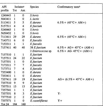
3.2.1. API results 66
3.2.2. Confirmatory testing 66
3.3. Typing of the enterococci 70
3.3.1. Biotyping 70
3.3.1.1. E.faecalis 70
3.3.1.2. E.faecium 71
3.3.1.3. E.durans 71
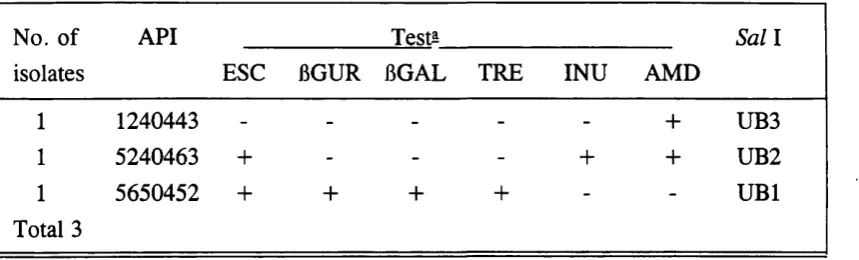
3.3.1.4. S.bovis 15
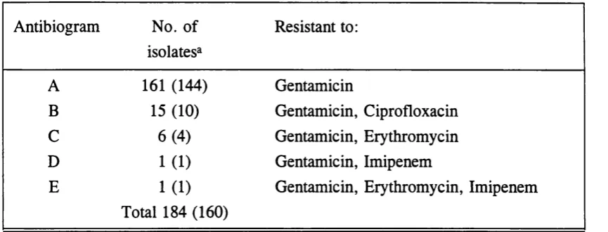
3.3.2. Antibiotic susceptibility testing 75
3.3.3. DNA finger-printing 81
3.3.3.1. DNA extraction 81
3.3.3.2. Choice of restriction endonuclease 82
3.3.3.3. E.faecium digestion with Sal I 92
3.3.3.4. E .durans digestion with Sal I 92
3.3.3.5. E.faecalis digestion with Sal I 96
3.3.3.6. Digestion of other species with Sal I 96
3.3.3.7. Summary of results with Sal I 96
3.3.4. Species identification by RFLP 98
3.3.5. Ribosomal RNA gene restriction patterns (ribotyping) 99
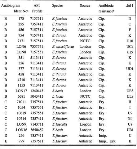
3.4.3. Antibiograms 111
3.4.4. Combined Grouping 112
3.5. Summary Table 112
4. DISCUSSION 119
4.1. Species identification 119
4.2. Clonality 123
4.3. Restriction fragment length polymorphism (RFLP) 126
4.4. Biotyping 129
4.5. Antibiograms 130
4.6. Ribotyping 133
4.7. S.bovis 134
4.8. Epidemiology 135
4.9. Conclusions 139
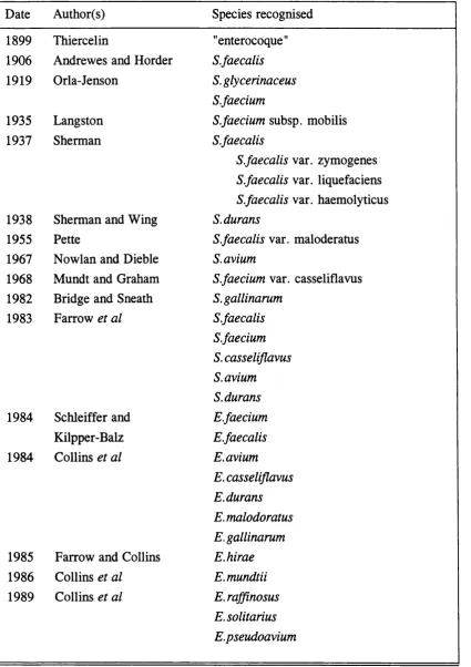
Table 1. Historical developments within the genus Enterococcus 19
Table 2. Phenotypic characteristics of enterococcal species 20
Table 3. API 20 Strep profiles and confirmatory tests 67
Table 4. Source and species distribution of isolates 69
Table 5. E.faecalis biotyping results 72
Table 6. E.faecium biotyping results 73
Table 1. E.durans biotyping results 74
Table 8. S.bovis biotyping results 76
Table 9. Individual API 20 Strep test variability for each enterococcal
species 77
Table 10. Antibiograms 78
Table 11. Isolates resistant to more than gentamicin alone. 79
Table 12. Distribution of antibiograms by species 80
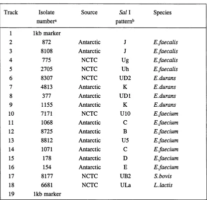
Table 13. Details of the pilot study isolates in Figures 3-7, 11 and 12 83
Table 14. Pilot study RFLP pattern results for Bam HI, Sac I and Sal la 90
Table 15. Summary of the RFLP patterns with S a il 91
Table 16. E.faecium API profile and Sal I patterns 94
Table 17. Ribotype patterns after digestion with Eco RIa and Hind III 102
Figure 2. British Antarctic Survey Base Signy, South Orkney Islands 53 Figure 3. Digestion of the pilot study isolates with the restriction
enzyme Bam HI 84
Figure 4. Digestion of the pilot study isolates with the restriction
enzyme Sac I 85
Figure 5. Digestion of the pilot study isolates with the restriction
enzyme Sal I 86
Figure 6. Digestion of the pilot study isolates with the restriction
enzyme Eco RI 87
Figure 7. Digestion of the pilot study isolates with the restriction
enzyme Hind III 88
Figure 8. RFLP patterns of Antarctic E.faecium isolates 93
Figure 9. RFLP patterns of Antarctic E.durans isolates 95
Figure 10. RFLP patterns of London E.casseliflavus, E.faecalis and
S.bovis isolates 97
Figure 11. Southern blot of the pilot study isolates digested with Eco RI 100
Figure 12. Southern blot of the pilot study isolates digested with Hind III 101
Figure 13. Temporal analysis of the Antarctic E.faecium RFLP results
using Sal I 104
Figure 14. Temporal analysis of the Antarctic E.faecium API 20 Strep
biotyping results 105
Figure 15. Temporal analysis of the Antarctic E.faecium antibiogram
results 107
Figure 16. Temporal analysis of the Antarctic E.durans isolates by
three typing methods 108
Figure 17. Temporal analysis of the Antarctic E.faecalis isolates
by three typing methods 109
Figure 18. Temporal analysis of the Antarctic E.faecium results using
This work was supported by the British Antarctic Survey (BAS) through
the National Environmental Research Council (NERC). I thank the logistics section
of BAS for the supplying of all equipment to the Antarctic and for the return of the
samples to Aberdeen. I am indebted to the the individuals wintering at Signy Base
in 1988 for their cooperation, and to Dr Chris Fenton for collection of the samples.
There are too many individuals within BAS to thank personally for their help and
support.
I would like to thank everyone involved with the British Antarctic Survey
Medical Unit based in the Survival Center, Robert Gordons Institute of Technology, Aberdeen. In particular. Professor Nelson Norman, Dr. Alan Milne, Dr. Sylvia
Wilcox and Dr. Wendy Haston.
This work was conducted within the Medical Microbiology Department of the University of Aberdeen, under the planning and guidance of Professor T.H.
Pennington. I am indebted to a large number of individuals within this department,
but especially to Dr. Zoe Jordens for her guidance and enthusiasm, to Dr. Tony
Maggs for his continued help and constructive suggestions, and to Audrey Innes for
her skilled technical help.
I thank the Department of Medical Illustration, University of Aberdeen for
their help in preparing the photographs, and Dr Tony Maggs for his help in
preparing the tables and figures.
I was fortunate in receiving periods of study leave from both the
Department of Anaesthesia, Ninewells Hospital, Dundee, (Tayside Health Board),
and from Lewis Hospital, Stornoway (Western Isles Health Board) whilst working
I am grateful to Professor J R Pattison for his helpful advice during the
preparation of this thesis.
SUMMARY
Restriction fragment length polymorphism (RFLP) of chromosomal DNA
for the intra-species characterisation of enterococci is reported. The bacterial DNA
was extracted by a rapid method and digested to provide a characteristic DNA
banding pattern after agarose gel electrophoresis. Five different restriction
endonucleases Bam HI, Sac I, Sal I, Eco RI and Hind III were assessed for their
ability to provide easily readable banding patterns that could be used to differentiate
between a representative sample of enterococcal isolates. One hundred and eighty
enterococcal isolates from three different sources were then examined using the
restriction endonuclease Sal I, and this provided 39 distinct DNA banding patterns;
this technique provided a discriminatory method of isolate characterisation for
E.faecalis, E.faecium and E.durans. Results were compared with biotyping with the API 20 Strep kit, and with antibiogram data using eight antibiotic impregnated
discs. The technique of restriction endonuclease analysis with Sal I was applied to
the temporal analysis of faecal enterococci from an isolated Antarctic community,
and the results compared with biotyping and antibiogram data. Southern blots were
probed with ribosomal RNA from Escherichia coli after digestion of the bacterial
DNA with either Eco RI or Hind III. This revealed some potential as a method for
differentiation within the enterococcal species, and also at the inter-species level
although E.faecium and E.durans were difficult to separate.
Recent clinical findings suggest that a typing scheme for S.bovis could be
important. These developments are discussed, and the methods above applied to
three S.bovis isolates.
The historical taxonomic changes within the enterococci are reviewed. The
pattern of antimicrobial resistance and to the growth in nosocomial infection.
Methods of typing the enterococci are reviewed, and the present position of genetic
1. INTRODUCTION
1.1. The genus Enterococcus in historical perspective
The historical taxonomy of the enterococci into a separate genus consisting
at the present time of twelve species has closely paralleled the development of new
identification techniques in bacteriology. The first use of the term "enterocoque"
was by Thiercelin (1899) for a gram-positive diplococcus of intestinal origin.
Andrewes and Horder (1906) in their paper giving an overview of the status of the
Streptococci at the time entitled "A study of the Streptococci pathogenic for man" found - "a group of streptococci so characteristic of the human intestine that the
term Streptococcus faecalis" may be justly applied to it. It is mostly short-chained,
rendering broth uniformly turbid, but in disease, at least, forms with long to
medium chains occur. It grows readily on gelatin at 20°C with rare exceptions and
is distinguished by its great chemical activity."" The species possesses a very great
resistance to desiccation, far greater than that of the long-chained throat forms."
Orla-Jenson (1919) suggested the names S.faecium and S.glycerinaceus,
and carefully defmed some of the physiological properties of what was to become
the enterococci. S.glycerinaceus was subsequently confirmed to correspond to the
description of S.faecalis.
In the subsequent decades there was a great deal of confusion concerning
what constituted an enterococcus, and upon the nomenclature which should be used.
Sherman (1937) while reviewing the literature felt that many workers had made
inadequate attempts to record isolate characteristics. He provided much greater
potential for order among the streptococci by suggesting their division into four
groups; pyogenic, viridans, lactic and enterococci. The characteristics of the
enterococci as suggested by Sherman were growth at 10°C and 45°C, growth in
ability to split aesculin. These characteristics have survived remarkably intact, and
are still considered among the basic properties of the enterococci (Schleifer, 1984).
Four different species of enterococci were discussed by Sherman (1937)
with liquefaction of gelatin (S.liquefaciens), and haemolysis in blood agar
(S.zymogenes) as important tests to differentiate the above from S.faecalis. Sherman
acknowledged that this sub-division was probably for insufficient reason, and
suggested the consideration of varietal status. S.durans was more easily classified as
a separate species due to its paucity of fermentation reactions. As reflected above
much of the earlier work wrestled with the questions about what did and did not
constitute an enterococcus, and where species lines were to be drawn.
The serological classification of Lancefield (1933) added the presence of
the group antigen D as a characteristic of what are now classified as the enterococci.
There was a striking association between the Lancefield serological groupings, and
the sources of the cultures. Subsequently, the group D antigen was identified as a teichoic acid and a component, not of the cell wall like those of the Lancefield
groups A, B, C, G, and F, but of the cytoplasm (Elliot, 1960; Jones and Shattock,
1960). Elliot also discovered a type specific antigen of carbohydrate situated in the
cell wall and which was analagous to the other Lancefield groups.
There was good agreement between the classifications of Lancefield and
Sherman for the broad group "streptococci", with minor discrepancies such as the
reaction of S.bovis with the Lancefield group D antisera.
Further serological work subdivided the enterococci (Sharpe and Shattoch,
1952), but not in a manner that correlated with the physiological characteristics of the emerging enterococcal species. It was not possible to define species in terms of
serology, although a broad division was made between S.faecalis and its varieties
The position of S.faecium and S.durans as species or sub-species was
unclear for many years. Orla-Jenson (1919) first suggested the species S.faecium,
this susequently corresponded with tellurite sensitive strains found during the
serological study of Sharpe and Shattock (1952), work which was also in support of
the species status for S.faecium.
Motility was a property which had long been associated with the
streptococci, and motile enterococci were observed early in the century as S.faecium
subsp. mobilis, while yellow motile isolates were called S.faecium var,
casseliflavus. S.faecium had become a repository for many isolates of uncertain
classification. S.malodoratus was isolated from gouda cheese, and S. avium from
chickens, the latter possessed both D and Q Lancefield antigens.
Paper chromatography of the hydrolysates of complete cell walls of
streptococci (Colman and Williams, 1965), did show 26 patterns from 197 strains,
but no overall relationship between taxa or species from the chromatography
patterns was evident. Similarly in 1973, Amstein and Hartman used gas
chromatography to demonstrate the relative fatty acid composition of 37 enterococci
in an attempt to clarify the position between different species. Differences were
observed in particular among the motile pigmented strains, however the differences
were inadequate for the purpose of species differentiation. The isoprenoid quinones
which have a role in electron transport were also examined and although the results
did agree in general with other forms of taxonomic information, alone they were
insufficient for use as a means of classification (Collins and Jones, 1979).
In 1968 Colman applied computers to the classification of the Streptococci;
with three different computer programmes analysing 75 different biochemical and physiological tests. He included 14 enterococci, 12 of which, a mixture of
E.faecalis and E.faecium formed a single cluster, however resolution to the species
Lancefield group D isolates with 45 different physiological and biochemical tests.
They found that the organisms clustered in five main groups consisting of
S.faecalis, S.faecium with S.durans (together considered to be one species),
S.avium, S.bovis and S.equinus. There were some ungrouped strains, and the
previous subdivision of S.faecalis isolates into the subspecies liquefaciens and
zymogenes was not upheld. Jacob et al 1975 showed that the ability to liquefy
gelatin (S.faecalis subsp. liquefaciens) and to haemolyse red blood cells (S.faecalis
subsp. zymogenese) are properties controlled by plasmids.
A much reduced group of physiological and biochemical tests was proposed
by Gross et al (1975) in a survey of over 4000 group D isolates. At this time there
was growing concern about the interchangable usage of the terms group D,
S.faecalis and the enterococci. In particular because of the different antimicrobial
treatment of diseases caused by the different group D organisms. Their scheme
considered pyruvate fermentation and arginine hydrolysis as useful tests to differentiate the enterococcal species. Using 5-7 initial tests, and 6-11 confirmatory
tests they were able to differentiate S.faecalis, S.faecium, S.faecium var. durans,
S.avium and S.bovis.
A numerical taxonomic study of the streptococci by Bridge and Sneath
(1983) found 28 different groups (phenons) covering a wide variety of species. The
methodology again involved a wide range of biochemical and physiological tests and
computer analysis. The enterococci formed four phenons, the system was able to
distinguish E.faecalis and E.faecium isolates, with the E.faecalis subsp. liquefaciens
and zymogenes forming no distinct separate grouping. E.durans isolates were
grouped with the E.faecium isolates, and a new enterococcal species was proposed
"S.gallinarum" isolated from the bowel of young chickens. There was also an
ungrouped strain "S.faecalis subsp. maloderatus". The results largely agreed with
Interesting results were presented by Plecas and Brandis (1974) who
studied enterococcal bacteriophages and found that some were species specific, they
were able to identify 370/411 strains as either S.faecalis, S.faecium or S.bovis.
Much of the confusion concerning the taxonomy of the enterococci was
clarified by the work of Farrow et al (1983) who examined the biochemical, fatty
acid, menaquinones and DNA of 50 group D isolates. The main contribution of this
work was in defining enterococcal species using DNA homology studies. The
varieties of S.faecalis had DNA homology values of greater than 70% indicating
that they should all be considered the same species. S.faecalis and S.faecium had
homologies of 20-30% confirming the latter as a separate species within the same
genus. S.durans, previously a sub-species of S.faecium was phenotypically similar
to S.faecium but homology studies suggested that separate species status was
appropriate. S.casseliflavus, the yellow motile species was confirmed to be a
separate species, and the suggested sub-species of S.faecium var. mobilis was found
to have high homology values (76-97%) with S. casseliflavus, and to be unrelated
except at the genus level to S.faecium. In summary, S.faecalis, S.faecium,
S.casseliflavus, S.durans, S.avium and S.gallinarum (Bridge and Sneath, 1982)
were different species of the same genus, and S.bovis was unrelated at the generic
level.
In the last ten years with the use of DNA/rRNA hybridisation studies and
comparative analysis of ribonuclease Ti-resistant oligonucleotides of the 16S rRNA (Schleifer and Kilpper-Balz, 1987) a clearer understanding of the streptococci as a
whole has emerged; the divisions of Sherman from 1937 continue to be very largely
upheld. Schleifer and Kilpper-Balz (1987) with other workers were instrumental in
the division of the streptococci into three genera, the Streptococci, Enterococci and Lactococci. This was not fully recognised in the 1984 Bergey's Manual of Bacterial
New species of enterococci have emerged, and there are now twelve
different enterococcal species (Table 1), with some enterococcal isolates still not
identified to this level (Murray, 1990a). Farrow and Collins (1985) described
E.hirae a species found from pig and chicken isolates. Collins et al (1986)
described E.mundtii a yellow pigmented non-motile strain from plants and soil.
E.pseudoavium, E.raffinosus and E.solitarius have been described by Collins et al
(1989).
Species identification still depends largely upon biochemical testing (Table
2), although DNA homology studies have been the initial basis for the defining of
new enterococcal species. Most recently polymorphism among penicillin-binding proteins have emerged as a taxonomic tool. Each of nine different species studied
by Williamson et al (1986) had a specific pattern of five or more penicillin-binding
proteins.
1.2. Present dav identification of species
The twelve enterococcal species have been defmed principally on genetic
DNA/DNA homology criteria. There is some confusion concerning the biochemical
differentiation between the enterococcal species. Facklam and Collins (1989) and
Rouff et al (1990) have both conducted studies to biochemically defme the species.
There are also many other published biochemical criteria for the distinction of
enterococcal species with some disagreements. There are both inconsistent
biochemical test results which vary within a species (Table 2) and isolates which fit
into no particular species (Facklam and Collins, 1989; Murray, 1990a). It can be
argued that it is not possible to confidently distinguish between all of the
Table 1. Historical developments within the genus Enterococcus
Date Author(s) Species recognised
1899 Thiercelin "enterocoque"
1906 Andrewes and Horder S.faecalis
1919 Orla-Jenson S.glycerinaceus
S.faecium
1935 Langston S.faecium subsp. mobilis
1937 Sherman S.faecalis
S.faecalis var. zymogenes
S.faecalis var. liquefaciens
S.faecalis var. haemolyticus
1938 Sherman and Wing S. durans
1955 Pette S.faecalis var. maloderatus
1967 Nowlan and Dieble S. avium
1968 Mundt and Graham S.faecium var. casseliflavus
1982 Bridge and Sneath S.gallinarum
1983 Farrow et al S.faecalis
S.faecium S. casseliflavus S. avium S. durans
1984 Schleiffer and E.faecium
Kilpper-Balz E.faecalis
1984 Collins et al E. avium
E.casseliflavus E.durans E.malodoratus E.gallinarum
1985 Farrow and Collins E.hirae
1986 Collins et al E.mundtii
1989 Collins et al E. raffinosus
Table 2. Phenotypic characteristics of enterococcal species
MAN SOR SBS ARG ARA RAF TEL MOT PIG SUC PYU Species
E. avium + + + - + - - - - + +
E. raffinosus + + + - + + - - - + +
E.malodoratus + + + - - + - - - + +
E.pseudoavium + + + + +
E.faecalis + + - + - - + - - +
E.solitarius + + - + - - - -kb
-E.gallinarum + - - + 4- + - + - +
-E.faecium + _b - + + _b - - - + b
-E. casseliflavus + _b - + - + _b + + + _b
E.mundtii + _b - + - + - - + +
-E.durans - - - + - - - - - -
-E.hirae - - - + - V - - - V
-E.faecalis - - - + - - + - - - V
(Facklam and Washington, 1991)
^ Abbreviations; MAN (mannitol), SOR (sorbitol), SBS (sorbose), ARG (arginine), ARA (arabinose), RAF (raffinose), TEL (0.04% tellurite), MOT (motility), PIG (pigmentation), SUC (sucrose), PYU (pyruvate).
+ : >90% positive; <10% positive; v: variable 60-90% positive,
The ability of the clinical laboratory to identify enterococci to the species
level is important as there are clinically significant differences in antimicrobial
susceptibility, for example the greater resistance of E.faecium isolates to
penicillin/aminoglycoside synergy when compared wih E.faecalis isolates
(Moellering et al, 1979). The work of Mackowiak (1989) has highlighted the
differences in the virulence between three enterococcal species {E.faecalis,
E.faecium and E. avium) suggesting that great precision is needed in their
identification.
A variety of commercial products for the identification of the streptococci
have been developed, usually based upon freeze-dried reagents. The names of these
products are confusing. In the present study the API 20-Strep has been used, this is manufactured in France by API system. La Balme les Grottes, France, and
distributed throughout Europe; in Britain by API-bioMerieux (UK) Ltd.,
Basingstoke, Hampshire. In the USA the same product is sold under a different
name - DMS-Rapid Strep., frequently written as simply the Rapid Strep system. A similar but different product is made by a another company in USA, and is only
distributed in the United States. It is not the same product as the API 20-Strep used
in this study but has a very similar name - API 20-S, the main difference between
the two kits involves the identification of the viridans streptococci. Another product
sometimes used for enterococcal studies is the API 50 CH also made by the French
company API-bioMerieux. This incorporates 50 carbohydrates, no buffer or media,
and can be applied to a wide variety of organisms.
Several studies have compared the various products (Appelbaum et al,
1984; Facklam et al, 1984; Facklam et al, 1985; Fertally and Facklam, 1987). The
overall conclusion has been that these products are good at identifying E.faecalis
Despite attempts to devise physiological and biochemical means of
confidently identifying the enterococci confounding problems continue to emerge.
Recently Vincent et al (1991) showed that the hitherto relied upon tests of yellow
pigment production and of motility, used for the identification of E.casseliflavus
and E.gallinarum can be misleading. They found the penicillin-binding protein
profiles and ultimately DNA homology results were required to confidently identify these species. Musher (1988) felt that; "Although DNA homology will almost
certainly provide the final word, the clinical laboratory must in the meantime identify streptococci with readily available techniques, and clinicians must utilize
these identifications to the best of their ability. "
1.3. The clinical importance of the enterococci
Despite their wide distribution in the environment and as part of the normal
human bowel, oral cavity, anterior urethra and vaginal flora, the enterococci do not
generally cause a significant amount of disease. It has been proposed that this is because they lack the virulence factors found in many other bacteria such as the
Staphylococci.
The types of infection that are caused by the enterococci include
endocarditis, bacteraemia, urinary tract infection, neonatal sepsis, intra-abdominal
sepsis, pelvic sepsis and soft tissue infections. Toala et al (1969) noted there was a
wide variety of clinical specimens from which enterococci were isolated, with urine
being the most common. There is frequently an association with general debility,
the extremes of age or the presence of invasive devices such as urinary catheters.
Wells et al (1990) have shown that E.faecalis can translocate across the mouse
intestinal tract and be recovered from liver, spleen and lymph nodes when
experimental intestinal overgrowth with enterococci is induced. Infections involving
the enterococci are also frequently polymicrobial and as a result it is difficult to
It is becoming clear that the influence of prior antibiotic administration is
also significant. The changing pattern of enterococcal infection may be related to the wide use of broad spectrum 6-lactam antibiotics encouraging enterococcal
overgrowth (Yu, 1981; Berk et al 1983; Morrison and Wenzel, 1986).
1.3.1. Endocarditis
Endocarditis is the most serious manifestation of enterococcal infection.
Enterococci are the third most common cause of endocarditis after the viridans
r
Streptococci and the staphylococci, and cause approximately 10-20% of cases
(Mandell et al, 1970). Parker and Ball (1975) found that 7.9% of 223 cases of
endocarditis were due to enterococci, and considered that this might be an
underestimate as E.faecalis is relatively easy to identify and so was less likely to
have been sent to the reference laboratory around which the work was based.
Enterococcal endocarditis is rare in children, and more common in men of the older age group than in women. The disease frequently runs a subacute rather than acute
course, with the genito-urinary tract being the commonest portal of entry (Koenig
and Kaye, 1961). It may be related to intravenous drug abuse, when it affects aortic
and mitral valves in particular, and is also more common if there is a prior history
of valvular heart disease (Koenig and Kaye, 1961). There is one report of human-
to-human transmission of enterococcal endocarditis in a couple sharing injection
equipment (Hall et al, 1976). The mortality rate varies between studies, but is high,
Moellering et al (1974) 47% of 15 cases, Mandell et al (1970) 16% of 36 cases.
There has been an interesting comparison between endocarditis due to
enterococci and that due to to S.bovis, both being Lancefield group D. In a review
of the cases at the Massachusetts General Hospital by Moellering et al (1974) the
two organisms were equally prevalent, clinical presentation was the same for each,
mortality of S.bovis disease is lower. This illustrates the need to differentiate
between these two organisms.
The usual treatment for enterococcal endocarditis is with the synergistic
combination of a penicillin or vancomycin with an aminoglycoside. This is under critical review with the advent of resistance in the enterococci to all of these agents.
The ad hoc group of the committee on Rheumatic fever, Endocarditis and Kawasaki
disease from the American Heart Association (Bisno et al, 1989) suggest that the
species identification of isolates is important as S.bovis and S.mutans can be treated
with penicillin alone. They also suggest where isolates have high level
aminoglycoside resistance further in vitro susceptibility testing is required, as some
of these isolates will be susceptible to streptomycin. Where isolates are B-lactamase
producers or where the patient has a well documented penicillin allergy treatment
should be with a combination of vancomycin and gentamicin. However such
combinations should be monitored regularly as drug toxicity is an important
consideration when therapy may be required for 4-6 weeks.
1.3.2. Bacteraemia
Bacteraemia without endocarditis is a less distinctive but more common
clinical problem than endocarditis alone. Parker and Ball (1976) found that 10.6%
of 250 bacteraemia isolates were enterococci. Maki and Agger (1988) have shown in a review of cases between 1970-1983 in the University of Wisconsin Hospitals
that the incidence of nosocomial enterococcal bacteraemia has increased since 1975.
They found a total of 153 enterococcal bacteraemias, with 65 cases involving the simultaneous isolation of other microorganisms, most commonly gram-negative
bacilli. The mean age of patients was 50 years, with men twice as common as
women. Serious underlying conditions were present in 133 cases, and nosocomial acquisition identified in 118 cases. As with endocarditis, disease of the urinary tract
of intravascular catheters (Maki and Agger 1988, Parker and Ball 1975). In a study
by Haslett et al (1988) on the microbiology of central venous catheters E.faecalis
was the second most common organism isolated after Staphylococcus epidermidis,
and was frequently present in combination with another organism. When detected
on the vascular catheter tip, E.faecalis was frequently also present in peripheral
blood and at the insertion site. Musher (1988) found that 23 out of 97 cases of
polymicrobial bacteraemia involved enterococci.
Fever may be the only clinical sign when the bacteraemia consists solely of
enterococci; this may be due to the lack of endotoxin production by the enterococci.
Mortality rates are reported to be between 34% and 46% (Shlaes et al, 1981;
Malone et al, 1986; Maki and Agger, 1988) at least in part due to the serious
associated underlying illness. In the study of Maki and Agger (1988) progression of
bacteraemia to endocarditis was more common in the solely enterococcal
bacteraemias, one suggested reason for this was that in the polymicrobial infections clinical signs were more apparent, and so prolonged bactericidal antimicrobial
treatment was more likely to have been instituted. These authors suggest treatment
of conditions likely to cause enterococcal bacteraemia because of the high
subsequent mortality. Specific therapy of such conditions has been shown to
improve survival (Hoge et al, 1991).
1.3.3. Urinarv tract infection
Urinary tract infections (UTI's) with enterococci are most frequent in men
who have had recent instrumentation or have a structural abnormality. Gross et al
(1976) in a study of elderly men in hospital used antibiograms as the typing method
and showed that the mode of spread was endogenous with the individuals own
faecal flora as the source of infection, the incidence of enterococcal infection was
21% and transfer by hospital personnel was not involved. In North America
Escherichia coli and Pseudomonas aeruginosa causing 14.7% of urinary tract
infections (Centre for Disease Control, 1986). In the United Kingdom the incidence
was 7.2% in 1980 (Meers et al, 1981). Savarino et al (1987) found that 11.8% of
1336 streptococcal UTI's were due to enterococci. Morrison and Wenzel (1986)
undertook a retrospective survey of nosocomial urinary tract infection due to
Enterococcus sp. in a single hospital in the United States. They defined nosocomial
as an infection that was neither present nor incubating when the patient was admitted to hospital. They demonstrated an increasing incidence of enterococcal
UTI, possibly related to cephalosporin use, although no information was obtained pertaining to whether nosocomial infections were due to isolates from the patients
own flora or whether there was spread between patients. The same authors also showed that nosocomial UTI's due to the enterococci had increased from 6-16%
over a ten year period, and that this closely parallelled the increase in cephalosporin
usage over the same time period.
1.3.4. Neonatal infections
Around 10% of neonatal infection is due to enterococci (Murray, 1990a).
The setting in which these infections occur has largely involved sick premature
infants frequently with invasive devices in situ. The source may be a combination of
maternal vaginal colonisation and possibly transmission on the hands of staff
members (Goudron et al, 1984; Luginbuhl et al, 1987). Enterococci are probably
more important in neonatal infection than has been thought, because of dissmisal in
the past of enterococci as bacteriological contaminants. Diagnosis of enterococcal
infection, as with many neonatal infections is difficult, with signs and symptoms frequently being non-specific.
1.3.5. Other enterococcal infections
Intra-abdominal and pelvic infections are likely to be endogenous with the
abdominal surgery. The pathogenicity of enterococci in abdominal infection is a
matter of debate. Some evidence suggests that polymicrobial infections with
Escherichia coli or Bacteroides fragilis are more likely to result in death or abscess
formation than infection with enterococci alone (Onderdonk et at, 1976). In an
animal study Ike et al (1984) showed that haemolysis by the strain of enterococcus
injected into mice was a determinant for abscess formation.
Soft tissue infections which involve enterococci are usually mixed and
affect previously injured tissue such as bums and diabetic or decubitis ulcers rather
than previously healthy tissue. Cellulitis does occur, predominantly in a clearly compromised patient. Skin and soft tissue infection is a common source of
enterococcal bacteraemia (Musher, 1988).
Pure enterococcal pneumonia was reported by Berk et al (1983) in two
patients receiving combination cephalosporin and aminoglycoside therapy. In the
study of Parker and Ball (1976) enterococci were the cause of 4.3% of 116 cases of
pumlent disease. Infection of the central nervous system is uncommon, occuring in
neonates and older patients. A serious underlying disorder may be present, or as
Bayer et al (1976) discussed an anatomical cause such as a skull fracture to result in
the enterococcal meningitis.
1.3.6. Polvmicrobial infection
Onkerdonk (1976) showed that pure enterococcal infection in rats cause
little problem, while in combination with gram-negative bacilli death or abscess
formation is more likely. One explanation of this is that the presence of E.faecalis
in a mixed culture has been shown to protect bacteroides strains from the killing
effect of a daptomycin and metronidazole combination even at a concentration of 4-
1.3.7. Superinfection
In 1981 Yu reported superinfection including two bacteraemias with
enterococci associated with a then new broad spectrum antibiotic moxalactam.
Several studies on enterococcal bacteraemia have reported data concerning the prior
use of broad-spectrum agents ineffective against the enterococci with a range of 42-
77% of patients previously exposed (Hoffman and Moellering, 1987). The study of
Morrison and Wenzel (1986) on UTI documented the increase in cephalosporin use
at the hospital from 64,000-114,000 g/year.
1.4. Nosocomial infection
Nosocomial infections are defined as those acquired while in hospital. The
source of such an infection may be from the patient i.e. endogenous, or from some
other external source i.e. exogenous. Nosocomial infections may be serious, particularly exogenous ones, as the organism is more likely to be multiply resistant.
The disease and increased hospital stay that are encountered result in significant
morbidity.
Nosocomial infection caused by the enterococci is of increasing
importance. The Centre for Disease Control (Atlanta) in 1986 listed enterococci as
the third most common nosocomial pathogen after E.coli and S.aureus. Indeed the
most recent American figures suggest that the enterococci are now a more common
nosocomial pathogen than S.aureus (Moellering, 1992). Ford-Jones et al (1989)
found that 4.9% of 4684 hospital acquired infections were due to enterococci,
commonly isolated from UTI’s and post-operative wound infections. That
enterococci are a major cause of nosocomial infection is incontrovertible, the
1.5. Endogenous versus exogenous
Evidence for an endogenous source of infection was provided by Gross et
al (1976) in a three month surveillance study on enterococcal urinary tract infection.
The workers used antibiograms as their epidemiological marker and found no
evidence for exogenous spread of organisms. In a review Kaye (1982) wrote, "All
available evidence indicates that in the vast majority of cases, enterococcal
infections of all types are acquired from the patients' own flora". During the 1980's
a variety of studies using a number of typing techniques challenged this statement.
In 1984 Goudron et al reported an outbreak of bacteraemia and meningitis
caused by E.faecium in a neonatal intensive care unit. Conventional biotyping with
26 different biochemical and physiological tests was found to be inadequate as a means of typing; improved discrimination was obtained by using commercially
available rapid identification products. Isolates from infected patients, clustered in
the neonatal intensive care unit and had different biotypes from 29
epidemiologically unrelated isolates with which they were compared.
Epidemiological information suggested a bowel source for the epidemic strain, and
possible transfer on the hands of hospital personnel.
Zervos et al (1986; 1987a) showed that nosocomial spread of enterococci
does occur. As an isolate marker they used the then uncommon feature of high level
gentamicin resistance and also plasmid content to show that environmental surfaces
and the hands of hospital personnel were implicated in the spread of enterococci.
There was temporal and geographical clustering, and the probability of spread
between two hospital sites. In the 1986 study three deaths were associated with
enterococcal infection.
In a retrospective study on neonatal enterococcal sepsis Luginbuhl et al
(1987) used a combination of phenotypic tests and plasmid content as
sepsis over a six month period were similar to each other and different from the
heterogenous endemic nursery strains, supporting the claim of a nosocomial
outbreak of infection, and also suggesting exogenous spread.
Uttley et al (1989) when first reporting high level vancomycin resistant
enterococci did so from an intensive care unit where the temporal and geographical
data suggested the exogenous transfer of bacteria. However two different
enterococcal species were involved, E.faecalis (two different serotypes) and
E.faecium. In general the typing information did not allow any firm conclusions about the method of spread of infection between patients. Both plasmid and
chromosomal sites for the vancomycin resistance gene were postulated, suggesting
the possibility of a transposon.
It has recently been shown that a B-lactamase-producing, high-level
aminoglycoside-resistant E.faecalis was rapidly disseminated among patients and
staff on an infant-toddler surgical ward (Rhinehart et al, 1990). There were no
infections attributable to the strain, but patients and staff were colonised.
Eradication was not possible despite careful infection control measures and a move
to different premises until one of the nursing staff colonised by this strain was
removed. A case-control study showed that children who acquired this organism
were 12 times more likely to have received care from this nurse. Characterisation of
the isolates by restriction enzyme digestion of plamid DNA showed the same pattern
from colonised patients and the colonised nurse. It is interesting to note that despite 78 children becoming colonised by this uncommon strain, some of whom had
serious underlying illnesses, none developed an associated infection. This
information strongly supports the exogenous transfer of enterococci, and contrasts
with the findings of Bingen et al (1991) who examined 17 vancomycin resistant
isolates collected over a one and a half year period from four different wards in one
only indistinguishable strains were from the same patient. One could argue that this
small group of isolates were quite widely dispersed in time and place, and so
evidence of spread would be unlikely, however vancomycin resistance is at present an uncommon phenotypic trait, and therefore probably a useful epidemiological
marker.
A common pattern can be detected from the above reports. Sick
immunocompromised patients such as neonates and renal patients, in environments
of high antibiotic usage and frequently with the presence of intravascular devices
can succumb to enterococcal infection, but may only be colonised by a new strain.
Most of the typing methods mentioned here have relied upon unusual phenotypic
characteristics, in particular antibiotic resistances. In order to demonstrate whether exogenous transmission of enterococcal strains which have no readily detectable
phenotypic markers is occurring a method of characterisation of these strains is
required.
1.6. Antibiotic susceptibilities of the enterococci
The genus enterococcus is already noted for possessing intrinsic resistance
to many antimicrobial agents, and in general being less susceptible than the other
"streptococci". Those antibiotics which are effective such as penicillin, ampicillin
and vancomycin are frequently not bactericidal in action, but can gain bactericidal
activity when used in combination with an aminoglycoside. This may not be
necessary for an uncomplicated UTI, but is of crucial importance for the more
serious infections such as endocarditis.
Toala et al (1969) noted a progressive decrease in susceptibility of strains
of enterococci to erythromycin, chloramphenicol, and streptomycin, but not to
penicillin over the years 1953-1969. In a more recent drug resistance surveillance
study Kling et al (1989) presented similar data with correlation between local
organisms. No correlation was observed for cefotaxime or metronidazole, and unfortunately isolates were not identified to the species level.
The range of antimicrobial agents effective in the treatment of severe
enterococcal infections has changed in the last 25 years as the organisms have
become more resitant. Many of these changes being noted in the last decade. The
mechanisms of antimicrobial resistance encountered in the enterococci will be
discussed below.
1.6.1. Penicillin resistance
The relative resistance of enterococci to 13-lactam antibiotics is an inherent feature related to the penicillin binding proteins (PBP's). The PBP's are the
enzymes involved in the manufacture of the bacterial peptidoglycan cell wall. 13-
lactams are analogues of the normal enzyme substrate (D-alanyl-D-alanine) and will interfere with the normal growth process to a certain degree. Cephalosporins and 13-
lactams in general have a lower affinity for the PBP's of enterococci than
streptococci and so are less active against the enterococci (Fontana et al, 1985;
Williamson et al, 1985). However among the 13-lactams, ampicillin remains a useful
treatment for ampicillin sensitive strains. There are also species differences with
E.faecium having a higher intrinsic resistance to 13-lactams than E.faecalis
(Mackowiak, 1989; Eliopoulos and Eliopoulos, 1990). Mutations in the structural
genes for the PBP's may lead to reduced affinity for the B-lactams. The affinities of
the PBP's for 13-lactams in enterococci have been shown to be directly related to
strain sensitivities. Only some of the PBP's are important for determining antibiotic
sensitivity (Williamson et al, 1985; Chen and Williams, 1987; Fontana et al, 1990).
PBP 5 in E.faecium has a low affinity for penicillin and may be responsible for high
level resistance; a novobiocin treated E.faecium isolate initially highly resistant to
PBP5 is present in low levels in most enterococci, and is likely to also be the
mechanism of the widely acknowledged natural low penicillin susceptibility.
A new development in the resistance of enterococci to the B-lactams is the
synthesis of 13-lactamase, a common feature of Pseudomonas and staphlococcal
isolates. This was first reported in E.faecium by Murray and Mederski-Samaroj
(1983) who also demonstrated that this was present on a transferable plasmid,
usually accompanying the genetic information confering high level gentamicin
resistance. Such strains have been reported from USA and Argentina, but in general
are fairly uncommon (Murray, 1990a; Patterson et al, 1988b). The plasmids
encoding this information are closely related to those encoding for the 6-lactamase of staphylococci, suggesting direct spread of this information between these genera
(Murray et al, 1986). Wanger and Murray (1990) showed there was extensive
homology between the 6-lactamase determinants from staphylococcal and enterococcal plasmids, but not for other portions of the plasmid suggesting the
presence of a transposon. Isolates that produce a 6-lactamase are still treatable with
a penicillin/aminoglycoside combination if a 6-lactamase inhibitor such as sulbactam
or clavulanic acid are included (Patterson et al, 1988b). However detection of these
strains can be difficult in the routine laboratory because a small inoculum of the
enterococci produces insufficient 6-lactamase to allow growth around a 6-lactam
antibiotic disk; this is known as the inoculum effect. Rhinehart et al (1990) have
demonstrated how quickly and widely 6-lactamase producing isolates can spread.
1.6.2. Aminoglvcosides
Enterococci have an intrinsic low level resistance to aminoglycosides.
Resistance to the aminoglycosides has altered dramatically over the last thirty years.
In 1960 strains in the New York Hospital were usually sensitive to streptomycin
(Koenig and Kaye, 1961), however by 1970 40% of enterococci from blood
streptomycin a related factor (Mandell et al 1970). The mechanisms for this are
either ribosomal gene mutation (streptomycin has a single ribosomal binding site as
its target) or enzymatic modification. A combination of penicillin and gentamicin
was then shown to be an effective treatment.
High level resistance to gentamicin is a more recent development; it was
first reported in E.faecalis isolates from France by Horodniceanu et al (1979) and is
now relatively common in many parts of the world. The first report from the USA
was by Mederski-Samoraj and Murray (1983). High level gentamicin resistance
occurs in 7-10% of enterococci in the UK and 30% of those from blood cultures.
Zervos et al (1987a) reported an incidence of 55% in a large medical centre in the
USA in 1985-6, with figures of 5-45% from other centres. The plasmid
characteristics using agarose gel electrophoresis after Eco RI digestion from high
level gentamicin resistance isolates from ten hospitals in nine geographical areas in
the USA were heterogeneous, which suggests that the spread of resistance has been
polyclonal (Patterson et al 1988a). One gene for high level gentamicin resistance
encodes an enzyme with both 6 '-acetyItransferase and 2 ''-phosphotransferase activity and there are other variants (Moellering, 1992). The nucleotide sequences
for this gene from E.faecalis and S. aureus are identical (Feretti et al, 1986; Rouch
et al, 1987). Hodel-Christian and Murray (1990), using plasmid restriction
endonuclease analysis and mating experiments, showed that the gentamicin
resistance determinant was present on a transposon designated Tn4001. To
differentiate the high-level gentamicin resistance from the intrinsic low-level
resistance, a single-concentration broth microdilution technique has been proposed
(Zervos et al, 1987b; Yagupsky et al, 1990).
High-level resistance to ' gentamicin is particularly important because
isolates are no longer sensitive to the gentamicin/penicillin synergy, although some
of these will still be sensitive to streptomycin (Murray, 1990a). Strains of E.faecium
With the documentation of transfer between hospitals (Zervos et al, 1987b)
emerges the question of screening and infection control measures that are required.
Nachamkin et al (1988) observed that "neither infection refractory to therapy nor
relapse of infection is a common sequelae of gentamicin resistant strains of
enterococci in hospitalised patients." At present the optimal treatment for the
enterococci resistant to both gentamicin and streptomycin is unknown, some will
respond to penicillin, ampicillin or vancomycin. Alternatives are discussed by
Eliopoulos and Eliopoulos (1990) and include: a continuous infusion of ampicillin, addition of a 8-lactamase inhibitor if required, the lipopeptide daptomycin,
rifampicin or a fluoroquinolone. There are drawbacks to all of these proposals, in
particular, they are largely bacteriostatic agents. A combination of ampicillin,
vancomycin and azithromycin has been used over an 18 month period in a patient
with an E.faecium endocarditis resistant to aminoglycosides (unpublished
communication, Oxford Public Health Laboratory). In refractory cases of
endocarditis early consideration should be given to valve replacement.
1.6.3. Vancomvcin
Vancomycin is a glycopeptide. These are cell wall active compounds affecting peptidoglycan synthesis, and like other agents acting at this site are
bacteriostatic. The range of minimum inhibitory concentrations (MIC's) for
enterococci is around 1-3 ug/ml (Poulson et al, 1989). Vancomycin is usually used
in combination with an aminoglycoside for serious enterococcal infections or where
the patient is allergic to penicillins (Besnier et al, 1990).
Vancomycin resistance in enterococci was first reported in Great Britain in
seven E.faecalis and 48 E.faecium isolates from a renal unit (Uttley et al, 1988) and
was possibly related to prior treatment of sepsis of unknown cause with a
combination of vancomycin and ceftazidime. Selection by prior antibiotic
ceftazidine, and 50% of isolates were from patients who had had prior vancomycin.
M IC's of greater than 4mg/l are generally considered to indicate isolates with
resistance. In the above report they were in excess of 64 mg/1. Leclercq et al (1988)
also reported resistance to high levels of vancomycin and also to teicoplanin in
enterococci, and showed that this was plasmid mediated. They compared two
resistant E.faecium strains, they were of different phenotypes and had different
plasmid restriction patterns, but hybridisation experiments demonstrated extensive
homology between the vancomycin resistant determinants. There was no cross
resistance with the related group of antibiotics the lipopeptides, although isolates
resistant to glycopeptides did appear to be prone to a one-step mutation to become
lipopeptide resistant. Since 1988 reports of vancomycin resistance with M IC's in the
range 64-2000 mg/L have been published from various European countries and the USA (Murray, 1990a). In the UK the Public Health Laboratory Service continues to
report vancomycin resistant enterococci; and now from more diverse locations than
the original London based reports (CDR Weekly).
Two mechanisms of resistance have been detected, a low level non-
transferable chromosomal resistance (MIC 32-64 mg/L) with cross-resistance with teicoplanin after induction by vancomycin, but not inducible by teicoplanin alone;
or a high level chromosomal or plasmid-bome resistance with cross resistance to,
and inducible by, teicoplanin. One group (Shlaes et al, 1989a; Shlaes, 1989b; Al-
Obeid et al, 1990) have studied the proteins associated with low level and high level
vancomycin resistance, and have described associated proteins with molecular
weights of 39.5 and 39 KDa respectively. It may be that both proteins function by
interference with the pentapeptide target of glycopeptides, although this mechanism
has not been confirmed. The protein associated with high level resistance was
different in E.faecium and E.faecalis, which would suggest that dissemination of a
plasmid or transposon is not the whole story. However the presence of a
A vancomycin resistant E.gallinarum strain has been recovered during
vancomycin prophylaxis (Kaplan et al, 1988), this strain was involved in a
polymicrobial infection which eventually resulted in the failure of the femoral
haemodialysis graft. Green et al (1990) studied vancomycin resistant gram-positive
cocci in faeces in a cross-sectional survey in a paediatric ward. They found that 14
isolates from 11 of 48 children were resistant, and 5 of these were enterococci; only
one child had previously received vancomycin. These results suggest that
vancomycin resistant enterococci may not be uncommon in the gastrointestinal tracts
of hospitalised children. Management of patients with vancomycin resistant
enterococci includes barrier nursing as with MRSA to control the spread of the organism. It is important to note that there is loss of synergy with gentamicin.
Organisms such as the Leuconostocs, Lactobacilli, Pediococci and
Erysipelothrix are inherently resistant to vancomycin, and can be confused with the
enterococci under certain circumstances. It is therefore important to ensure that
vancomycin resistant enterococci are indeed enterococci (Green et al, 1990).
Teicoplanin is also a glycopeptide and initially it was hoped that a once
daily regime might be possible with this compound. This has not proved to be the
case, and the doses suggested initially have been found to be inadequate (Sullam et
al, 1985). In general high level vancomycin resistant strains are also resistant to
teicoplanin, so the place of teicoplanin in enterococcal infection has, as yet, not
been defmed.
1.6.4. Ouinolones
The fluoroquinolones are broad spectrum bactericidal agents which
influence the function of DNA gyrase. This group of antimicrobials have recently become more important. Nalidixic acid, the early example in this group has been
used mainly in urinary tract infections (Doulder and Snodgrass, 1989).
including its use in the more severe invasive salmonella infections. Ciprofloxacin in general is not greatly active against the enterococci, but has been useful in the
Dulwich outbreak (Uttley et al, 1989). A new quinolone WIN 57273 is similar to
ciprofloxacin, and has good in vitro activity against gram-positive pathogens
including MRSA and gentamicin resistant strains of E.faecalis (Kaatz et al, 1990).
1.6.5. Other antibiotics
Plasmid or transposon-bome resistance has been recorded for
chloramphenicol and tetracycline, and also for lincomycin, streptogramin B and
erythromycin (Engel et al, 1980).
Imipenem is a member of the thienamycins, an antimicrobial group which
contains the B-lactamase ring. It is administered in combination with cilastin sodium
an inhibitor of dehydropeptidase I, the enzyme which metabolises imipenem in the
normal human kidney. Imipenem appears to be effective against the enterococci at a
lower MIC than cefotaxime, cefoperazone or piperacillin (Tutlane et al, 1981).
Maki and Agger (1988) and others have suggested that E.faecium are resistant to
imipenem.
Daptomycin, a new lipopeptide, a drug family related to the glycopeptides
has good activity against high level gentamicin-resistant strains, B-lactamase
producers and vancomycin-resistant strains, however neurotoxicity has been
reported (Stratton et al 1987).
1.6.6. Svnergv
The phenomenon of increased susceptibility of enterococci to a penicillin
and an aminoglycoside in combination when compared to the effects of the
compounds separately has been explained as being due to the facilitated entry of
aminoglycosides by penicillins damaging the cell wall, and loss of this synergy
(Zimmerman et al, 1971). The magnitude of this synergistic effect has been shown
to be species specific (Moellering et al, 1979).
In summary, the more recent developments concerning antimicrobial agents
and the enterococci have been the emergence of high level transferable gentamicin
resistance (Horodniceanu et al, 1979; Courvalin et al, 1980; Patterson et al,
1988a), reports of 6-lactamase production (Murray and Mederski-Samoraj 1983) on
two or more self transferable plasmids (Patterson et al, 1988b), alterations in
penicillin binding proteins (Fontana et al, 1985; Chen and Williams, 1987) and of
resistance to vancomycin (Kaplan et al, 1988; Leclercq et al, 1988; Uttley et al,
1989).
1.7. Plasmids and transposons in the enterococci
Genetic elements which can move between isolates are central to the problem of antibiotic resistance in the enterococci. The first discovery of the
association between plasmids and antibiotic resistance in the enterococci was by
Courvalin et al (1972, in Clewell, 1990). He showed that two different plasmids
were associated with the resistances to erythromycin and to tetracycline. Jacob and
Hobbs (1974) showed that conjugal transfer of plasmid-bome antibiotic resistance
was possible. The conjugal transfer is of two forms, high fequency in broth, and
low frequency on solid surfaces. These are also different in that in the former, the
transferable plasmids also code for a response to sex pheromones produced by the
recipient strain (Engel et al, 1980). E.faecalis is unique at present in having
pheromone-inducible plasmid transfer. Each pheromone will induce a response only
in cells carrying a particular plasmid (Dunny, 1990). Once the recipient acquires a copy of the plasmid it shuts down production of only the related sex pheromone,