Case Report
Myotonic Dystrophy-1 Complicated by
Factor-V (Leiden) Mutation
Josef Finsterer
1and Claudia Stöllberger
21Krankenanstalt Rudolfstiftung, 1030 Vienna, Austria
22nd Medical Department with Cardiology and Intensive Care Medicine, Krankenanstalt Rudolfstiftung, 1030 Vienna, Austria
Correspondence should be addressed to Josef Finsterer; [email protected]
Received 18 January 2015; Revised 10 March 2015; Accepted 12 March 2015
Academic Editor: Di Lazzaro Vincenzo
Copyright © 2015 J. Finsterer and C. St¨ollberger. This is an open access article distributed under the Creative Commons Attribution License, which permits unrestricted use, distribution, and reproduction in any medium, provided the original work is properly cited.
Objectives. Presence of a factor-V Leiden mutation in a patient with myotonic dystrophy type 1 (DM1) has been reported only once.
Here we report the second DM1 patient carrying a factor-V mutation who died from long-term complications of this mutation.
Case Report. A 66-year-old DM1 patient with multi-organ-disorder syndrome developed a first deep venous thrombosis (DVT) and
consecutive pulmonary embolism (PE) at age 50 y. Acetyl-salicylic acid was given. One year later he experienced a second DVT; that is why phenprocoumon was started. Despite anticoagulation, he experienced a third DVT bilaterally and a second PE bilaterally at 61 y; that is why a vena cava filter was additionally deployed. Despite therapeutic anticoagulation, he experienced a vena cava filter thrombosis at age 62 y. Genetic workup revealed a heterozygous factor-V mutation in addition to a CTG-repeat expansion of 500. As a consequence of PE he developed chronic obstructive pulmonary disease and experienced recurrent pulmonary infections, which were lastly responsible for decease at age 66 y despite intensive care measures.Conclusion. The heterozygous Leiden mutation may severely affect DM1 patients to such a degree that they die from its complications. If DM1 patients present with unusual manifestations, search for causes other than a CTG-repeat expansion is indicated.
1. Introduction
Double genetic troubles are rare or underdiagnosed and have been only rarely reported in patients with myotonic dystrophy type 1 (dystrophia myotonica (DM1)) [1,2]. Here we present a DM1 patient with heterozygosity for the factor-V (Leiden) mutation, which lastly was responsible for the fatal
outcome of this patient.
2. Case Report
The patient is a 66- year-old Caucasian male, with height 176 cm and weight 77 kg, with a history of cholecystectomy at age 24 y, hypogonadotropic hypogonadism since age 26 y (onset of disease), and steatosis hepatis, hyperuricemia, and hyperlipidemia since at least age 42 y (Table 1). Since age 42 y, mild cognitive impairment developed. Since age 45 y, bilateral ptosis became apparent. Since at least age 49 y, hepatopathy was evident and he developed chronic obstructive pul-monary disease (COPD). At age 50 y, he experienced a first
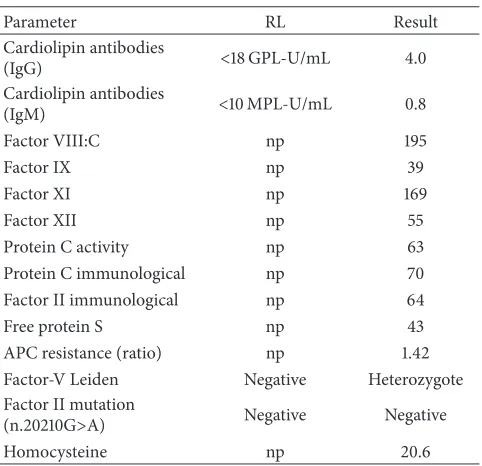
spontaneous deep venous thrombosis (DVT) with consec-utive pulmonary embolism (PE). Acetyl-salicylic acid was given for secondary prophylaxis because of cognitive impair-ment. Cerebral CT (CCT) showed leukoencephalopathy and pneumosinus dilatans. At age 51 y, he experienced a second spontaneous DVT. Since then oral anticoagulation with phenprocoumon was given. Neurologic exam revealed ptosis exclusively. Diagnostic workup for the cause of recurrent DVT revealed the factor-V Leiden mutation in its heterozy-gous form (Table 2). Since age 53 y, weakness of the limb muscles developed. Muscle biopsy from the left brachial biceps muscle at age 54 y revealed only nonspecific alterations with some central nuclei and type-II-fiber predominance. Genetic testing at age 55 y revealed a heterozygous CTG-repeat expansion of 500 CTG-repeats in theDMPKgene on chro-mosome 19q13.3. Prostate hyperplasia became apparent and a synthetic growth factor was applied for growth retardation and short stature during 1 y, without success. At age 59 y, he experienced pneumonia two times. Thrombocytopenia became apparent (Table 1). Further downstream writing Volume 2015, Article ID 271639, 4 pages
2 Case Reports in Medicine
Table 1: Blood chemical values.
Parameter RL 6/99 7/01 7/02 2/07 10/07 11/07 2/08 12/09 12/11 4/12
Thrombocytes 150–300 G/L nd nd nd 90 99 108 111 124 117 157
Uric acid 3.5–7.0 mg/dL nd 7.7 6.6 nd 6.6 7.3 nd nd 6.5 6.5
GOT 0–18 U/L 11 11 28 75 59 36 nd 39 50 36
GPT 0–23 U/L 11 11 14 84 65 53 nd 34 79 43
GGT 6–28 U/L 47 32 32 217 181 158 nd 170 125 69
TG 50–172 mg/dL 299 168 180 nd 256 189 249 205 340 277
CHE 150–200 mg/dL 239 225 220 nd 200 195 255 225 256 320
CK 0–80 U/L 39 60 nd 351 151 132 nd 269 123 136
IgG 700–1600 mg/dL nd nd nd nd nd 647 nd nd nd nd
RL: reference limits, GOT: glutamate-oxalate transaminase, GPT: glutamate-pyruvate transaminase, GGT: gamma-glutamyl transpeptidase, TG: triglycerides, CHE: cholesterol, CK: creatine-kinase, and nd: not done; since 2/07 other reference limits were used for GOT, GPT, GGT, and CK, and abnormal values are in bold.
Table 2: Coagulation parameters.
Parameter RL Result
Cardiolipin antibodies
(IgG) <18 GPL-U/mL 4.0
Cardiolipin antibodies
(IgM) <10 MPL-U/mL 0.8
Factor VIII:C np 195
Factor IX np 39
Factor XI np 169
Factor XII np 55
Protein C activity np 63
Protein C immunological np 70
Factor II immunological np 64
Free protein S np 43
APC resistance (ratio) np 1.42
Factor-V Leiden Negative Heterozygote
Factor II mutation
(n.20210G>A) Negative Negative
Homocysteine np 20.6
RL: reference limits, np: not provided.
became increasingly impaired. Echocardiography showed aortic insufficiency grade 1 and mitral insufficiency grade 1. Cerebral MRI revealed marked general atrophy and marked leukoencephalopathy (Figure 1). At age 60 y, he experienced vestibular neuronitis but search for acoustic nerve neurinoma was negative. Clinical neurologic exam at age 60 y revealed myopathic face, bilateral hypoacusis, dysarthria, weak head anteflexion (M5-), diffuse weakness (M5-) of the upper limbs, cogwheel rigidity bilaterally, bilateral postural tremor, weak-ness for hip flexion (M5-), weakweak-ness for foot extension (M5-), general diffuse wasting, generally reduced tendon reflexes, and recurrent hyper-CKemia (Table 1). During hospitalisa-tion, ascites from hepatopathy developed. At age 61 y, he expe-rienced a second PE bilaterally with right-sided infarction pneumonia and a third DVT bilaterally. The INR value was
outside the therapeutic range at this event. As a conse-quence, a vena cava filter was implanted. CCT showed atro-phy and leukoencephalopathy. Abdominal CT revealed an atrophic pancreas, a left-sided renal cyst, and prostate hyper-plasia. At age 62 y, he experienced thrombosis of the vena cava filter under an INR of 3.0. At age 63 y, he underwent cataract surgery bilaterally. At age 64 y, an AV-block I was documented for the first time. At age 65 y, an event recorder was implanted because of suspected arrhythmias. Recurrent interrogations revealed paroxysmal AFIB. The MMSE was 25. Shortly afterwards he was admitted to the intensive care unit because of severe dyspnea caused by pneumonia with an oxygen saturation of 63% and a pH of 6.92 requiring mechan-ical ventilation. Since hd1 he developed a striking restless-leg syndrome. On hd12 he underwent tracheostomy. On hd47 he received a percutaneous endoscopic gastrostomy (PEG). One day later he was transferred to the neurologic rehabilitation unit. Three days later he again required mechanical ventila-tion because of respiratory failure. He died on hd78 because of sepsis from pneumonia. The patient was a nonsmoker and nonalcoholic. His family history was positive for cataract and glaucoma (father), diabetes (brother of mother), and myocar-dial infarction (mother). He had one healthy son with gamma-globulin deficiency.
3. Discussion
The presented patient is interesting for several reasons. First, he is only the second patient with a double genetic trouble comprising a CTG-repeat expansion in theDMPKgene and the Leiden mutation [1]. The first DM1 patient with a Leiden mutation additionally had protein C deficiency [1]. In a further DM1 patient the CTG-repeat expansion was asso-ciated with a de novo shortened D4Z4 repeat fragment at the 4q35 locus giving rise to a facioscapulohumeral muscular dystrophy phenotype [3].
Figure 1: MRI of the cerebrum showing diffuse atrophy gliotic spots and leucoaraiosis. Pneumosinus dilatans can be also seen.
Possibly, the presence of theDMPKmutation enhanced the pathologic effect of the Leiden mutation. It can be speculated that expression of proteins required for the autochthon thrombolysis system was impaired to such a degree that it resulted in a gross prothrombotic effect. It is also possible that the mutated factor-V mRNA was further modified at the post-translational level by sequestered RNA-binding proteins. It is also conceivable that liver disease favoured the development of DVT, PE, and cava filter thrombosis [4]. However, history and laboratory investigations were negative for hepatitis, HIV, cholangitis, cholestasis, and storage disease, and abdominal CT did not show liver disease. Secret alcohol consumption was rather unlikely given the absence of red blood cell stig-mata. Thus, hepatopathy was most likely attributable to liver involvement in DM1. Though impaired mobility from DM1 has been reported to favour the development of thrombotic events [2] and the patient was lazy, it is rather unlikely that muscle involvement contributed to the etiology of recurrent DVT, since he displayed only mild muscle impairment, also in the leg compartment, and had rather normal mobility.
Third, the patient is interesting for marked central ner-vous system (CNS) involvement in the disease. He presented not only with dysarthria but also with cognitive decline, extrapyramidal manifestations, cogwheel rigidity, tremor,
cerebral atrophy, and leukoencephalopathy, a combination of abnormalities previously described [5–8]. Of particular inter-est are the Parkinson’s features (cogwheel rigidity, impaired writing) in the presented patient since, contrary to DM2 [9], only few DM1 patients have been reported with manifesta-tions of Parkinson’s syndrome [10].
Fourth, the patient is interesting for multiorgan disorder syndrome (MODS) affecting the skeletal muscles (ptosis, limb weakness, and weakness of respiratory muscles), the CNS (leukoencephalopathy, cognitive decline, dysarthria, cogwheel rigidity, postural tremor, impaired writing, and cerebral atrophy), the ears (hypoacusis, vestibular neuroni-tis), the liver (hepatopathy, steatosis, and cholecystolithiasis), the endocrine system (hypogonadism, prostate hyperplasia), the bones (pneumosinus dilatans [11]), and the immune system (reduced IgG, reduced IgM, and recurrent pulmonary infections) [12].
4 Case Reports in Medicine
that small PE occurred even after anticoagulation and place-ment of the vena cava filter, favouring recurrent pulmonary infections (infarction pneumonia). It is also conceivable, that PE had been mistaken as pneumonia. Since the patient never complained about dysphagia, microaspiration was rather excluded as the cause of recurrent pulmonary infections.
In conclusion, this case shows that a Leiden mutation, even if present in a heterozygous form, may severely affect DM1 patients to such a degree that they die from its long-term complications. If DM1 patients present with uncommon phenotypic manifestations, search for causes other than a CTG-repeat expansion including other mutations is indi-cated. Neurologists should be alert to the possibility that individuals having two separate mutations in unrelated genes may develop unusually severe phenotypes.
Conflict of Interests
The authors declare that there is no conflict of interests regarding the publication of this paper.
References
[1] J. A. Hodapp, G. T. Carter, H. P. Lipe, S. J. Michelson, G. H. Kraft, and T. D. Bird, “Double trouble in hereditary neuropathy: concomitant mutations in the PMP-22 gene and another gene produce novel phenotypes,”Archives of Neurology, vol. 63, no. 1, pp. 112–117, 2006.
[2] E. Cobos, M. Phy, and Y. K. Keung, “Thrombotic risk of muscu-lar dystrophy: protein C deficiency, factor V Leiden, and myo-tonic dystrophy,”Clinical and Applied Thrombosis/Hemostasis, vol. 5, no. 3, pp. 185–186, 1999.
[3] M. Masciullo, E. Iannaccone, M. L. E. Bianchi et al., “Myotonic dystrophy type 1 and de novo FSHD mutation double trouble: a clinical and muscle MRI study,”Neuromuscular Disorders, vol. 23, no. 5, pp. 427–431, 2013.
[4] B. Hewing, L. Ghaeni, H. Dreger et al., “Echocardiographic diagnosis, management and monitoring of pulmonary embolism with right heart thrombus in a patient with myotonic dystrophy: a case report,”Cardiovascular Ultrasound, vol. 8, no. 1, article 18, 2010.
[5] J. R. Wozniak, B. A. Mueller, C. J. Bell, R. L. Muetzel, K. O. Lim, and J. W. Day, “Diffusion tensor imaging reveals widespread white matter abnormalities in children and adolescents with myotonic dystrophy type 1,”Journal of Neurology, vol. 260, no. 4, pp. 1122–1131, 2013.
[6] D. T. Franc, R. L. Muetzel, P. R. Robinson et al., “Cerebral and muscle MRI abnormalities in myotonic dystrophy,”
Neuromus-cular Disorders, vol. 22, no. 6, pp. 483–491, 2012.
[7] M. Kobayakawa, N. Tsuruya, A. Takeda, A. Suzuki, and M. Kawamura, “Facial emotion recognition and cerebral white matter lesions in myotonic dystrophy type 1,”Journal of the
Neurological Sciences, vol. 290, no. 1-2, pp. 48–51, 2010.
[8] G. Meola and V. Sansone, “Cerebral involvement in myotonic dystrophies,”Muscle & Nerve, vol. 36, no. 3, pp. 294–306, 2007. [9] A. Annie, D. Devos, A. Dest´ee et al., “Early dopasensitive Parkinsonism related to myotonic dystrophy type 2,”Movement
Disorders, vol. 23, no. 14, pp. 2100–2101, 2008.
[10] Y. Celik, N. Turgut, K. Balci, and L. Kabayel, “Proximal myotonic dystrophy associated with parkinsonism,”Journal of
Clinical Neuroscience, vol. 13, no. 2, pp. 275–276, 2006.
[11] J. Finsterer, C. St¨ollberger, G. M¨olzer, and E. Prager, “Pneumosi-nus dilatans and hypercalcification of the falx and ligamentum petroclinoideum in myotonic dystrophy 1,”Neurologist, vol. 16, no. 2, pp. 125–128, 2010.
Submit your manuscripts at
http://www.hindawi.com
Stem Cells
International
Hindawi Publishing Corporationhttp://www.hindawi.com Volume 2014
Hindawi Publishing Corporation
http://www.hindawi.com Volume 2014
INFLAMMATION
Hindawi Publishing Corporation
http://www.hindawi.com Volume 2014
Behavioural
Neurology
Endocrinology
International Journal of Hindawi Publishing Corporationhttp://www.hindawi.com Volume 2014
Hindawi Publishing Corporation
http://www.hindawi.com Volume 2014
Disease Markers
Hindawi Publishing Corporation
http://www.hindawi.com Volume 2014
BioMed
Research International
Oncology
Journal ofHindawi Publishing Corporation
http://www.hindawi.com Volume 2014
Hindawi Publishing Corporation
http://www.hindawi.com Volume 2014 Oxidative Medicine and Cellular Longevity Hindawi Publishing Corporation
http://www.hindawi.com Volume 2014
PPAR Research
The Scientific
World Journal
Hindawi Publishing Corporationhttp://www.hindawi.com Volume 2014
Immunology Research
Hindawi Publishing Corporation
http://www.hindawi.com Volume 2014
Journal of
Obesity
Journal ofHindawi Publishing Corporation
http://www.hindawi.com Volume 2014
Hindawi Publishing Corporation
http://www.hindawi.com Volume 2014
Computational and Mathematical Methods in Medicine
Ophthalmology
Journal ofHindawi Publishing Corporation
http://www.hindawi.com Volume 2014
Diabetes Research
Journal ofHindawi Publishing Corporation
http://www.hindawi.com Volume 2014
Hindawi Publishing Corporation
http://www.hindawi.com Volume 2014
Research and Treatment
AIDS
Hindawi Publishing Corporation
http://www.hindawi.com Volume 2014
Gastroenterology Research and Practice
Hindawi Publishing Corporation
http://www.hindawi.com Volume 2014
Parkinson’s
Disease
Evidence-Based Complementary and Alternative Medicine
Volume 2014 Hindawi Publishing Corporation