1535-9778/07/$08.00⫹0 doi:10.1128/EC.00020-07
Copyright © 2007, American Society for Microbiology. All Rights Reserved.
The
MAT1
Locus of
Histoplasma capsulatum
Is Responsive in a
Mating Type-Specific Manner
䌤
Meggan Bubnick
1,2and A. George Smulian
2,3*
Department of Pathology1and Infectious Disease Division,2University of Cincinnati College of Medicine, 231 Albert Sabin Way,
Cincinnati, Ohio 45267-0560, and Cincinnati VA Medical Center, 3200 Vine Street, Cincinnati, Ohio 452203
Received 17 January 2007/Accepted 14 February 2007
Recombination events associated with sexual replication in pathogens may generate new strains with altered
virulence. Histoplasma capsulatumis a mating-competent, pathogenic fungus with two described phenotypic
mating types,ⴙandⴚ. The mating (MAT) locus ofH. capsulatumwas identified to facilitate molecular studies
of mating in this organism. Through syntenic analysis of theH. capsulatumgenomic sequence databases, a
MAT1-1idiomorph region was identified inH. capsulatumstrains G217B and WU24, and aMAT1-2idiomorph region was identified in the strain G186AR. A mating type-specific PCR assay was developed, and two clinical
isolates of opposite genotypic mating type, UH1 and VA1, were identified. A knownⴚmating type strain, T-3-1
(ATCC 22635), was demonstrated to be ofMAT1-2genotypic mating type. The clinical isolates UH1 and VA1
were found to be mating compatible and also displayed mating-type-dependent regulation of theMAT
tran-scription factors in response to extracts predicted to contain mating pheromones. These studies support a role
for the identifiedMAT1locus in determining mating type inH. capsulatum.
The mating process has the potential to play a role in the virulence of human pathogens. Recombination between two strains can result in a new strain with increased virulence. This has been documented in the parasiteToxoplasma gondii(12, 25) and also may have occurred in the fungusCryptococcus
gattiiduring the Vancouver Island outbreak (9). Populations of
the pathogenic fungus Histoplasma capsulatum have been shown to recombine in nature (5, 13); however, little is known about mating on a molecular level in this organism.
H. capsulatumis a dimorphic ascomycete found worldwide.
Inhalation of the conidia may result in pulmonary disease and, in some cases, severe disseminated disease and death (27).H.
capsulatumis a heterothallic organism with two mating types,
⫹and⫺, defined by phenotype (14). Mating between freshly isolated⫹and⫺strains occurs in the mycelial phase and has been demonstrated in the laboratory (14); however, strains lose the ability to mate with continuous culture (14, 15, 18). Strains of each mating type are not equally represented among clinical isolates ofH. capsulatum. In two separate studies, ⫺ mating type strains were dominant among the clinical samples tested (17, 18). In contrast, strains isolated from the soil rep-resented both mating types equally (18). It was also shown that the mating type disequilibrium occurs among strains isolated from patients with the pulmonary form of the disease but not the severe disseminated form (17). It is unknown whether these observations can be attributed to virulence differences between strains of opposite mating type.
While an organism’s mating type can be determined pheno-typically, it can also be determined by the genes present at the mating (MAT) locus (6). In filamentous ascomycetes, theMAT
locus is a region of low sequence similarity between two or-ganisms of opposite mating type, with each organism contain-ing a different idiomorph of theMATlocus (6, 26). TheMAT
locus usually contains genes for one or more transcription factors with structural motifs such as␣1 domains, high-mobil-ity-group (HMG) DNA binding domains, amphipathic␣ -heli-ces, and metallothioneins (6, 26). In a filamentous ascomycte, the first identified MAT idiomorph region encoding an ␣1 domain transcription factor is often designatedMAT1-1, while the first identifiedMATidiomorph region encoding an HMG DNA binding domain is often designatedMAT1-2(26).
The aim of this study was to identify theMAT1locus inH.
capsulatumand correlate the previous phenotypic mating type
designations with genotypic mating type designations. Mating loci have been identified in other filamentous ascomycete fungi, includingNeurospora crassa(11, 23),Aspergillus nidulans
(8), and Aspergillus fumigatus (21). Using characteristics of these knownMATloci, predictedMAT1-1and MAT1-2 idio-morph regions were identified in the genome sequences of three different strains of H. capsulatum. Genotypic mating types were assigned to strains based on the structure of theMAT1
idiomorph regions, and a known⫺ mating type tester strain was shown to be of MAT1-2genotype. Mating compatibility was confirmed betweenH. capsulatumstrains of opposite ge-notypic mating type. Additionally, message levels of predicted
MAT1locus transcription factors were differentially regulated, depending on the genotypic mating type, in response to pher-omone extracts. Taken together, these studies support a role for the predictedMAT1locus in determining mating type inH.
capsulatum.
MATERIALS AND METHODS
Strains and conditions.H. capsulatumstrains G217B (ATCC 26032; a kind gift from George Deepe, University of Cincinnati, Cincinnati, OH), G186A (ATCC 26029; a kind gift from William Goldman, Washington University, St. Louis, MO), G184A (ATCC 26027; from William Goldman), T-3-1 mating type ⫺(ATCC 22635), UH1 (a clinical isolate obtained from a transplant patient with * Corresponding author. Mailing address: Infectious Disease
Divi-sion, University of Cincinnati College of Medicine, 231 Albert Sabin Way, Cincinnati, OH 45267-0560. Phone: (513) 861-3100, ext. 4425. Fax: (513) 475-6415. E-mail: [email protected].
䌤Published ahead of print on 23 February 2007.
616
on September 8, 2020 by guest
http://ec.asm.org/
disseminated histoplasmosis), and VA1 (a clinical isolate obtained from a human immunodeficiency virus/AIDS patient with disseminated histoplasmosis) were grown inHistoplasmamacrophage medium (HMM) broth or on HMM agarose plates. H. capsulatum strains WU8 (G186A ura5⌬32) and WU15 (G217B
ura5⌬42), gifts from William Goldman and Russ Osguthorpe, were grown in HMM broth supplemented with 0.2 mg/ml uracil (Sigma-Aldrich, St. Louis, MO).
Yeast-phase organisms were grown at 37°C in liquid media on an orbital shaker or on plates at 37°C under 5% CO2in a humidified incubator. Mycelial-phase organisms were grown in liquid culture at 25°C on an orbital shaker in HMM or yeast extract Medium (YEM) (14), with uracil supplementation as appropriate. Mating assays on solid media were performed at 25°C on Alphacel yeast extract medium (A-YEM) agarose plates (14).
Analysis of fungal mating loci.TheH. capsulatumgenome sequence is avail-able at the websites of the Washington University Genome Sequencing Center (http://genome.wustl.edu/genome_group_index.cgi) (strains G217B and G186AR) and the Broad Institute (http://www.broad.mit.edu/annotation/fgi) (strain WU24). Using BLASTX or TBLASTN,H. capsulatum MAT1-1-1andMAT1-2-1proteins and genes were compared to the fungal genomes at the Broad Institute or the database of the National Center for Biotechnology Information.
MATlocus PCR.DNA was isolated from selectedH. capsulatumstrains grown in liquid culture by using the MasterPure yeast DNA kit (Epicentre Inc., Mad-ison, WI). PCR was performed using sense primers MAT1-1S (5⬘-CGTGGTT AGTTACGGAGGCA-3⬘) and MAT1-2S (5⬘-ACACAGTAGCCCAACCTCT C-3⬘) and antisense primers MAT1-1AS (5⬘-TGAGGATGCGAGTGATGGG A-3⬘) and MAT1-2AS (5⬘-TCGACAATCCCATCCAATACCG-3⬘), designed based on theMAT1-1andMAT1-2idiomorph sequences. PCR was performed under standard conditions with a 60°C annealing temperature by using Jump-StartTaqpolymerase (Sigma-Aldrich). Products were analyzed by agarose gel electrophoresis.
Mating compatibility test.The organisms to be tested were streaked onto HMM plates and grown at 25°C until mycelial growth was observed. A plug of each organism was transferred onto A-YEM agarose (14). Plugs were placed approximately 8 mm apart. Organisms were grown at 25°C for 3 weeks and then examined microscopically. Mycelial organisms were lifted from the plates by using clear Petri-Seal tape. Organisms on the tape were mounted on slides by using lactophenol mounting media and covered with a coverslip. Slides were observed using a Nikon E600 microscope equipped with a Spot RT slider camera.
Isolation of a and␣pheromones.Putative pheromone-containing suspen-sions were isolated using a protocol described by Strazdis and MacKay (24). Briefly, hydrophobic polystyrene resin Amberlite XAD2 was prepared by washing in water, followed by either 1:3 methylene chloride-methanol (for␣ factor isolation) or 1:3 dichloropropane–n-propanol (forafactor isolation), and stored in propanol. Prior to use, the resin was washed in excess water and sterilized by autoclaving in water. Fifty milliliters of the appropriate resin preparation was added to a 500-ml coculture ofH. capsulatumstrains WU8 and WU15. The culture was grown for 1 to 2 weeks in uracil-supplemented YEM at 25°C. The resin was then removed from the culture and washed extensively with water, and bound proteins were eluted from the beads by incubation in propanol (afactor) or 40% methanol (␣factor) at 40°C for 2 h. The alcohol was evaporated by vacuum centrifugation, and the remaining protein was reconstituted in 100 l dimethyl sulfoxide (DMSO). Eluted proteins were analyzed by sodium dodecyl sulfate-polyacrylamide gel elec-trophoresis on 4 to 20% acrylamide gels in a Tris-Tricine buffer system and observed following silver staining.
Quantitative real-time PCR (qRT-PCR) of mating-related gene expression.H. capsulatumstrains VA1 and UH1, growing at 25°C on HMM agar, were used to start liquid cultures in HMM. Liquid cultures were grown to saturation at 25°C with shaking. Organisms were then transferred to YEM and grown at 25°C overnight with shaking. Five microliters of␣orapheromone-containing suspen-sions, extracted fromH. capsulatumculture as described above, or DMSO was added to the cultures in YEM. RNA was extracted after 30 min by using the MasterPure yeast RNA kit (Epicentre). cDNA synthesis, primed from antisense primers specific toMAT1-1-1,MAT1-2-1, andGAPDH, was performed using Superscript II reverse transcriptase (Invitrogen, Carlsbad, CA). The gene-spe-cific antisense primers were MAT1-1-1 (5⬘-GTAATACGACTCACTATAGGG TTGTCCATGCTCACAGCCAATT-3⬘), MAT1-2-1 (5⬘-GTAATACGACTCA CTATAGGGGGTGATGCCGGCGATACAAAAT-3⬘), and GAPDH (5⬘-GTA ATACGACTCACTATAGGGGACAGCCTTGCCATTGACGGTC-3⬘).
cDNA samples were diluted 1:10 in water, and qRT-PCR was performed using SYBR green PCR master mix (Applied Biosystems, Foster City, CA) in the Applied Biosystems 7500 real-time PCR system (Applied Biosystems Inc.).
qRT-PCR was performed in triplicate for each cDNA sample. The primer pairs for each qRT-PCR were designed such that one primer in each pair spanned an intron, preventing amplification from genomic DNA. The specificity of each primer pair for cDNA was confirmed by the lack of amplification, as evi-denced by the lack of SYBR green incorporation and verified by analysis with agarose gel electrophoresis, using genomic DNA as the template. The prim-ers and sequences used were as follows: MAT1-1-1-S, 5⬘-TTCGTTCATAG CCTTCAGAAGCTTC-3⬘; MAT1-1-1-AS, 5⬘-GGCCAGCATGACTGTCAC GAAT-3⬘; MAT1-2-1-S, 5⬘-AAAATCAAAGACCGCTTGAGCGCA-3⬘; MAT1-2-1-AS, 5⬘-AACAACGGCAGCATCGACAATCCC-3⬘; GAPDH-S, 5⬘-ATTGGGC GTATTGTCTTCC-3⬘; and GAPDH-AS, 5⬘-TTGAGCATGTAGGCAGCATA-3⬘. The annealing temperatures forMAT1-1-1,MAT1-2-1, andGAPDHprimers were 57°C, 62°C, and 60°C, respectively. cDNA for a relative standard curve was generated using a pool of RNA produced by pooling an aliquot of RNA from each sample collected. The standard cDNA was diluted 1:10, and a standard curve was generated using four samples, serially diluted 1:4 from the original 1:10 dilution. Relative expression levels of genes of interest were calculated using the standard curve method for relative quantification (2). Outliers were removed using Grubb’s test for detecting outliers, and RNA levels under the different conditions were compared using analysis of variance (GraphPad Instat; Graph-Pad, San Diego, CA).
Nucleotide sequence accession numbers.The sequences of theMAT1-1and
MAT1-2loci have been deposited in GenBank and are available under the accession numbers EF433757 and EF433756, respectively.
RESULTS
Identification ofMAT1locus inH. capsulatum. To identify
theMAT1-1idiomorph inH. capsulatum, a TBLASTN analysis
was performed to compare the␣1 region of a previously iden-tifiedA. nidulans MAT1-1peptide (8) with genome sequences
of H. capsulatum strains G217B, G186AR, and WU24.
HISTO_EA.Contig33 of strain G217B contained a region of 55% sequence identity and 71% similarity to theA. nidulans␣1 region. Similarly, supercontig 1-1 of the WU24 genome se-quence contained a region of 45% sese-quence identity and 63% similarity to theA. nidulans␣1 region. No significant sequence similarity to the A. nidulans ␣1 region was noted in the G186AR genome sequence. TheH. capsulatum␣-box region was predicted to be located within a 1,256-bp gene, which was designatedMAT1-1-1(Fig. 1A) (26). The 1,206-bpMAT1-1-1
open reading frame, interrupted by one 50-bp intron, was pre-dicted to encode a 401-amino-acid protein. The position of the intron was confirmed by sequencing and alignment of MAT1-1-1PCR products generated from both DNA and cDNA (data not shown). The position of the intron was identical to that of introns found in other ascomycete␣1MATtranscription fac-tors (Fig. 1B) (6).
Since strain G186AR showed no sequence similarity to the
A. nidulans␣1 region, the G186AR genome was searched for
aMAT1-2idiomorph. When two ascomycetes of opposite
mat-ing type are compared, theMATidiomorph regions generally contain little sequence similarity between the two but are flanked by regions of homology between the two (1, 6). Based on this, a BLASTN analysis was performed to compare the
MAT1-1-1gene sequence, and 10 kb of G217B sequence
up-stream and downup-stream of the gene, to the G186AR genome sequence. A region of approximately 5 kb with only 33.9% sequence identity was identified, flanked by regions with 95.8% and 96.4% sequence identity, respectively. In addition to se-quence disparity, the regions of dissimilarity varied in length, containing 5,702 bp in the G217B strain and 4,997 bp in the G186AR strain, respectively. The MAT1-1 idiomorph con-tained only the one putative open reading frame described
on September 8, 2020 by guest
http://ec.asm.org/
above. The 4,997-bpMAT1-2idiomorph in G186AR was lo-cated in contig 30-9 and also contained a single predicted open reading frame. The open reading frame was designated MAT1-2-1(26), as it was predicted to encode a 348-amino-acid pro-tein with an HMG DNA binding domain (Fig. 1A). The
MAT1-2-1open reading frame was interrupted by two introns,
which were confirmed in the same manner as theMAT1-1-1
intron (data not shown). Both introns were in the same relative position as introns found in other ascomycete MAT HMG transcription factors (Fig. 1C) (6, 22). Based on this informa-tion, the mating type of strain G186AR was designated
MAT1-2, while the mating type of strains G217B and WU24
was designatedMAT1-1(26).
MAT locus PCR. PCR primers were designed to amplify
either theMAT1-1or theMAT1-2region specifically. Ampli-fication of theMAT1-1region would result in a 440-bp prod-uct, while amplification of theMAT1-2region would result in a 528-bp product. This MAT locus PCR assay was used to determine the genotypic mating type of a known⫺mating type tester strain, T-3-1, to correlate genotypic and phenotypic mat-ing type. The phenotypic⫺mating type strain was shown to be of theMAT1-2genotypic mating type (Fig. 2). The assay was also used to determine the genotypic mating type of two clin-ical isolates, UH1 and VA1. UH1 and VA1 were shown to be of opposite mating type, MAT1-1and MAT1-2, respectively (Fig. 2). An additional laboratory strain in common use, G184A, was similarly analyzed and shown to be of theMAT1-2
mating type (data not shown).
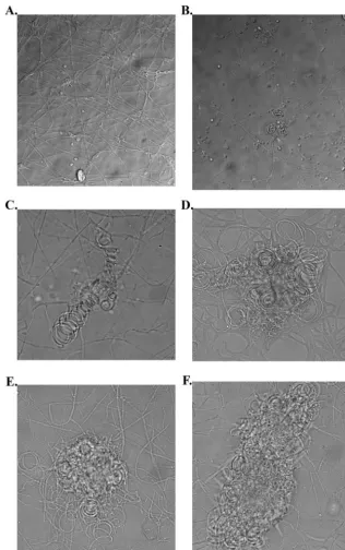
Mating compatibility test. Phenotypic confirmation was
sought for the assigned opposite genotypic mating types of
clinical isolates UH1 and VA1. Mating compatibility assays were performed between UH1 ⫹ and VA1 ⫺ strains, with self-crosses of UH1 or VA1 as controls. After 10 to 14 days, a confrontation border appeared between the mycelial plugs of opposite mating type where the advancing hyphal colonies met (14). After an additional 7 days, the mycelia in this region were viewed microscopically. The formation of the ascocarp inH.
cap-sulatumhas been described previously (14), and several structures
indicative of the mating process in this organism were observed. The structures observed included hyphal coiling and cleistothecia covered by short branching hyphae. These structures were not seen when strains were tested as self-crosses (Fig. 3).
FIG. 1.MAT1locus inH. capsulatum. (A) Representation of theMAT1-1andMAT1-2idiomorph regions, as identified in strains G217B and G186AR, respectively. Hatched boxes indicate regions with 96 to 98% sequence identity between the two strains. Black lines indicate theMAT1
idiomorph regions, with only 34% sequence identity. Predicted open reading frames are represented by arrows, white forMAT1-1-1and black for
MAT1-2-1, with introns designated by asterisks. (B and C) Alignment ofH. capsulatum␣1 (B) and HMG (C) regions withMAT1-1andMAT1-2
protein sequences in other ascomycete fungi. The positions of conserved introns are noted by arrows. The organism abbreviations are as follows: A. nid,Aspergillus nidulans; A. fum,Aspergillus fumigatus; P. mar,Penicillium marneffei; H. cap,Histoplasma capsulatum; C. zei,Cercospora zeina; G. fuj,Gibberella fujikuroi; C. imm,Coccidioides immitis; M. gra,Mycosphaerella graminicola.
FIG. 2. Results ofMAT1locus PCR assay. PCR was performed using primers specific to either theMAT1-1or theMAT1-2idiomorph region. Both primer pairs were used to test DNA isolated from various
H. capsulatumstrains. Lane 1, molecular weight marker; lanes 2 to 6,
H. capsulatumstrains as indicated under each lane; lane 7, “no tem-plate” control (NTC).
on September 8, 2020 by guest
http://ec.asm.org/
Differential expression ofMATlocus transcription factors.
Regulation of the expression of theMAT1-1andMAT1-2 tran-scripts in H. capsulatum was examined under mating condi-tions to examine the association ofMAT1gene expression with mating. Mating pheromones have not been identified or iso-lated inH. capsulatum; however, based on the assumption that the pheromones produced byH. capsulatumresemble the pep-tide and lipopeppep-tide pheromones of other ascomycetous fungi, procedures enriching for each class of pheromone produced by
Saccharomyces cerevisiae were performed on mixedMAT1-1
and MAT1-2mycelial cultures of H. capsulatum. When
ana-lyzed by sodium dodecyl sulfate-polyacrylamide gel electro-phoresis and examined by silver staining, the resulting suspen-sions contained only a few (⬃5 to 10) bands, all with molecular masses below 20 kDa (data not shown).
Clinical isolates of opposite genotypic mating type, UH1
(MAT1-1) and VA1 (MAT1-2), were stimulated either with
each of the putative pheromone-containing extracts or with FIG. 3. Results of mating compatibility test between UH1 and VA1. Organisms were plated on A-YEM and grown for 3 weeks. (A) UH1 self-cross. (B to F) UH1 crossed with VA1. (B and C) Coiled hyphae, consistent with younger mating structures. (D to F) Coiled hyphae covered by short, branching hyphae, consistent with more mature mating structures.
on September 8, 2020 by guest
http://ec.asm.org/
vehicle controls. The expression levels of the predictedMAT1
locus transcription factors were then measured by RT-PCR. Stimulation of UH1 with an extract predicted to contain the␣ pheromone resulted in a fivefold increase inMAT1-1-1 expres-sion compared to stimulation with DMSO alone. This was not seen when UH1 was stimulated with an extract predicted to contain theapheromone (Fig. 4). The opposite response was seen in VA1, the organism ofMAT1-2genotype. Stimulation of VA1 with an extract predicted to contain theapheromone resulted in a 4.4-fold increase inMAT1-2-1 expression com-pared to stimulation with DMSO alone. Stimulation of VA1 with an extract predicted to contain the␣pheromone caused no significant change in MAT1-2-1 expression compared to stimulation with DMSO (Fig. 4).
DISCUSSION
Similarities between MATloci of several ascomycete fungi have made the identification ofMATloci possible in organisms whose genomes have been sequenced. In this manner, a
MAT1-1 region was identified in the homothallic fungus A.
nidulans(8), andMAT1-1andMAT1-2idiomorphs were
iden-tified in A. fumigatus (21, 22). The predicted MAT1 locus, identified inH. capsulatumthrough the current study, shared common characteristics with MAT loci in other ascomycete fungi (6, 11, 21, 23, 26). TheH. capsulatum MAT1-1idiomorph contained a predicted transcription factor characterized by the
presence of an␣1 domain. Using standard nomenclature, this is a common feature ofMAT1-1idiomorphs found in ascomy-cete fungi (6, 26). Similarly, theMAT1-2idiomorph contained a transcription factor characterized by an HMG domain, which is common amongMAT1-2idiomorph regions in ascomycete fungi (6, 26). Heterothallism, with strains containing either a
MAT1-1 or a MAT1-2 idiomorph, appears to be common
among the dimorphic ascomycetes. Using a similar genomic approach, MAT1-2 regions can be identified in Coccidioides
immitisstrains RS and RMSCC 2394 (10), while a MAT1-1
region can be identified inC. immitisstrain 4538.4, using ge-nome sequences available at the Broad Institute website (http: //www.broad.mit.edu/annotation/fgi) (data not shown). MAT1-2
regions can also be identified inCoccidioides posadasii(10) and
Blastomyces dermatitidis, using genome sequences available from
The Institute for Genomic Research (http://www.tigr.org/tdb /fungal/index.shtml) and the Washington University Genome Se-quencing Center (http://genome.wustl.edu/genome_group_index .cgi) websites, respectively (data not shown). Additionally, ascomycete␣1 and HMG domains each contain introns at con-served positions within the domains (6), which were also identi-fied in the sequences for the predictedMATtranscription factors
inH. capsulatum.
H. capsulatum has a heterothallic mating system, and the
mating morphology of this fungus has been described in great detail using strains of opposite phenotypic mating type (14). Through the present study, mating compatibility was con-firmed morphologically in two organisms of opposite genotypic mating type. Asci and ascospores, the results of mating, were not sought in this study; however, many structures were ob-served that are associated with the formation of the ascocarp. This supports the genomic evidence that the predictedMAT1
locus is associated with mating type in H. capsulatum. This assertion is further supported by the mating type-specific reg-ulation of theMATtranscription factors in response to pher-omone extracts. As the pherpher-omone extracts were not purified to homogeneity, it is feasible that other proteins, unrelated to the pheromone response in this organism, may have caused the changes in levels of MAT1-1-1and MAT1-2-1expression. If this were the case, however, one would have expected to see the␣pheromone extract affect both UH1 and VA1, with the same holding true for thea pheromone extract. The mating type-specific regulation suggests that the effect was due to pheromones present in the semipurified extracts.
This study was carried out with the assumption that in H.
capsulatum, organisms of opposite mating type produce different
mating pheromones: a peptide␣-type pheromone and a lipopep-tidea-type pheromone. This is the case forS. cerevisiae(3, 4); however, this is not the case for the fungusCryptococcus neofor-mans, which produces two lipid-modified pheromones (7, 20). The techniques used to purify the putative pheromones rely on the differences in structure between the two pheromone types, allowing differential purification of peptide and lipopeptide pher-omones. The fact that up-regulation ofMATtranscription factors in the present study occurred only in one mating type organism for each pheromone extract argues that theH. capsulatum cocul-ture produced lipopeptide a-type and peptide ␣-type phero-mones. This is further supported by the existence of predicted pheromone-processing machinery for both pheromone types as well as a predicted␣-type pheromone sequence in theH.
capsu-FIG. 4. Expression levels ofMAT1transcription factors.H. capsu-latumstrains UH1 (A) and VA1 (B) were stimulated with DMSO,a pheromone extract, or␣pheromone extract for 30 min. RNA levels of
MAT1-1-1(A) andMAT1-2-1(B) were determined by qRT-PCR and
normalized to RNA levels ofGAPDH.*,Pⱕ0.05;**,Pⱕ0.01.
on September 8, 2020 by guest
http://ec.asm.org/
latumgenome (data not shown). The genes predicted to encode pheromone-processing proteins and the␣-type pheromone are unlinked to theMAT1locus or to each other.
Characterization of the MAT locus in H. capsulatum will allow a molecular comparison of⫹and⫺mating type organ-isms. Mating type disequilibrium exists among clinical isolates of this organism, with ⫺ mating type organisms dominating (17, 18). This disequilibrium was not seen in environmental isolates (18). While the mating type disequilibrium in clinical isolates may suggest differences in virulence between mating types, prior studies ofH. capsulatumtesting the virulence po-tentials of⫹and⫺mating type strains demonstrated no dif-ference in a mouse model of disease (19). These findings may require reevaluation, as the studies were performed using un-related strains ofH. capsulatum and utilized an intravenous inoculation model of disease. Mating type disequilibrium is found among clinical isolates ofC. neoformans, with organisms of the␣mating type dominating (16). For theC. neoformans
cases, this may be explained partly by the predominance of␣ mating type organisms in the environment (16). Characteriza-tion of theMATlocus inH. capsulatumwill allow the possi-bility of constructing congenic mating type strains in this or-ganism to reexamine questions regarding virulence and mating type. This will prove challenging, however, as few chromo-somal markers are known for this organism and the rate of targeted homologous integration in this organism is low (27). Characterization of theMATlocus inH. capsulatumwill also allow a molecular characterization of the mating process in this organism. Not only will this provide a molecular means of comparing organisms of opposite mating type but this will allow a molecular comparison of mating-competent and non-mating-competent organisms. While it has been noted that, with time,H. capsulatumloses the ability to mate when grown in culture (14, 15, 18), the molecular mechanisms of this loss of mating competence have not been explored. This is an obstacle to the development of classical genetic systems in this organ-ism. Laboratory strains G186AR and G217B did not generate a confrontation reaction or form mating structures in the mat-ing compatibility assay described above (data not shown). However, cocultivated derivatives of these strains yielded the pheromone extracts used to stimulateMAT1gene expression in a regulated manner, implying that the inability of laboratory strains to mate is not due to the loss of the ability to produce pheromone. Additional studies will attempt to determine the level of the defect resulting in the loss of mating competence. In summary, genomic studies, mating compatibility tests, and the regulation of predictedMATlocus transcription fac-tors in response to pheromone extracts together support the conclusion that the identified MAT1 locus in H. capsulatum
plays a role in determining mating type in this organism. The results of these studies will be useful in answering questions regarding the virulence of this organism as well as in develop-ing new tools to study the organism.
ACKNOWLEDGMENTS
We thank William Goldman and Russ Osguthorpe forH. capsula-tum strains, George Deepe for H. capsulatum strains, advice, and support, Judith Rhodes for advice and assistance, and Jeff Demland, Reiko Tanaka, Debbie Spaulding, Reta Gibbons, and Holly Allen for technical assistance.
REFERENCES
1.Astell, C. R., L. Ahlstrom-Jonasson, M. Smith, K. Tatchell, K. A. Nasmyth, and B. D. Hall.1981. The sequence of the DNAs coding for the mating-type loci of Saccharomyces cerevisiae. Cell27:15–23.
2.Bookout, A. L., C. L. Cummins, D. J. Mangelsdorf, J. M. Pesola, and M. F. Kramer.2006. High-throughput real-time quantitative reverse transcription PCR, p. 15.8.1–28.InF. M. Ausubel, R. Brent, R. E. Kingston, D. D. Moore, J. G. Seidman, J. A. Smith, and K. Struhl (ed.), Current protocols in mo-lecular biology. John Wiley and Sons, Hoboken, NJ.
3.Bussey, H.1988. Proteases and the processing of precursors to secreted proteins in yeast. Yeast4:17–26.
4.Caldwell, G. A., F. Naider, and J. M. Becker.1995. Fungal lipopeptide mating pheromones: a model system for the study of protein prenylation. Microbiol. Rev.59:406–422.
5.Carter, D. A., A. Burt, J. W. Taylor, G. L. Koenig, and T. J. White.1996. Clinical isolates ofHistoplasma capsulatumfrom Indianapolis, Indiana, have a recombining population structure. J. Clin. Microbiol.34:2577–2584. 6.Coppin, E., R. Debuchy, S. Arnaise, and M. Picard.1997. Mating types and
sexual development in filamentous ascomycetes. Microbiol. Mol. Biol. Rev. 61:411–428.
7.Davidson, R. C., T. D. Moore, A. R. Odom, and J. Heitman.2000. Charac-terization of the MF␣pheromone of the human fungal pathogen Cryptococ-cus neoformans. Mol. Microbiol.38:1017–1026.
8.Dyer P. S., M. Paoletti, and D. B. Archer.2003. Genomics reveals sexual secrets ofAspergillus. Microbiology149:2301–2303.
9.Fraser, J. A., S. S. Giles, E. C. Wenink, S. G. Geunes-Boyer, J. R. Wright, S. Diezmann, A. Allen, J. E. Stajich, F. S. Dietrich, J. R. Perfect, and J. Heitman.2005. Same-sex mating and the origin of the Vancouver Island
Cryptococcus gattiioutbreak. Nature437:1360–1364.
10.Fraser, J. A., and J. Heitman.2006. Sex,MAT, and the evolution of fungal virulence, p. 13–33.InJ. Heitman, S. G. Filler, J. E. Edwards, Jr., and A. P. Mitchell (ed.), Molecular principles of fungal pathogenesis. ASM Press, Washington, DC.
11.Glass, N. L., J. Grotelueschen, and R. L. Metzenberg.1990.Neurospora crassa Amating-type region. Proc. Natl. Acad. Sci. USA87:4912–4916. 12.Grigg, M. E., S. Bonnefoy, A. B. Hehl, Y. Suzuki, and J. C. Boothroyd.2001.
Success and virulence inToxoplasmaas the result of sexual recombination between two distinct ancestries. Science294:161–165.
13.Kasuga, T., J. W. Taylor, and T. J. White.1999. Phylogenetic relationships of varieties and geographical groups of the human pathogenic fungus His-toplasma capsulatumDarling. J. Clin. Microbiol.37:653–663.
14.Kwon-Chung, K. J.1973. Studies onEmmonsiella capsulata. I. Heterothal-lism and development of the ascocarp. Mycologia65:109–121.
15.Kwon-Chung, K. J.1975. Perfect state (Emmonsiella capsulata) of the fungus causing large-form African histoplasmosis. Mycologia67:980–990. 16.Kwon-Chung, K. J., and J. E. Bennett.1978. Distribution of alpha and alpha
mating types ofCryptococcus neoformansamong natural and clinical isolates. Am. J. Epidemiol.108:337–340.
17.Kwon-Chung, K. J., M. S. Bartlett, and L. J. Wheat.1984. Distribution of the two mating types amongHistoplasma capsulatumisolates obtained from an urban histoplasmosis outbreak. Sabouraudia22:155–157.
18.Kwon-Chung, K. J., R. J. Weeks, and H. W. Larsh.1974. Studies on Em-monsiella capsulata(Histoplasma capsulatum). II. Distribution of the two mating types in 13 endemic states of the United States. Am. J. Epidemiol. 99:44–49.
19.Kwon-Chung, K. J., and W. B. Hill.1981. Virulence of the two mating types ofEmmonsiella capsulataand the mating experiments withEmmonsiella capsulatavar.duboisii, p. 48–56.InC. De Vroey and R. Vanbreuseghem (ed.), Sexuality and pathogenicity of fungi. Masson, Paris, France. 20.McClelland, C. M., J. Fu, G. L. Woodlee, T. S. Seymour, and B. L. Wickes.
2002. Isolation and characterization of theCryptococcus neoformans MATa pheromone gene. Genetics160:935–947.
21.Paoletti, M., C. Rydholm, E. U. Schwier, M. J. Anderson, G. Szakacs, F. Lutzoni, J. P. Debeaupuis, J. P. Latge, D. W. Denning, and P. S. Dyer.2005. Evidence for sexuality in the opportunistic fungal pathogenAspergillus fu-migatus. Curr. Biol.15:1242–1248.
22.Po¨ggeler, S.2002. Genomic evidence for mating abilities in the asexual pathogenAspergillus fumigatus. Curr. Genet.42:153–160.
23.Staben, C., and C. Yanofsky.1990.Neurospora crassa amating-type region. Proc. Natl. Acad. Sci. USA87:4917–4921.
24.Strazdis, J. R., and V. L. MacKay.1982. Reproducible and rapid methods for the isolation and assay ofa-factor, a yeast mating hormone. J. Bacteriol. 151:1153–1161.
25.Su, C., D. Evans, R. H. Cole, J. C. Kissinger, J. W. Ajioka, and L. D. Sibley. 2003. Recent expansion ofToxoplasmathrough enhanced oral transmission. Science299:414–416.
26.Turgeon, B. G., and O. C. Yoder.2000. Proposed nomenclature for mating type genes of filamentous ascomycetes. Fungal Genet. Biol.31:1–5. 27.Woods, J. P.2002.Histoplasma capsulatummolecular genetics, pathogenesis,
and responsiveness to its environment. Fungal Genet. Biol.35:81–97.