Abstract
We monitored changes in caliber, position and branching of blood vessels in fetuses of 4 - 9 months of intrauterine life. By precise dissection we prepared starting parts of common carotid artery and internal jugular vein in 40 ca-daver fetuses. The vessels were injected with Telebrix and subjected to postmortem angiography at the Institute of Radiology Clinics Center in Sarajevo. Thereafter, arteries obtained were compared and analyzed. In preparations of few months old fetal material we observed arteries of fair-ly straight course, low caliber and with no observable ramification. When preparations of more mature stillborn infants were examined, we detected arteries of undulating course, more expressed ramification and higher caliber. In stillborn babies, all three arteries are of high caliber with rich branching. Considering that in this phase of brain de-velopment sulcuses are relatively wide, we can say that course of arteries is partially tortuous. Analysis of venous vessels shows good distinction of venous sinuses and sub-arachnoidal cisterns. We can conclude with great certain-ty that the changes occur in position, caliber and relation-ship among vessels in fetus during the period of brain sul-ci and gyri formation.
Key words:brain, fetuses, cerebral blood vessels.
Introduction
Large cerebral arteries develop concurrently with cerebral hemispheres reaching their utmost development in the second and third month of human fetal life (1). Moniz's presentation of cerebral angiography as a new diagnostic method for localizing pathological changes enabled clini-cal researchers to estimate the adequacy of collateral cir-culation. Upgrade in this technique, i.e. introduction of se-rial angiography, enables visualization of cerebral arteries and veins and thus provides more anatomic details regard-ing course, caliber, direction and possible anastomoses be-tween cerebral arteries. Kier (2) considers that inadequa-cies of joint roentgengraphy-anatomical studies in fetal pe-riod can be attributed to technical obstacles. Cerebral fetal blood course is fragile and partially myelinated with high water content which considerably hampers handling. Newton and Potts (3) emphasize that normal prenatal an-gio-architecture is more understandable and pathogenesis of safe congenital anomalies may be illuminated if data on
prenatal growth and topographic change of different arter-ies are known. These authors have prepared a detailed study of the cerebral arterial system in fetus, which in-cluded methods such as vascular injections, molding, dis-section and roentgenography-anatomical analyses. Mate-rial they used for their research consisted of aborted hu-man fetuses, ranging from 10 to 36 weeks in gestation age. The age was determined by measuring occipitotem-poral diameter and cranio-coccygeal length. Numerous fe-tal blood vessels correspond to the adult configuration al-ready by the end of the first trimester. Hoyt (4) reports that enlargement of cranium is not followed by the growth of hemispheres, so that gyri are formed on hemispheres that are separated by temporary sulci. These temporary sulci disappear during the fourth month of intrauterine life, probably as the result of somewhat faster growth of the cranium. Takashi (5) reported that the increase in the width of cerebral hemispheres and the brain as a whole changes the course of the first segment of middle cerebral artery from the slanting into the more horizontal direction. Padget (6) reports that the branches of middle cerebral ar-tery appear as straight arteries at first and that develop-ment of the operculum causes characteristic winding of its branches. Until the 24th week, the arteries flow mostly in a straight line upwards; after the 32nd week the artery starts to take tortuous course as the result of gyri growth. Streeter (7) reports that development of corpus callosum has a great impact on development of anterior cerebral ar-tery. Through the development of corpus callosum, anteri-or cerebral artery gradually loses its vertical course and becomes bent more forward.
Material And Methods
Blood vessels of cerebrum were studied by serial postmortem angiography in 30 foetuses of gestation age from 16 -36 weeks intrauterine life and 10 cadavers of stillborn ba-bies. Opening the frontal thoracic wall and preparing neck regions provided access to the common carotid artery, through which we carefully inserted a thin needle into the internal carotid artery and fixed it. We injected the arteries with the contrast medium (75% Telebrix solution). Radiog-raphy was made simultaneously with the injection of con-trast medium. Thus we received angiograms displaying blood vessels which vascularise the cerebrum. We analyzed the angiograms by following developmental changes in course, position and anastomoses of cerebral arteries.
MONTHS INTRAUTERINE LIFE OLD BY POSTMORTEM
ANGIOGRAPHY METHOD
Amela Kulenovi}*, Faruk Dilberovi}
Results
Anterior cerebral artery, middle cerebral artery and pos-terior cerebral artery are displayed on angiograms of fe-tuses of 16 weeks intrauterine life. These arteries are ob-served as thin and of straight course. The horizontal seg-ment of middle cerebral artery is placed aslant and up-wards. The right and left hemispheres are filled symmetri-cally. Although the contrast medium was injected through internal carotid artery, retrograde filling occurred though cerebral arterial circle of Willis and basilar artery and ver-tebral arteries. (Fig. 1)
Angiograms of fetuses of 20 weeks intrauterine life show arteries of somewhat higher caliber. Arteries do not lose their straight course although their length increases con-siderably, so that blood vessels reach almost to the interi-or outlines of cranium. The initial part of middle cerebral artery is oriented aslant and upwards. Also, ramification of main cerebral arteries occurs in this stadium. It is ob-served that branches of pericallosal artery incline toward convexity of the brain, where they meet middle cerebral artery branches, which implies the possibility that cortical anastomoses already exist in this stadium (Fig. 2). Angi-ograms of 28 weeks intrauterine life fetuses show pro-gressive changes in all of the three cerebral arteries, which from the straight assume ever more winding course. The initial part of middle cerebral artery is placed almost hor-izontally. Distance of insular branches increases toward lateral position as shown in the AP projection. Both cere-bral arteries are clearly shown. Posterior communicating artery is also shown, through which is vertebral artery ret-rogradely filled and observed (Fig. 3).
From the 32nd week of fetal life onwards, the arteries as-sume more winding course, as well as a greater number of branches shown as a dense arterial net. Series of angiog-rams show that insular part of middle cerebral artery is considerably distant from anterior cerebral artery at this gestation age. Anastomoses of cortical branches of ante-rior cerebral artery and middle cerebral artery can also be observed. In several cases anastomoses of anterior cere-bral artery and posterior cerecere-bral artery are established in splenium corpus callosum region (Fig. 4).
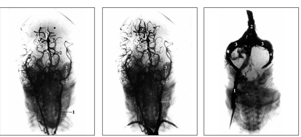
No essential changes are observed from 32 to 40 weeks intrauterine life. Analysis of stillborn babies' angiograms show clearly expressed winding course of cerebral arter-ies. All of the three cerebral arteries are considerably strong, with winding course and rich branches. Vertebral artery is filled retrogradely through Willis' circle (Fig. 5). Arteries that vascularise the basal ganglia, hypothalamus and choroid plexus appear in the form of wide blood ves-sels. Lateral angiogram (Fig. 5) shows an abundance of blood vessels. Middle cerebral artery has an expressed slant position and its insular part is well shown. It is es-tablished that distribution of blood vessels and their ram-ification is similar to that seen on angiograms of the adults. Lateral projection shows well all cerebral arteries (Fig. 5). Certain deviations are noticed in some cases. In
our research material we have encountered a rather inter-esting case of 36 weeks intrauterine life fetus. In the early filling phase (Fig. 6), the AP projection clearly shows an-terior cerebral artery and middle cerebral artery. Asymme-try of the right and left cerebral arteries is visible. Dis-tance of anterior cerebral artery from insular branches of middle cerebral artery is smaller and matches the distance found in the adults.
In the late filling phase, long, high caliber cerebral arteries are visible. Blood vessels are longer and do not match with gyri. Arteries are very tortuous, so that it can be said that blood vessels "spare gyri" exist. A strong anastomo-ses exist that enable retrograde filling of arteries. The slowed down filling of the right side is also characteristic, as well as the widened subarachnoid space (Fig. 6). We have succeeded to partially show dural sinuses also by injecting contract medium through interior jugular vein (Fig. 7). Internal jugular vein, superior sagittal sinus and transverse sinus are shown.
Confluence of sinuses is well shown as well as the abun-dance of vein vessels that can be observed in the region of foramen magnum; sigmoid sinus is also shown.
Discussion
ar-Fig. 1. Carotid angiogram in foetus of 16 weeks I.U. life. AP projection.
1. internal carotid artery 2. anterior cerebal artery 3. middle cerebal artery 4. posterior cerebal artery 5. basilar artery
6. vertebral artery
Fig. 2. Carotid angiogram in foetus of 20 weeks I.U. life. AP projection.
1. internal carotid artery 2. anterior cerebal artery 3. middle cerebal artery
4. cortical anastomosis between anteri-or cerebal and middle cerebal artery 5. posterior cerebal artery
6. basilar artery 7. vertebral artery
Fig. 3. Carotid angiogram in foetus of 28 weeks I.U. life. AP projection.
1. internal carotid artery 2. anterior cerebal artery 3. middle cerebal artery 4. posterior cerebal artery 5. basilar artery
6. vertebral artery
Fig. 4. Carotid angiogram in foetus of 32 weeks I.U. life. Lateral projection.
1. anterior cerebal artery 2. middle cerebal artery
3. posterior communicating artery 4. Anastomosis ACA and ACP in
sple-nium corporis callosi region
Fig. 5. Carotid angiogram of a stillborn baby. AP and lateral projections
1.internal carotid artery 1. internal carotid artery 2. anterior cerebal artery 2. anterior cerebal artery 3. middle cerebal artery 3. middle cerebal artery
4. posterior cerebal artery 4. middle cerebal artery (insular part) 5. basilar artery
teries of significantly higher caliber; they branch abun-dantly, while due to their relatively wide sulci, it can be said that their course is tortuous. It should be noted that also in the early fetal life we have ascertained anastomo-ses between anterior cerebral artery and posterior cerebral artery in fetuses of 32 weeks intrauterine life. Also, cer-tain deviations have been observed. In fetuses old 36 weeks intrauterine life, we have ascertained that brain ar-teries are of high caliber and great length. Length of blood vessels does not match with gyri, while arteries are very tortuous, so that it can be said that blood vessels "spare gyri" exist. Comparing our findings on fetal blood vessels with the results of other authors occupied to some meas-ure with the subject issues, we can say that our findings do not deviate from those of Hoyt (4), and Streeter (7).
Icardo (8), Kaplan (9) and Van Overbeeke (10), reports that posterior cerebral artery tree moves more backwards as the brain hemispheres grow over the thalamus and mid-brain thus vascularising ever greater number of observa-ble structures, what is also in accordance with our find-ings.
Conclusion
Our findings show clearly that position, course and rela-tionship of brain arteries change concurrently with the de-velopment of adjacent brain structures and gyri appear-ance, what is in accordance with scarce literature data in this field.
Fig. 6. Carotid angiogram in foetus of 36 weeks I.U. life. AP and lateral projections.
1. internal carotid artery 2. anterior cerebral artery 3. meddle cerebral artery
Fig. 7. Dural sinuses AP projection.
References
(1) Hodes P.J. et all. Cerebral angiography. Fundamentals in anatomy and physiology. Am. J. Roentgenol. Radium Their Nucl. Med. 1953; 70: 61-82.
(2) Kier L. Development of cerebral vessels. Section I. Fetal cerebral arteries: a phylogenetic and ontogenetic study. In: Newton T.H. and Potts G.D. (eds): Radiology of the skull and brain. Angiography. Mosby Co., Saint Louis, 1974.
(3) Newton T.H. and Potts D.G.Radiology of the scull and brain. Angiography. Mosby Co., Saint Louis, 1974.
(4) Hoyt W.F. et all. The posterior cerebral artery. Section I. Embryology and developmental anomalies. In: Newton T.H. and Potts D.G. (eds): Radiology of the Skull and Brain. Angiography. Mosby Co. Saint Louis, 1974.
(5) Takashi M. et all. Roentgenographic anatomy of the cerebral artery. Radiology 1986; 8/90: 271-297.
(6) Padget, D.H. The development of the cranial arteries in the human embryo, Contrib. Embryol. 1948; 32:205-262.
(7) Streeter, G.L. The development alterations in the vascular system of the brain of the human embryo. Contrib. Embryol. 1918; 8:5-38.
(8) Icardo J.M. The cerebral arteries: Cortical patterns and vascularisation of the cerebral nuclei. Acta anat. 1982. 113; 108-116.
(9) Kaplan H.A. Anatomy and embriology of the arterial system of the forebrain In: Vinken P.J. and Bruyan G.W. (eds): Handbook of Clinical Neurology. North - Holland Publishing Co., Amsterdam, 1975.