University of Pennsylvania
ScholarlyCommons
Publicly Accessible Penn Dissertations
1-1-2015
Development and Characterization of Tool
Compounds Targeting the Runt Domain’s
interaction With Cbfβ
Zaw Min Oo
University of Pennsylvania, [email protected]
Follow this and additional works at:
http://repository.upenn.edu/edissertations
Part of the
Cell Biology Commons
, and the
Molecular Biology Commons
This paper is posted at ScholarlyCommons.http://repository.upenn.edu/edissertations/1924 For more information, please [email protected].
Recommended Citation
Oo, Zaw Min, "Development and Characterization of Tool Compounds Targeting the Runt Domain’s interaction With Cbfβ" (2015).
Publicly Accessible Penn Dissertations. 1924.
Development and Characterization of Tool Compounds Targeting the
Runt Domain’s interaction With Cbfβ
Abstract
RUNX1 and CBFβ, which encode subunits of the core binding factor, are frequent targets of chromosomal aberrations in hematological malignancies. We previously determined that CBFβ (encoded by CBFB) is important for the transforming activity of the chimeric protein AML1-ETO protein (RUNX1-RUNX1T1) generated by the t(8;21), and other studies showed that normal Runx1 functions are essential for survival and maintenance of some leukemias lacking RUNX1 or CBFB mutations. Thus, we hypothesized that we could achieve therapeutic efficacy in multiple leukemias by targeting the Runx1:CBFβinteraction with small molecules. Using the structural information of the DNA binding Runt domain (RD) of Runx1 and its interface with CBFβ, we employed a computational screen for a library of 78,000 drug-like compounds, and further optimized our leads. The Runt domain inhibitors (RDIs) bind directly to the RD and disrupt its interaction with CBFβ. We showed that the RDIs reduced growth and induced apoptosis of t(8;21) acute myeloid leukemia (AML) cell lines, and reduced the progenitor activity of mouse and human leukemia cells harboring the t(8;21), but not normal bone marrow cells. The RDIs had similar effects on murine and human T cell acute lymphocytic leukemia (T-ALL) cell lines that did not harbor the t(8;21). Furthermore, our inclusion of a structurally related and weakly active compound as a control strongly support that the efficacies we observed were due to on target inhibition of RUNX functions. Our results confirmed that the RDIs might prove efficacious in various AMLs, and that a therapeutic window is available to specifically target malignant cells. We developed a pro-drug AI-9-59 with improved solubility and pharmacokinetic properties and assessed whether it has any in vivo efficacies in mouse leukemia models. Our results showed that the pro-drug was toxic to mice at dosage above 50 mg/kg and had no observable growth inhibitory effect on leukemia cells, suggesting that the concentration of the pro-drug necessary to inhibit endogenous core binding factor activity exceeds the maximum tolerated dose in mice. However, the expansion of granulocyte macrophage
progenitors, and the gastrointestinal toxicity phenotype we observed suggested that the effects could be from on-target repression of RUNX proteins functions.
Degree Type Dissertation
Degree Name
Doctor of Philosophy (PhD)
Graduate Group
Cell & Molecular Biology
First Advisor Nancy A. Speck
Subject Categories
DEVELOPMENT AND CHARACTERIZATION OF TOOL COMPOUNDS TARGETING THE RUNT DOMAIN’S INTERACTION WITH CBFβ
Zaw Min Oo
A DISSERTATION
in
Cell and Molecular Biology
Presented to the Faculties of the University of Pennsylvania
in
Partial Fulfillment of the Requirements for the Degree of Doctor of Philosophy
2015
Supervisor of Dissertation
Nancy A. Speck
Professor of Cell and Developmental Biology
Graduate Group Chairperson
Daniel S. Kessler
Associate Professor of Cell and Developmental Biology
Dissertation Committee:
Celeste M. Simon, Professor of Cell and Developmental Biology Xianxin Hua, Professor of Cancer Biology
ACKNOWLEDGEMENT
First and foremost, I would like to thank my advisor Dr. Nancy A. Speck for her
mentorship and support over the years. I have learned so much from Nancy, and would
like to take this opportunity to express my deepest gratitude.
I would also like to thank members of my thesis committee, Dr. Celeste M.
Simon, Dr. Xianxin Hua, Dr. Warren Pear, and Dr. Wei Tong for their advice and help
throughout my graduate career.
I am very grateful to my collaborators Dr. John Bushweller and Dr. Anuradha
Illendula from University of Virginia. Much of my graduate work would not have been
possible without their contribution.
I would like to thank past and present members of the Speck lab: Dr. Michael JF
Chen, Dr. Liya Roudaia, Dr. Jing-mei Hsu, Dr. James Mangan, Dr. Xiongwei Cai, Dr.
Joanna Tober, Dr. Yan Li, Chung-Tsai Lee, Amanda Yzaguirre, and Dana Bellissimo. I
am fortunate to have you as colleagues, and I will always treasure our years in the
Speck lab.
On a personal note, I would like to especially thank my wife Marybeth Tong, for
her kind support, patience and her unwavering love. I would also like to thank my
ABSTRACT
DEVELOPMENT AND CHARACTERIZATION OF TOOL COMPOUNDS
TARGETING THE RUNT DOMAIN’S INTERACTION WITH CBFβ
Zaw Min Oo
Nancy A. Speck
RUNX1 and CBFβ, which encode subunits of the core binding factor, are
frequent targets of chromosomal aberrations in hematological malignancies. We
previously determined that CBFβ (encoded by CBFB) is important for the transforming
activity of the chimeric protein AML1-ETO protein (RUNX1-RUNX1T1) generated by the
t(8;21), and other studies showed that normal Runx1 functions are essential for survival
and maintenance of some leukemias lacking RUNX1 or CBFB mutations. Thus, we
hypothesized that we could achieve therapeutic efficacy in multiple leukemias by
targeting the Runx1:CBFβ interaction with small molecules. Using the structural
information of the DNA binding Runt domain (RD) of Runx1 and its interface with CBFβ,
we employed a computational screen for a library of 78,000 drug-like compounds, and
further optimized our leads. The Runt domain inhibitors (RDIs) bind directly to the RD
and disrupt its interaction with CBFβ. We showed that the RDIs reduced growth and
induced apoptosis of t(8;21) acute myeloid leukemia (AML) cell lines, and reduced the
progenitor activity of mouse and human leukemia cells harboring the t(8;21), but not
normal bone marrow cells. The RDIs had similar effects on murine and human T cell
acute lymphocytic leukemia (T-ALL) cell lines that did not harbor the t(8;21).
control strongly support that the efficacies we observed were due to on target inhibition
of RUNX functions. Our results confirmed that the RDIs might prove efficacious in
various AMLs, and that a therapeutic window is available to specifically target malignant
cells. We developed a pro-drug AI-9-59 with improved solubility and pharmacokinetic
properties and assessed whether it has any in vivo efficacies in mouse leukemia models.
Our results showed that the pro-drug was toxic to mice at dosage above 50 mg/kg and
had no observable growth inhibitory effect on leukemia cells, suggesting that the
concentration of the pro-drug necessary to inhibit endogenous core binding factor
activity exceeds the maximum tolerated dose in mice. However, the expansion of
granulocyte macrophage progenitors, and the gastrointestinal toxicity phenotype we
observed suggested that the effects could be from on-target repression of RUNX
TABLE OF CONTENTS
ACKNOWLEDGEMENT ...
………
.
……
II
ABSTRACT
……
...
………
.
…
.... III
LIST OF TABLES ...
………
...
…
.
…
.. VII
LIST OF ILLUSTRATIONS .
………
.. VIII
INTRODUCTION
………
... 1
CHAPTER I:
DEVELOPMENT AND CHARACTERIZATION OF THIAZOLE COMPOUNDS TARGETING TO DISRUPT THE RUNT DOMAIN: CBFβ INTERACTION…
.
………
... 24
ABSTRACT
……
...
………
..
………
..
25INTRODUCTION
………
..
………
...
26MATERIALS AND METHODS
…
...
………
..
29RESULTS
……
...
………
...
35DISCUSSION
…
...
………
..
………
...
57APPENDIX
………
...
…
.
…
...
59SUPPLEMENTARY METHODS
………
...
………
...
………
63CHAPTER II: CHARACTERIZATION OF THE RUNT DOMAIN INHIBITOR’S IN
VIVO EFFICACY BY MOUSE LEUKEMIA MODELS .
……
.
……
.
………
. 81
INTRODUCTION
………
..
………
..
82MATERIALS AND METHODS
…
...
………
...
85DISCUSSION
…
...
………
..
………
.
100CHAPTER III: CONDITIONAL EXPRESSION OF AML1-ETO IN
HEMATPOIETIC CELLS
………
..
…
.
………
... 103
INTRODUCTION
………
..
………
..
…………
..
104MATERIALS AND METHODS
…
...
………
..
…
...
106RESULTS
……
...
………
..
…
...
108DISCUSSION
…
...
………
..
………
.
115CLOSING REMARKS AND FUTURE DIRECTIONS
…
.
………
..
…
117
LIST OF TABLES
INTRODUCTION:
Table 1. Knockout phenotypes of the core binding factor genes in mouse .…...… 11
Table 2. Conditional knockout phenotypes of the core binding factor genes in mouse
……...………...………..………...14
Table 3. Chromosomal translocations and mutations affecting Runx1 and CBFβ in
various hematological diseases
.
……...……….……….……...16CHAPTER I:
Table 1. Structures and FRET IC50 values for selected inhibitors ……….………….…… 41
Table 2. A list of 87 unique transcript IDs that are differentially expressed as shown in
LIST OF ILLUSTRATIONS
INTRODUCTION:
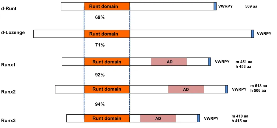
Figure 1. The core binding factor subunits and genes …...……...……… 7
Figure 2. A diagrammatic representation of the structure of RUNX1, RUNX2 and RUNX3
together with Drosophila runt and lozenge…...……...………..….……… 8
Figure 3. Schematic diagram of an E10.5 mouse embryo …...………...….… 9
Figure 4. Impact of loss of function Runx1 mutations in hematopoietic stem cells (HSCs)
...…...……...………..….………...………..….……….…...…… 15
Figure 5. Runt inhibitors reduce cell growth and induce apoptosis in leukemia cells .… 18
Figure 6. Survival curves of core binding factor leukemias compared to other leukemia
types ………...……….…...……… 19
Figure 7. Mutations that disrupt DNA or CBFβ binding impair AML1-ETO’s leukemogenic
CHAPTER I:
Figure 1. Schematic diagram of FRET assay…...……...……….……… 36
Figure 2. Library of analogs synthesized …...……….……… 37
Figure 3. NMR STD and FRET data for Runt domain inhibitors …...……….… 40
Figure 4. Pharmacokinetics data for AI-9-54 and the AI-9-59 prodrug in mice ...…..… 43
Figure 5. Runt inhibitors reduce cell growth and induce apoptosis in leukemia cells .… 45 Figure 6. FACS/FRET analysis strategy for assessing the efficacy the Runt domain inhibitors ………...……….……… 47
Figure 7. FACS/FRET analysis of the Runt domain inhibitors …...………...… 48
Figure 8. Effect of Runt inhibitors on colony formation by normal and leukemic mouse bone marrow cells, and in human AML samples ……...……….………….…… 50
Figure 9. Microarray analysis of gene expression changes induced by RDIs ...…...… 53
Figure 10. Runt domain inhibitor reduced luciferase activity of Runx1 in transient co-transfection experiments ………...……….………….…… 60
Figure 11. Effect of the Runt domain inhibitors on CBFβ-RUNX1 binding at 16 hours in 720 T-ALL cells, measured by co-immunoprecipitation ……….………….…… 61
CHAPTER II:
Figure 1. Flow cytometry analysis of peripheral blood from mice transplanted with
AML1-ETO9a + NRASG12D bone marrow cells ……...………..………...…...…………...88
Figure 2. Assessing in vivo efficacy of the pro-drug for the Runt domain inhibitors …...89
Figure 3. Assessing in vivo efficacy of the pro-drug for the Runt domain inhibitors …... 91
Figure 4. Efficacy of RDI in AML1-ETO9a; NRASG12D mouse model ………...….……...93
Figure 5. Determining the maximum tolerated dose of NSG mice to the pro-drug AI-9-59
……...……...…….……...……...…….……...……...…….……...……...……...……...94
Figure 6. End point analysis of NSG mice engrafted with 720 T-ALL cells and treated
with DMSO or 50 mg/kg AI-9-59 ……...………...………...95
Figure 7. Flow cytometry analysis of peripheral blood and bone marrow cells from NSG
mice engrafted with 720 T-ALL cells and treated with DMSO or 50 mg/kg AI-9-59 ...97
Figure 8. Toxicity analysis of peripheral blood and bone marrow cells from NSG mice
CHAPTER III:
Figure 1. Lineage analysis of bone marrow cells from Vav1-Cre; AML1-ETO and WT
mice ……...………..………...………...……...108
Figure 2. Phenotypic HSC analysis of bone marrow cells from Vav1-Cre; AML1-ETO and
WT mice ……...………..………...……..109
Figure 3. Competitive limited dilution transplant to assess the frequency of functional
HSCs in AML1-ETO expressing mice ……...…….………...…...111
Figure 4. Expression of AML1-ETO lead to lower functional HSCs ...……...112
Figure 5. Co-expression of AML1-ETO and activated NRAS (NRASG12D) in
hematopoietic compartments leads to development of hematological disorder ...113
Figure 6. Malignant hematopoietic cells from Vav1-Cre; AML1-ETO; NRASG12D are
Introduction
Targeting Protein-protein interaction by small molecules
The past two decades have witnessed major advances in our understanding of
the molecular mechanisms of diseases. As a consequence, drug discovery research has
transitioned from searching for compounds with desired efficacy but against unknown
targets to a target therapy approach. The aim of targeted therapy is to use our
understanding of the cellular programs associated with the pathology of disease to
design treatments with improved therapeutic efficacy. Although the majority of such
studies have focused on targeting activated components of cytokine receptor signaling
pathways, interest in targeting protein-protein interactions (PPIs) has grown in the past
decade.
Protein-protein interactions play important roles in all aspects of cellular
processes, particularly in the regulation of transcription where the assembly of
protein-protein complexes is essential for appropriate gene regulation. PPIs, especially those
involving transcription factors are highly attractive targets for developing inhibitors due to
their biological importance, as many cancers either directly involve transcription factors,
or indirectly modulate transcription factor activity. A variety of transcription factors have
been identified as driving agents promoting tumorigenesis and cancer progression (1).
Therefore, inhibiting PPIs involving transcription factors has a high therapeutic potential
(2). However, until recently PPIs were considered undruggable due to several
challenges. First, the contact surfaces involved in PPIs are large and lack the grooves
and pockets for small molecule binding. Second, PPIs do not have natural small
Despite these challenges, research in identifying small molecule inhibitors of
PPIs has made considerable progress in the past decade. Several recent technological
advances facilitate this trend and provide hope for finding small molecules that target
PPIs. We now have a better understanding of binding energetics at the macromolecular
interface of PPIs. Although the protein-protein interfaces are large, mutational studies
show that only a small subset of the residues involved contributes to most of the free
energy of binding (3-5). The presence of such “hotspots” makes PPIs amenable to small
molecule perturbations, and disrupting the interactions mediated by these hotspot
residues proves to be effective in inhibiting PPIs. The identification of hotspot residues in
turn allows for structure-based virtual screening to identify novel bioactive molecules. In
virtual screening, large libraries of drug-like compounds that are commercially available
are computationally screened against targets of known structure, and those that are
predicted to bind well are experimentally tested. Structure-based virtual screening
provides a more efficient and cost-effective approach over high-throughput screening
(HTS) for identifying new lead compounds as it utilizes the knowledge of the
three-dimensional (3D) structure of the biological target. I will provide several examples of
small molecules targeting PPIs in distinct biological pathways.
MDM2-p53 inhibitors: a breakthrough in targeting PPI
One recent success story in targeting PPI is the development of small molecules
that inhibit the interaction of MDM2 with p53 (6). p53 is a tumor suppressor gene that
normally triggers growth arrest, senescence or apoptosis in response to cellular stress.
p53 activation increases the expression of E3 ligase MDM2, which in turn binds p53,
directly represses p53 DNA binding ability and tags p53 for proteasomal degradation (7).
transcriptional activity (8). Structurally, the N-terminal domain of MDM2 has a
well-defined hydrophobic pocket that mediates p53 binding, suggesting that the pocket could
be targeted with small molecule drugs to block the MDM2-p53 interaction (9). Nutlins, a
family of cis-imidazoline analogs, were identified by screening for compounds that
disrupt the MDM2-p53 interaction (10). Nutlins inhibit the interaction of MDM2 with p53,
leading to stabilization of p53 levels, and restore p53 mediated apoptosis (10-13). The
success of MDM2-p53 PPI inhibitors has significantly accelerated studies to target other
PPIs with small chemical compounds as anticancer drugs.
STATs inhibitors
Signal transducers and activators of transcription (STATs) are a family of
cytoplasmic transcription factors. One of the key structural features of STATs is the Src
homology (SH) 2 domain, the phosphorylation of which activates STATs. Activated
STATs form dimers that translocate to the nucleus and regulate transcription of target
genes controlling cell growth, differentiation, and survival (14). In normal cells, STAT
signaling is transient and tightly regulated. However, cancer cells frequently have
persistent STATs activation from hyper-activation, overexpression of upstream tyrosine
kinases, or loss of function of negative regulators. As activated STATs, particularly
STAT3, drive the expression of genes involved in cell proliferation and survival, blocking
STAT3 activity presents a useful strategy for drug discovery (14). The availability of the
three-dimensional (3D) structure of phosphorylated STAT3 (15), and the existence of a
clearly defined binding pocket for small molecules in the SH2 domain allowed virtual
screening of chemical databases for the identification of small molecule inhibitors of the
S3I-STAT3-dependent cellular effects in transcription, transformation, and apoptosis.
Furthermore, S3I-201 is effective against a tumor cell line displaying constitutive STAT3
activity in a mouse model xenograft model (17).
BCL6 inhibitor
BCL6 (B cell lymphoma 6) is the most frequently involved oncogene in diffuse
large B cell lymphomas (DLBCLs) (18). BCL6 is required for B cell maturation, during
which BCL6 represses genes involved in sensing DNA damage or their downstream
checkpoints (19-21). BCL6 is constitutively expressed in the majority of patients with
aggressive B cell lymphomas (18), and mice engineered to constitutively express BCL6
in B cells develop DLBCL similar to the human disease (22, 23). BCL6 knockdown or
peptide inhibitors kill DLBCL cells, demonstrating that BCL6 is required for survival of
lymphoma cells and suggesting that BCL6 is a bone fide therapeutic target for DLBCL
(19, 24, 25).
BCL6 is a member of the BTB/POZ family of transcription factors (26). BCL6 acts
as a sequence specific transcription repressor by recruiting SMRT, N-CoR, and BCOR
corepressors through its BTB domain (27, 28). Using structured-based virtual screening,
a potent inhibitor of BCL6 BTB domain repressor activity was identified. The inhibitor
binds to the BCL6 BTB domain, specifically inhibits BCL6, disrupts BCL6 transcriptional
complexes and reactivates BCL6 target genes. The inhibitor selectively kills
BCL6-dependent DLBCL cells, and suppresses human DLBCL xenografts in mice (29).
Menin-MLL inhibitors
Translocations involving the mixed lineage leukemia (MLL) gene are frequently
The MLL protein is a histone H3 lysine 4 (H3K4) methyltransferase required for the
expression of HOX family genes during hematopoietic differentiation (32, 33). Fusion of
MLL with one of numerous partner genes results in chimeric proteins that upregulate
MLL target gene expression, block hematopoietic differentiation, and promote
proliferation (33-35). The function of the wildtype MLL as well as MLL fusion requires
interaction between MLL and menin, a tumor suppressor encoded by MEN1 (multiple
endocrine neoplasia 1) (36). Loss of menin binding eliminates the oncogenic potential of
MLL fusions (37, 38), and therefore targeting the menin-MLL interaction represents a
potential therapeutic approach for the treatment of AML with the MLL translocation.
A single point mutation in the high-affinity menin-binding motif 1 (MBM1) in MLL
is sufficient to abolish the menin-MLL interaction, suggesting the existence of a hotspot
residue for this interaction (39, 40). Subsequently, Grembecka et al (41) reported the
identification of a family of small molecules that inhibit the menin-MLL interaction. One of
the compounds, MI-2 binds to menin, disrupts the menin-MLL interaction and
downregulates the expression of the MLL target genes essential for leukemogenesis.
MI-2 inhibits the proliferation of several leukemia cell lines harboring MLL translocation
but has minimal anti-proliferation effect on leukemia cell lines without MLL fusions.
Subsequent structure-based design resulted in MI-2-2 with improved binding affinity for
menin and enhanced cellular potency (42). Therefore, the inhibitor represents a
promising lead that may be developed into a new antileukemic drug for MLL leukemia.
CBFβ-SMMHC inhibitor
The chromosome inversion 16 inv(16)(p13;q22) is one of the most common
generates the chimeric transcription factor fusion CBFβ-SMMHC. CBFβ-SMMHC
cooperates with activating mutations in components of cytokine signaling pathways in
leukemia transformation (43-46). Studies in mice and patient samples support the
concept that inv(16) is a driver mutation that generates preleukemic progenitor cells that,
upon acquisition of additional cooperating mutations, progress to leukemia (44, 45, 47,
48). CBFβ-SMMHC outcompetes wildtype CBFβ for binding to transcription factor
RUNX1 (49), deregulates RUNX1-mediated transcription in hematopoiesis, and induces
AML. Most recently, Illendula et al. reported the development of a PPI inhibitor that
selectively binds to CBFβ-SMMHC and disrupts its binding to RUNX1 (50). Using a
fluorescent resonance energy transfer (FRET) assay, the authors identified a lead
compound that blocks the interaction of CBFβ-SMMHC with the Runt domain of RUNX1.
As CBFβ-SMMHC is oligomeric in solution, the authors designed a bivalent analog of the
lead compound with a seven-atom polyethylene glycol linker, resulting in the drug
AI-10-49. AI-10-49 specifically binds to CBFβ-SMMHC, restores RUNX1 transcriptional activity
and delays leukemia progression in mice. Treatment of primary inv(16) AML patient
blasts with AI-10-49 triggers selective cell death (50). The work provides additional
support for targeted therapy against transcription factor drivers of cancers.
These results demonstrate that targeting transcription factors with small
molecules is feasible, and provide validation for our proposal to develop a novel class of
small molecule inhibitors that target the RUNX proteins, a small family of DNA binding
transcription factors, by interfering with their interaction with the non-DNA binding
Core-binding factor in development
RUNX proteins are members of the core-binding factor (CBF), a family of
heterodimeric transcription factors that play major roles in hematopoiesis. The CBF
family consists of three distinct DNA binding CBFα subunits (RUNX1, RUNX2, and
RUNX3), and a common non-DNA binding CBFβ subunit (encoded by CBFB) (Figure 1).
Figure 1. The core binding factor subunits and genes.
Shown is the structure of the DNA binding Runt domain of Runx1 (gray) and the CBFβ subunit (blue) bound to DNA.
RUNX1, also known as acute myeloid leukemia 1 (AML1) or core-binding factor
subunit alpha-2 (CBFA2), was initially identified from chromosome 21 in the t(8;21)
(q22;q22) chromosomal translocation, frequently found in acute myeloid leukemia (AML)
(51). RUNX proteins are evolutionally conserved (Figure 2). There are two well-studied
RUNX homologs in Drosophila: runt and lozenge (52, 53). runt is a pair-rule gene
involved in embryonic patterning and also plays a role in sex determination,
segmentation and neural development (54, 55). lozenge is involved in hematopoiesis
Runx CBF
YGYGGY
Cbfb
and eye development (56). Subsequent work established that the RUNX proteins are
sequence-specific DNA-binding proteins that bind to the polyomavirus enhancer and
Moloney murine leukemia virus enhancer core sites, hence the name core-binding
factors (57-61). All RUNX proteins have a highly conserved 128-amino-acid Runt
domain in their N-terminal portion, which mediates both DNA binding and CBFβ
heterodimerization (Figure 2). Through the Runt domain, RUNX proteins recognize the
consensus sequence PyGPyGGT. The CBFβ subunit does not touch DNA but enhances
the affinity of RUNX proteins for DNA (62), and protects them from ubiquitination and
proteasomal degradation (63).
Figure 2. A diagrammatic representation of the structure of RUNX1, RUNX2 and RUNX3 together with Drosophilarunt and lozenge.
The Runt domain, the activation domain (AD), and C terminal VWRPY motif, along with the size of each protein are indicated. m: mouse RUNX, h: human RUNX
Runt domain d-Runt
Runx1
Runx2
Runx3
VWRPY
VWRPY
VWRPY AD
AD AD
Runt domain
Runt domain
Runt domain
VWRPY 509 aa
m 451 aa h 453 aa
m 513 aa h 506 aa
m 410 aa h 415 aa
69%
92%
94%
d-Lozenge Runt domain VWRPY 828 aa
Numerous studies established that both Runx1 and CBFβ are critical for
hematopoietic development. Runx1 is constitutively expressed in all hematopoietic
lineages, except mature erythroid cells (64). In the conceptus, Runx1 is expressed in all
sites of hematopoietic cell formation, including the vitelline and umbilical arteries, the
yolk sac, the placenta, and the ventral portion of the dorsal aorta in the
aorta-gonad-mesonephros (AGM) region (65-67) (Figure 3). In the embryo, hematopoietic stem and
progenitor cells (HSPCs) emerge from Runx1-expressing
vascular-endothelial-cadherin-positive endothelial cells called hemogenic endothelium, and Runx1 activity is essential
for HSPC formation from these cells (65, 68-72). Thus RUNX1 is recognized as a
mandatory transcription factor in embryonic hematopoiesis.
Figure 3. Schematic diagram of an E10.5 mouse embryo.
Sites of Runx1 expression in hemogenic endothelium are colored in red. These include the ventral aspect of the dorsal aorta in the aorta/gonad/mesonephros region, the umbilical artery, vitelline artery, and in areas of the yolk sac.
Homozygous deletion of Runx1 or CBFβ in mice results in nearly identical
developmental defects including extensive hemorrhaging within the central nervous
placenta
umbilical artery (U) vitelline artery (V)
dorsal aorta
yolk sac
Runx1 expression
system, and mid-gestation embryonic lethality between embryonic day 12.5-13.5
(73-76). Runx1 or Cbfb deficient mouse embryos lack definitive hematopoietic progenitors,
definitive enucleated erythrocytes, and mature myeloid-lineage cells (73-77), and the
lack of definitive hematopoietic cells is observed in all sites of hematopoiesis (65, 67, 69,
70, 78) (Table 1). In addition, no transplantable hematopoietic stem cell (HSC) activity is
found in the AGM region of Runx1 deficient embryos (79). Chimeric animals made from
Runx1 or Cbfb deficient embryonic stem (ES) cells and wild type mouse blastocysts
contain no ES-derived cells in adult hematopoietic tissues, which indicates that both
genes are required in a cell-autonomous fashion (73, 75, 80). Therefore, both Runx1
and CBFβ are required to establish HSPCs during embryonic development.
Runx2, another member of the RUNX family, is primarily associated with
osteoblast differentiation and bone formation (81, 82) (Table 1). Runx2 null mice died
soon after birth, and the embryos and newborns showed a complete lack of bone
formation and ossification, and the development of cartilage in Runx2 null mice was
delayed (81, 82). Further studies established RUNX2 as a master transcription factor of
osteoblast differentiation that controls both osteoblast differentiation and expression of
osteoblast specific genes (83, 84).
The knockout of RUNX3 caused phenotypes in several different tissues (Table
1). Germline Runx3 knockout led to mice with defects in T-cell development, as well as
gastrointestinal and neural disorders (85-88). During T-lymphocyte development, Runx3
is essential for silencing of CD4 expression and maturation of single positive CD8+ T
cells. The absence of Runx3 results in defective cytolytic activity of T lymphocytes (86,
89). As Runx3 deficient mice spontaneously develop tumors in the intestine, lung and
breast (90), RUNX3 may play a tumor suppressor role during the early stages of solid
Table 1. Knockout phenotypes of the core binding factor genes in mouse.
Core-binding factor in adult hematopoiesis
As will be discussed later, we developed small molecule inhibitors of the RUNX
proteins that disrupt the Runt domain’s interaction with CBFβ. These function as
pan-RUNX/CBFβ inhibitors. In order to anticipate and interpret the inhibitors’ on-target
activities, it is worth reviewing the murine phenotypes associated with Runx and CBFβ
mutations, alone or in combinations. Conditional deletion of Runx1 in hematopoietic cells
using the interferon inducible Mx1-Cre or pan-hematopoietic Vav1-Cre shows that once
definitive hematopoiesis develops in the embryos, RUNX1 expression is not required to
maintain HSCs (Table 2). Conditional knockout (cKO) of Runx1 does not cause
hematopoietic failure in adult mice (94-96) but results in multi-lineage blocks in
Gene Phenotype References
Runx1 -/- Lethal at embryonic day (E) 11.2-12.5. Absence of definitive hematopoiesis. (73, 74)
Absence of intra-aortic hematopoietic clusters. (65)
Runx2 -/- Lethal at birth, failure of osteoblast differentiation and bone
formation. (81, 82)
Perturbation of chondrocyte differentiation. (91, 92)
Runx3 -/- Hyperplastic gastric epithelia due to excessive proliferation. (87)
Loss of dorsal-root ganglion proprioceptive neuron function
and ataxia. (85, 88)
Loss of homeostatic control of dendritic-cell function and lung
inflammation. (93)
lymphocyte and megakaryocyte development, and increase in the number of committed
erythroid/myeloid progenitors. Runx1 deletion in the adult results in expansion of the
lineage negative Sca1+ Kit+ (LSK) phenotypic HSPC population in the bone marrow (94,
97, 98) in a cell autonomous manner and reduces the frequency of functional long-term
repopulating hematopoietic stem cells (LT-HSCs) in the bone marrow by 3-fold without
affecting their self-renewal properties (97-99).
Runx1 loss in adult mice does not cause acute myeloid leukemia (AML) but
establishes a pre-leukemic state that predisposes to AML following the acquisition of
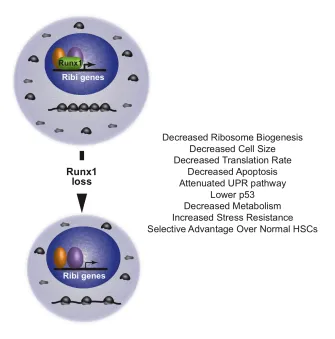
secondary mutations (99, 100). At the cellular level, Runx1-deficient HSPCs have slow
growth, low biosynthesis and markedly reduced ribosome biogenesis (Ribi) (Figure 4).
Runx1-deficient HSPCs have lower p53 levels, reduced apoptosis, an attenuated
unfolded protein response, and are resistant to genotoxic and endoplasmic reticulum
(ER) stress. The low biosynthetic activity and corresponding stress resistance provide a
selective survival advantage to Runx1-deficient HSPCs, allowing them to expand in the
bone marrow and outcompete normal HSPCs (101).
Young Runx3 cKO mice do not display gross hematopoietic abnormalities, and
aged Runx3 cKO mice show a mild expansion of the LSK compartment, partially
phenocopying Runx1 cKO mice (102) (Table 2). In contrast, Runx1;Runx3 double
knockout (DKO) mice exhibited much more severe phenotypes. The DKO mice had
decreased white blood cell and platelet counts, developed anemia, and died within 18
weeks post deletion. The DKO mice showed differentiation blocks in all hematopoietic
lineages, and a 48-fold expansion in the LSK fraction that is followed by subsequent
exhaustion in the HSPC compartment (103). Transplant experiments showed that the
differentiation blocks and stem cell exhaustion in the DKO mice are cell autonomous
hematopoietic defects than Runx1 cKO mice, suggesting that RUNX family genes have
compensatory mechanisms in hematopoiesis.
Conditional deletion of CBFβ, which should affect the activity of all three RUNX
proteins, also shows more pronounced defects in hematopoiesis than Runx1 cKO mice
(Table 2). Cbfb cKO mice have marked differentiation blocks in all hematopoietic
lineages and significant expansion of LSK and short-term HSCs (ST-HSCs) in the bone
marrow (BM). Cbfb cKO mice showed progressive decreases in leukocyte, hemoglobin,
and platelet counts, and died by 6 months of age due to bone marrow failure (104).
Although fetal liver and BM cells from Cbfb cKO mice show significant increase in colony
forming activity, recipient mice transplanted with Cbfb deficient cells have extremely low
donor chimerism in the hematopoietic tissues, including HSPC fractions in the BM.
These results showed that while the progenitors in the Cbfb cKO mice possess greater
proliferative capacity, Cbfb-deficient HSCs were incapable of long-term engraftment
(104, 105). In addition, Cbfb cKO mice are not born at Mendelian ratios. The fact that
CBFβ deficient HSCs are much more severely compromised than Runx1 deficient HSCs
suggests Runx2 and/or Runx3 contribute substantially to HSC functions and also
suggests the existence of functional compensation by the remaining two Runx genes for
Table 2 . Conditional knockout phenotypes of the core binding factor genes in mouse.
Targeted
Gene Phenotype of conditional knockout References
Runx1 Mx1-Cre
Normal HSC numbers.
Normal myeloid cell compartment. Defective B and T cell differentiation.
(95)
Mx1-Cre Reduced competitive repopulating ability.
Myeloid expansion in spleen and liver. (94)
Mx1-Cre Vav1-Cre
Expands phenotypic stem and progenitor population. Three-fold reduction in frequency of long term
repopulating HSC but showed no exhaustion. Runx1 loss slowed HSC proliferation and reduced apoptosis.
(97)
Vav1-Cre
Runx1 deficient HSPCs have slow growth, low biosynthesis, and markedly reduced ribosome biogenesis.
(101)
Runx3 Mx1-Cre Old Runx3 KO mice shows expanded LSK
compartment. (102)
Runx1 and Runx3
Mx1-Cre
The majority of double knock out mice developed bone marrow failure (BMF) and died within 18 weeks.
BMF preceded by drastic expansion of the LSK compartment, followed by exhaustion.
Differentiation blocks in all hematopoietic lineages.
(103)
Cbfb Mx1-Cre
Vav1-Cre
Differentiation blocks in all hematopoietic lineages. Significant expansion of LSK and ST-HSC
populations.
Stem cells incapable of long engraftment.
(104)
Vav1-Cre
No significant perturbation in E14.5 fetal liver LSK and phenotypic long-term HSCs populations. Long term repopulating ability severely diminished.
Figure 4. Impact of loss of function Runx1 mutations in hematopoietic stem cells (HSCs).
Runx1 directly occupies genes involved in ribosome biogenesis (Ribi). Loss of Runx1 results in decreased ribosome biogenesis, and other phenotypes thought to be secondary to decreased ribosome biogenesis that are listed. From Cai et al. (101).
Core-binding factors in hematological diseases
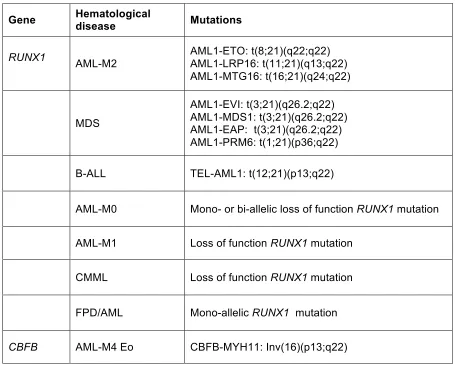
The importance of the core binding factors in hematopoiesis is further
underscored by the fact that both Runx1 and CBFβ are frequent targets of mutations and
chromosomal aberrations in various hematological malignancies (Table 3).
respectively. Each defines subgroups within the category of recurrent cytogenetic
abnormalities, and confers a favorable prognosis.
Table 3. Chromosomal translocations and mutations affecting Runx1 and CBFβ in various
hematological diseases.
Abbreviations: AML, acute myeloid leukemia; AML-M0, minimally differentiated acute myeloid leukemia; AML-M1, acute myeloid leukemia without maturation; AML-M2, acute myeloid leukemia with maturation; AML-M4Eo, acute myeloid leukemia with bone marrow eosinophilia; CMML, chronic myelomonocytic leukemia; FPD/AML, familial platelet disorder with predisposition to acute myeloid leukemia; MDS, myelodysplastic syndrome.
Gene Hematological disease Mutations
RUNX1 AML-M2 AML1-ETO: t(8;21)(q22;q22) AML1-LRP16: t(11;21)(q13;q22)
AML1-MTG16: t(16;21)(q24;q22)
MDS
AML1-EVI: t(3;21)(q26.2;q22) AML1-MDS1: t(3;21)(q26.2;q22) AML1-EAP: t(3;21)(q26.2;q22) AML1-PRM6: t(1;21)(p36;q22)
B-ALL TEL-AML1: t(12;21)(p13;q22)
AML-M0 Mono- or bi-allelic loss of function RUNX1 mutation
AML-M1 Loss of function RUNX1 mutation
CMML Loss of function RUNX1 mutation
FPD/AML Mono-allelic RUNX1 mutation
t(8;21) AML and AML1-ETO
The 8;21 translocation t(8;21)(q22;q22) is one of the most common chromosomal
aberrations in de novo AML, occurring in 10% of adult and 12% of pediatric AMLs (96,
106, 107). The t(8;21) breaks the RUNX1 gene in intron 5, resulting in fusion of the
N-terminal 177 amino acids of Runx1 (including the runt domain) to ETO (eight twenty-one,
encoded by RUNX1T1) (108-112). This generates the chimeric protein AML1-ETO
(Figure 5). ETO has no known role in hematopoiesis: homozygous loss of Runx1t1 in
mice resulted in gastrointestinal defects, but no hematopoietic deficiencies (113, 114).
ETO contains four domains conserved with its Drosophila homologue nervy. Thus the
chimeric protein AML1-ETO consists of the intact Runt domain and almost the entire
coding region of ETO, including 4 functional domains named Nervy homology region
(NHR) 1, NHR2, NHR3, and NHR4, the structures for all of which, along with their
interacting proteins or peptides from those proteins, have been solved (115-120).
Although AML1-ETO is an essential causative factor of t(8;21)-positive AML, the full
length chimeric protein is not leukemogenic by itself, and can only induce AML in mice
when combined with an additional oncogene such as an activated kinase, including
Fms-like tyrosine kinase 3 internal tandem duplication (FLT3-ITD), TEL-PDGFBR, or
activated KIT (121-123). AML1-ETO confers phenotypes different from loss of function
Runx1 mutations. Genetic experiments in Drosophila showed that AML1-ETO acts as a
constitutive repressor of Runx1 homologue lozenge (124). The phenotype of conditional
knock-in mice in which AML1-ETO expression is activated in the adult bone marrow
resembles a milder version of Runx1 loss, including increased numbers of
Figure 5. Schematic for AML1-ETO and its interacting proteins.
On top is a schematic diagram of RUNX1 in grey and ETO sequences in gold and orange. t(8;21) generates AML1-ETO which fuses the Runt domain to nearly all of ETO. Structures of conserved domains (grey or orange) and their interacting proteins or peptides from those proteins (blue), and DNA (purple) are shown below.
Clinically, t(8;21)-positive leukemia is associated with favorable prognosis, with
70% of patients achieving complete remission following standard therapy (126, 127)
(Figure 6). However, many patients retain AML1-ETO expressing cells in their bone
marrow due to incomplete eradication of the leukemic cells (128, 129). As a result
35-40% of these patients relapse within five years and have poor long-term survival (126,
Figure 6. Survival curves of core binding factor leukemias compared to other leukemia types.
Favorable prognosis is associated with t(15;17), t(8;21), or inv(16) whether alone or in combination with other chromosomal abnormalities, with the possible exception of inv(16) or t(8;21) with complex karyotype. From Gulley et al. 2010 (128).
Mutations that specifically disrupt the interaction between individual domains in
AML1-ETO and their associated proteins revealed that the Runt domain and the NHR2
domain (also known as hydrophobic heptad repeat or HHR) are essential for
AML1-ETO’s leukemogenic activity (130-133). The Runt domain of AML1-ETO mediates DNA
binding and CBFβ binding, and both interactions are essential for AML1-ETO’s
leukemogenic activities (130, 132, 133). Amino acid substitutions in the Runt domain
that either disrupt AML1-ETO’s DNA binding or AML1-ETO’s interaction with CBFβ
severely impaired AML1-ETO’s ability to transform hematopoietic cells, and abolished
AML1-ETO’s ability to initiate leukemia in cooperation with activated kinase
oligomerization and both DNA and CBFβ binding are required for AML1-ETO’s
leukemogenic activity.
Figure 7. Mutations that disrupt DNA or CBFβ binding impair AML1-ETO’s leukemogenic activity. From Roudaia et al, 2009 (132).
A. Structures of the Runt domain and CBFβ are shown in gray and blue respectively, and the DNA is purple. The R174 residue in the Runt domain is shown in green, and the T161 and Y113 residues are in orange. B. Schematic of transplantation. Bone marrow mononuclear cells harvested from 5-fluorouracil treated C57BL/6 mice were co-infected with MigR1 expressing AML1-ETO (or its mutated derivatives) and TEL-PDGFβR. IRES-mediated expression of EGFP marked AML1-ETO expressing cells while hCD4 marked TEL-PDGFβR expressing cells. One million transduced cells were transplanted along with 200,000 normal bone marrow cells into lethally irradiated mice.
C. Kaplan-Meier survival curve of mice after transplantation with retroviruses expression AML1-ETO (AE) or its mutated derivatives and TEL-PDGFβR (TP).
A
C
0 10 20 30 40 50 60
0 20 40 60 80 100
AML1-ETO
AML1-ETO Y113A/T161A AML1-ETO R174Q
Mantel-Cox test p<0.0001
Days post-transplant
P
er
ce
n
t
su
rv
iv
The NHR2 mediates tetramer (dimer of dimers) formation, and mutations that
reduced the tetramer to a dimer abrogated AML1-ETO’s leukemogenic activity (116,
131). Substitution of NHR2 by an oligomerization domain from the forkhead binding
protein retained AML1-ETO’s ability to confer serial replating activity to primary bone
marrow cells, demonstrating that oligomerization per se is important for AML1-ETO’s
transforming ability (134).
On the other hand, NHR1, NHR3 and NHR4 are not essential for AML1-ETO’s
leukemogenic activity. The NHR1 (also known as the eTAFH domain) is homologous to
several TATA binding protein-associated factors (TAFs) and interacts with E proteins
(E2A and HEB). It has been proposed that AML1-ETO mediated silencing of E protein
functions is important for t(8;21) leukemogenesis (119, 135). However, amino acid
substitutions that disrupt NHR1’s association with HEB did not impair AML1-ETO’s
ability to confer serial replating to primary mouse bone marrow cells (117), and deletion
of the entire NHR1 domain had no effect on AML1-ETO’s leukemogenic activity (130).
The NHR3 (also known as the Nervy domain) shares homology with A-Kinase Anchoring
Proteins (AKAPs) and interacts with the regulatory subunit of type II cAMP-dependent
Protein Kinase (PKA RIIα) (136). Amino acid substitutions that disrupt the interaction of
NHR3 with PKA RIIα did not affect AML1-ETO’s ability to transform primary mouse bone
marrow cells nor its leukemogenic activity (136).
The NHR4 (also known as the myeloid-Nervy-DEAF-1, MYND) appears to
restrain ETO’s leukemogenic activity, as mutations of NHR4 promote
AML1-ETO’s activity (137). NHR4 binds the silencing mediator of retinoid and thyroid hormone
receptor (SMRT) and nuclear receptor co-repressor (N-CoR) complexes, as well as the
(141). Interestingly, an alternative splice form of AML1-ETO that results in the formation
of a C-terminal truncated protein called AML1-ETO9 (142). AML1-ETO9a is found in
human leukemia cells, and introducing AML1-ETO9a alone into mouse hematopoietic
cells results in the rapid development of leukemia in transplanted mice, suggesting that
AML1-ETO9a does not require additional cooperating mutations (142).
Therefore, AML1-ETO has several interactions that may be targeted with small
molecule inhibitors: the Runt domain:DNA interface, the Runt domain:CBFβ interface,
and NHR2 mediated oligomerization. The interaction of CBFβ with the Runt domain is
essential for the ability of TEL-AML1 (ETV6-RUNX1), frequently found in B-ALL, to
promote the serial replating of B cell progenitors in vitro (132). This suggests that the
interaction with CBFβ may be important for the activity of multiple chimeric Runx1
proteins.
Interestingly, no loss of function (LOF) RUNX1 mutations in the remaining
RUNX1 allele were found in the favorable risk group with the characteristic genetic
abnormality (8;21) (143, 144). Similarly, no LOF RUNX1 mutations were found in AMLs
containing the inv(16) (143, 144). These data suggest either that the t(8;21) and inv(16)
were redundant with LOF RUNX1 mutations, or that they were synthetically lethal. In
support of the latter interpretation, recent studies have provided evidence that wildtype
CBF functions are required for the maintenance and survival of leukemia cells.
Specifically, knock down of wild type Runx1 reduced growth and induced apoptosis in
t(8;21) cell lines, and also in AML1-ETO transformed human CD34 positive cells (145,
146). Knock down of Runx1 also induced apoptosis of MLL-AF9 transformed cells, and
deletion of Runx1 and CBFβ extended the disease latency in a mouse MLL-AF9 model
may rely on continuous CBF function. T cell acute lymphocytic leukemia (T-ALL) also
appears to rely on sustained Runx1 activity. A small molecule targeting cyclin-dependent
kinase 7 displayed activity in a subset of cancer cell lines, including T-ALL, and its
mechanism of action appeared to involve the down regulation of Runx1 expression
(147). Taken together, these findings strongly suggest that, despite the proposed tumor
suppressor function of Runx1 in normal hematopoiesis, a continued low level of normal
Runx1 function is required to maintain cell growth or viability in a certain subset of
leukemia. We hypothesize that these leukemias may be more sensitive to perturbations
in the RUNX proteins’ functions, and therefore small molecules interfering with
Runx1:CBFβ interaction may achieve therapeutic efficacy in a wide range of leukemia.
In summary, we established the RD:CBFβ interface as viable drug target. In a
previous study, we carried out structure-based mutagenesis study at the Runt domain of
Runx1 to determine the energetic contribution of the amino acids in the Runt domain that
contact CBFβ for heterodimerization. We identified two energetic hot spots at the
heterodimerization interface of the Runt domain that contribute to the bulk of binding
energy for heterodimerization with CBFβ (148). Herein we describe the identification
small molecule inhibitors by screening for a library of compounds against the hot spot
residues at the Runt domain:CBFβ interface, and the development of a tool compound
Chapter I: Development and characterization of thiazole compounds targeting to
disrupt the Runt Domain:CBFβ interaction
Zaw Min Oo1, Anuradha Illendula2, Charles Schmidt2, Yunpeng Zhou2, Chung-Tsai Lee1,
Roger A. Rajewski3, Nancy A. Speck1, and John H. Bushweller2
1Abramson Family Cancer Research Institute and Department of Cell and Molecular
Biology, University of Pennsylvania, Philadelphia, Pennsylvania, USA, 19104
2Department of Molecular Physiology and Biological Physics, University of Virginia,
Charlottesville, Virginia, USA, 22908
3Department of Pharmaceutical Chemistry, University of Kansas, Lawrence, Kansas,
USA, 66047
Data for Figures 2-3 was contributed by Anuradha Illendula, Charles Schmidt, and
Yunpeng Zhou.
Data for Figure 4 was contributed by Roger A. Rajewski.
Data for Figure 11 was contributed by Chung-Tsai Lee.
Data for Figures 5, 7-10, and 12 were contributed by Zaw Min Oo.
The text of this chapter is a result of a collaborative effect by Zaw Min Oo, Nancy A.
Abstract
RUNX1 and CBFB, which encode subunits of the core binding factor, are
frequent targets of chromosomal aberrations in hematological malignancies. We
previously determined that CBFβ (encoded by CBFB) is important for the transforming
activity of the chimeric protein AML1-ETO protein (RUNX1-RUNX1T1) generated by the
t(8;21), and other studies showed that normal Runx1 functions are essential for survival
and maintenance of some leukemias lacking RUNX1 or CBFB mutations. Thus, we
hypothesized that we could achieve therapeutic efficacy in multiple leukemias by
targeting the Runx1:CBFβ interaction with small molecule inhibitors. Using the structural
information of the DNA binding Runt domain (RD) of Runx1 and its interface with CBFβ,
we employed a computational screen for a library of 78,000 drug-like compounds, and
further optimized our lead compounds. The Runt domain inhibitors (RDIs) bind directly to
the RD and disrupt its interaction with CBFβ compounds reduced growth and induced
apoptosis of t(8;21) acute myeloid leukemia (AML) cell lines, and reduced the progenitor
activity of mouse and human leukemia cells harboring the t(8;21), but not normal bone
marrow cells. The RDIs had similar effects on murine and human T cell acute
lymphocytic leukemia (T-ALL) cell lines that did not harbor the t(8;21). Our results
confirmed that the RDIs might prove efficacious in various AMLs, and that a therapeutic
Introduction
Acute myeloid leukemia (AML) often harbors non-random clonal chromosomal
aberrations. Among them, the 8;21 translocation t(8;21)(q22;q22) is one of the most
common in de novo AML, occurring in 10% of adult and 12% of pediatric AMLs (96, 106,
107). The translocation fuses the N-terminal 177 amino acids of Runx1 (also known as
AML1, encoded by RUNX1) to ETO (eight twenty-one, encoded by RUNX1T1),
generating the chimeric protein AML1-ETO, an essential causative factor of
t(8;21)-positive AML (107). Runx1 is a sequence-specific DNA binding transcription factor and
member of heterodimeric core binding factors (CBFs) that play important roles in
hematopoiesis (106, 107). Loss of Runx1 during embryonic development results in a
failure of hematopoietic stem cell (HSC) emergence, whereas loss in adult HSCs leads
to a pre-leukemic state (79, 94, 96, 97, 99, 100). ETO, on the other hand, has no known
role in normal hematopoiesis. Homozygous loss of Runx1t1 in mice resulted in
gastrointestinal defects, but no hematopoietic deficiencies (113, 114).
Clinically, t(8;21)-positive leukemia is associated with favorable prognosis, with
70% of patients achieving complete remission following standard therapy (126, 127).
However, many patients retain AML1-ETO expressing cells in their bone marrow due to
incomplete eradication of the leukemic cells (128, 129). As a result 35-40% of these
patients relapse within five years and have poor long-term survival (126, 127). We
hypothesize that direct therapeutic targeting of the chimeric protein AML1-ETO may
reduce the rate of relapse and improve long-term survival.
AML1-ETO has five domains conserved with its Drosophila homologues: the
Runt domain (RD) from Runx1, and four from ETO (eTAFH, HHR, Nervy, MYND) (107).
and introduced amino acid substitutions to assess their contribution to AML1-ETO’s
transforming ability (62, 115-118, 120, 132, 134, 149-151). We determined that the
interaction between the Runt domain of AML1-ETO and CBFβ, the non-DNA binding
partner of all three RUNX proteins, is important for AML1-ETO mediated
leukemogenesis (132). We introduced a single amino acid substitution into the Runt
domain that disrupted DNA, but not CBFβ binding (R174Q), and a pair of mutations that
impaired CBFβ but not DNA binding (Y113A/T161A). Both sets of mutations severely
impaired AML1-ETO’s ability to transform hematopoietic cells. Most importantly, both
DNA and CBFβ binding were essential for AML1-ETO’s ability to cooperate with the
activated kinase TEL-PDGFβR to promote leukemia in mice. Collectively these data
demonstrated that both DNA and CBFβ binding are critical for AML1-ETO’s activity, and
validated the AML1-ETO:CBFβ interaction as a viable drug target.
In addition, we determined that the interaction of CBFβ with the Runt domain is
essential for the ability of TEL-AML1 (ETV6-RUNX1), frequently found in B-ALL, to
promote the serial replating of B cell progenitors in vitro (132). This suggests that CBFβ
may be important for the activity of multiple chimeric Runx1 proteins.
Recent studies have provided additional evidence that normal CBF functions are
required for the maintenance and survival of certain leukemia cells. Specifically, knock
down of wild type Runx1 reduced growth and induced apoptosis in t(8;21) cell lines, and
also in AML1-ETO transformed human CD34 positive cells (145, 146). Knock down of
Runx1 also induced apoptosis of MLL-AF9 transformed cells, and deletion of Runx1 and
CBFβ extended the disease latency in a mouse MLL-AF9 model (146), suggesting that a
subset of AMLs that do not harbor mutations in the CBF genes may rely on continuous
sustained Runx1 activity. A small molecule targeting cyclin-dependent kinase 7
displayed activity on a subset of cancer cell lines, including T-ALL, and its mechanism of
action appeared to involve the down regulation of Runx1 expression (147).
Taken together, these findings strongly suggest that normal CBF function is
required for the maintenance of leukemic stem cell functions in a certain subset of
leukemia. We hypothesize that these leukemias may be more susceptible to
perturbations in normal CBF functions. Therefore, small molecules interfering with
Runx1:CBFβ interaction may achieve therapeutic efficacy in a wide range of leukemia.
We have established the RD:CBFβ interface as viable drug target and have developed
small molecules interfering with the RD:CBFβ interaction.
Herein we describe the development of a tool compound that binds to the Runt
domain of RUNX proteins and inhibits their interaction with CBFβ. We also developed a
pro-drug version of this tool compound to improve solubility and thereby potential utility
in vivo. These Runt domain inhibitors (RDIs) inhibit growth in culture as well as
clonogenic potential of AML1-ETO and T-ALL leukemia cell lines. The tool compounds
show clear effects on the expression of well-characterized RUNX1 target genes.
Analysis of genome-wide changes in gene expression identified lipid and sterol
biosynthesis and ribosome biogenesis pathways, which are RUNX regulated (152, 153),
Materials and Methods
Virtual screening
Details of the virtual screen are described in the Supplemental Methods. In brief, the
computer program LUDI/InsightII (154) was applied for virtual screening of CAP
(Chemicals Available for Purchase, 78,000 compounds) library to the CBFβ binding
interface on the Runt domain structure. Compounds from CAP library were docked and
ranked by the scoring function (Energy Estimate 1) implemented in the LUDI program
(155). The 500 best-scored hits (compounds with the predicted binding affinity < 300 µM
as evaluated by LUDI’s empirical scoring function) were subjected to visual inspection of
their potential interactions with the Runt domain. Compounds with diverse scaffolds and
involved in at least two hydrogen bonds with the Runt domain were selected for
experimental evaluation.
Fluorescence resonance energy transfer (FRET) assays
Cerulean-Runt domain and Venus-CBFβ were expressed, purified and used in FRET
assays as described previously (50, 156). Cerulean-Runt domain and Venus-CBFβ
proteins were used at a concentration of 100 nM for all assays.
Saturation transfer difference NMR
Saturation transfer difference (STD) NMR experiments (157, 158) were performed with
30 µM Cerulean-Runt domain or Venus-CBFβ, 800 µM AI-7-54 or AI-8-45, 10% D2O,
and 5% DMSO in 50 mM KPi, 100 mM KCl, 10 mM K2SO4, 2 mM MgSO4, pH 7.5 in a
NMR spectrometer at 25°C with saturation times of 500, 750, 1000, 1500, and 2000 ms.
Samples were irradiated at 0.4 ppm (protein) and 30 ppm (off-resonance control) and
the difference spectra calculated using MestReNovaChemical
Chemical synthesis
Details of the chemical synthesis including relevant NMR and mass spec data are
provided in Supplementary Information.
Pharmacokinetics
Detailed methods are provided in the Supplemental Methods.
Mice
All mouse procedures were approved by the University of Pennsylvania University
Animals Resource Center (ULAR) and Institutional Animal Care and Use Committees
(IACUC) of the University of Pennsylvania and the University of Kansas. C57BL/6 mice
were used in all studies.
Human samples
Patient AML specimen pheresis and bone marrow mononuclear cells were obtained
from the University of Pennsylvania Stem Cell and Xenograft Core, under the approval
from the University of Pennsylvania Institutional Review Board (IRB).
Mouse and human cell lines
Kasumi-1 (ATCC), Jurkat E6-1 (ATCC), 8946 T-ALL, and 720 T-ALL cell lines were
(FBS) and 1% penicillin/streptomycin at 37°C under 5% CO2. K562 (ATCC) cell line is
cultured in Iscove's Modified Dulbecco's Medium (IMDM) supplemented with 10% FBS.
720 T-ALL cells were derived from a Tcf12+/- mouse expressing a Tal1 transgene under
the control of the Lck promoter (159). 8946 T-ALL cells are derived from a murine T-ALL
induced with a doxycycline-repressible human c-MYC transgene (160).
MTT Cell Proliferation Assay
Mouse and human leukemia cell lines (104 cells/200 µl) were plated in a 96-well
flat-bottom plate and cultured with DMSO (vehicle), 1 µM Staurosporine, or RDIs (12.5, 25,
50, or 100 µM) for 24, 48, and 72 hours. After treatment, 10 µl of 5 mg/ml MTT solution
(3-(4, 5-dimethylthiazolyl-2)-2, 5-diphenyltetrazolium bromide) (Sigma-Aldrich) was
added to each well cultured at 37°C for 4 hours. The plate was centrifuged at 300 g for 5
minutes, the media removed, and 100 µl of DMSO (Sigma-Aldrich) was added to
solubilize the resulting reagent formazan and incubated at room temperature for 10
minutes. The plate was then analyzed by measuring absorbance at 540 nm wavelength
in a SpectraMax plate reader (Molecular Devices). Data are plotted as percentage of
viable cells relative to DMSO.
CFU-C Assay
Frozen human AML samples, and mouse leukemic bone marrow cells were thawed and
cultured in RPMI 1640 with 10% FBS for two hours. Live cells were washed and
recovered, and subsequently plated in Human Methylcellulose Complete Media HSC003
(R&D Systems) in 5% CO2 at 37°C for 14 days, or in Methocult GF M3434 (Stem Cell
cells were counted according to manufacturer’s recommendations.
Western blotting
Murine leukemic cells treated with RDIs or DMSO were harvested and lysed with RIPA
buffer (25mM Tris•HCl pH 7.6, 150mM NaCl, 1% NP-40, 1% sodium deoxycholate, 0.1%
SDS). Total protein lysates were resolved on 4-12% SDS-PAGE gels, transferred to a
nitrocellulose membrane (GE Life Sciences), and probed with primary antibodies.
Proteins of interest were visualized by chemi-luminescence (Pierce). The following
antibodies were used for immunoblotting: caspase-3 (Cell Signaling, #9662), p53 (Santa
Cruz, DO-1; Leica, CM5), and actin (Santa Cruz, N21).
Flow cytometry
Cells were stained with fluorochrome-conjugated antibodies for 30 minutes at 4°C and
washed with 2% FBS in PBS prior to analysis. Apoptosis analysis (Annexin V-APC; BD
Biosciences) was performed according to the manufacturer’s recommendations on a
LSR II flow cytometer (BD Biosciences). The data were analyzed using FlowJo v.9.8
(TreeStar).
Gene expression analysis (quantitative real-time PCR and microarrays).
RNA for quantitative real-time PCR (qRT-PCR) was isolated with the RNeasy Kit
(QIAGEN), and total RNA was reverse-transcribed using cDNA Reverse Transcription
Kit (Applied Biosystems). The cDNA produced was used for quantitative real-time PCR