Estimating the Probability of Abusive Head Trauma:
A Pooled Analysis
WHAT’S KNOWN ON THIS SUBJECT: In recent debate in
medicolegal circles, clinical basis for a diagnosis of abusive head trauma (AHT) has been questioned. Studies have been
underpowered to address the key clinical questions. Estimates of the association of single clinical variables with AHT are
recognized.
WHAT THIS STUDY ADDS: This study provides an estimate of probability of AHT based on 6 clinical features. In a child younger than 3 who has an intracranial injury andⱖ3 of these features, the positive predictive value of AHT is⬎85% (odds
ratio:⬎100).
abstract
CONTEXT AND OBJECTIVE:To determine which combinations of clini-cal features assist in distinguishing abusive head trauma (AHT) from nonabusive head trauma.
METHODS:Individual patient data from 6 comparative studies of chil-dren younger than 3 years with intracranial injury were analyzed to determine the association between AHT and combinations of apnea; retinal hemorrhage; rib, skull, and long-bone fractures; seizures; and head and/or neck bruising. An aggregate analysis of data from these studies used multiple imputation of combined clinical features using a bespoke hotdeck imputation strategy, which accounted for uncertainty arising from missing information.
RESULTS:Analyzing 1053 children (348 had AHT), excluding nonsignif-icant variables (gender, age, skull fractures), for a child with an intra-cranial injury and 1 or 2 of the 6 features, the positive predictive value (PPV) of AHT varies from 4% to 97% according to the different combi-nations. Although rarely recorded, apnea is significantly associated with AHT (odds ratio [OR]: 6.89 [confidence interval: 2.08 –22.86]). When rib fracture or retinal hemorrhage was present with any 1 of the other features, the OR for AHT is⬎100 (PPV⬎85%). Any combination of 3 or more of the 6 significant features yielded an OR of⬎100 (PPV for AHT⬎ 85%).
CONCLUSIONS:Probabilities of AHT can be estimated on the basis of different combinations of clinical features. The model could be further developed in a prospective large-scale study, with an expanded clinical data set, to contribute to a more refined tool to inform clinical deci-sions about the likelihood of AHT.Pediatrics2011;128:e550–e564
AUTHORS:Sabine Ann Maguire, MRCPI, MRCPCh, FRCPI,a Alison Mary Kemp, MRCP, DCH, FRCPCH, FRCP,aRebecca Caroline Lumb, BA,aand Daniel Mark Farewell, MMath, PhDb
Departments ofaChild Health andbPrimary Care and Public
Health, School of Medicine, Cardiff University, Cardiff, United Kingdom
KEY WORDS
abusive head trauma, child abuse, diagnostic tool, pooled estimates of probability of AHT
ABBREVIATIONS
AHT—abusive head trauma ICI—intracranial injury nAHT—nonabusive head trauma PPV—positive predictive value OR—odds ratio
RH—retinal hemorrhage CI—confidence interval
Dr Maguire had the initial study idea and was principle investigator for primary systematic review, drafted and revised the article, and gave final approval for publication; Ms Lumb entered all anonymized data into the database, formatted the original article, communicated with all primary authors, and edited the article; Dr Farewell designed and conducted all statistical analyses, wrote the statistical component of article, and contributed to overall editing; and Prof Kemp was program director for primary systematic review and contributed to the conception of analysis, drafts of the article, and subsequent editing.
www.pediatrics.org/cgi/doi/10.1542/peds.2010-2949
doi:10.1542/peds.2010-2949
Accepted for publication May 5, 2011
Address correspondence to Sabine Ann Maguire, MRCPI, MRCPCh, FRCPI Wales College of Medicine, Cardiff University, UHW, Heath Site, Cardiff CF14 4XN, United Kingdom. E-mail: [email protected]
PEDIATRICS (ISSN Numbers: Print, 0031-4005; Online, 1098-4275).
Copyright © 2011 by the American Academy of Pediatrics
Abusive head trauma (AHT) remains the commonest form of fatal child abuse and predominantly affects in-fants.1These children classically
pres-ent with an intracranial injury (ICI) that is evident on neuroimaging or at post mortem, where there is no explanation of trauma, or a history that is inconsis-tent with the severity of injury or the developmental level of the child. The challenge for the clinician is to deter-mine, firstly, which children should be investigated to exclude AHT; secondly, when concerning features are found, how confident one can be that AHT is the cause rather than nonabusive head trauma (nAHT) or an organic dis-ease. After a full clinical assessment, the multidisciplinary team must piece together all available (eg, clinical, so-cial, forensic) information and deter-mine the likelihood of an abusive etiol-ogy. These decisions are difficult, and the pressures to make the correct di-agnosis are considerable. A child with missed AHT may later present with a fatal injury2; on the other hand, an
in-correct diagnosis of AHT will have dev-astating consequences for the child and family.
When cases of suspected child abuse enter the legal child protection pro-cess, the medical evidence contributes heavily to decisions about the future risks to the child, and child care pro-ceedings, regardless of whether the evidence informs a criminal prosecu-tion. In a recent law review article, Turkheimer questioned the diagnosis of AHT on the basis of combinations of clinical features.3This stimulated
con-troversial argument in The New York Times: Tuerkheimer suggested that ex-perts are questioning the scientific ba-sis for shaken baby syndrome (sic),4
which was roundly refuted by re-sponses from M. Barr and J. Lev-enthal,5among others, thus
highlight-ing the need for an agreed scientific basis to contribute to this diagnosis. In
their role as expert witnesses in court, clinicians in the United Kingdom have also come under pressure to support their opinion with scientific evidence, as highlighted by Baroness Kennedy: “A doctor can be convinced based on his or her experience that a defendant is guilty, but unless there is compelling evidence, supported scientifically, he or she should not express that view in criminal proceedings.”6 As such, an
evidence-based calculation that assim-ilates the clinical data and contributes to determining the probability of AHT would be a valuable adjunct for the professionals concerned.
Although many studies have evaluated the diagnostic utility of individual fea-tures in identifying children with AHT, only a small number have explored how combined features predict the condition7,8; given that AHT is relatively
uncommon in any given population,9
individual studies are frequently un-derpowered to draw statistically valid conclusions for multiple combinations of features.10Previously, we conducted
a meta-analysis of high quality studies that derives the positive predictive val-ues (PPV) and odds ratios (ORs) for AHT for individual features.11However,
in a clinical scenario, the diagnosis of AHT is based on an interpretation of the combined findings. In this study we aim to combine individual patient data from 6 large population-based com-parative studies of AHT and nAHT to es-timate the probability of AHT, given different combinations of features identifiable during the initial evalua-tion of such cases. In so doing, it may provide a statistical estimate to assist clinicians.
METHODS
We conducted a systematic review of the clinical features of AHT and nAHT11
(apnea; retinal hemorrhages [RHs]; rib, skull, and long-bone fractures; sei-zures; and head and/or neck bruising)
and identified 14 high quality compar-ative studies. We contacted the au-thors of the most recent larger studies that encompassed the majority of the clinical indicators of AHT and nAHT in children younger than 3.11Of the 9
au-thors we contacted, 2 did not respond, 1 was unable to help, and the authors of the remaining 6 studies, all pub-lished since 2003,9,12–16generously
pro-vided anonymized, individual-level pa-tient information. We have used these data to perform an individual-patient data pooled-analysis to relate combi-nations of clinical features to AHT or nAHT.
All studies included children younger than 3 who were admitted to hospital with an ICI, defined as any combination of subdural hemorrhage, subarach-noid hemorrhage, extradural hemor-rhage, intraparenchymal injury, cere-bral contusion, diffuse axonal injury, hypoxic ischemic injury and/or associ-ated cerebral edema. The inclusion cri-teria with respect to ICI type varied be-tween studies; cases of isolated skull fracture with no ICI were excluded. In 2 instances,15,16the authors provided us
with additional data over and above that which they had published in the referenced studies. All extra cases had been ascertained in the same manner as that described in the published study.
re-corded as “missing.” “Seizure” in-cluded documented seizures, either single or, in some cases, status epilep-ticus. “Apnea” and “head/neck bruis-ing” were simply recorded as “pres-ent,” “abs“pres-ent,” or “missing.”
We defined AHT (terminology as recom-mended in the recent American Acad-emy of Pediatrics’ recommenda-tions17) as ICI where abuse had been
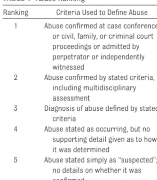
confirmed as a cause. We only included confirmed cases of AHT, ie, those ranked 1 or 2 for abuse, according to our previously published ‘rank of abuse’ (Table 1). This was to ensure that the decision was based on a thor-ough child protection assessment and multidisciplinary decision or court proceedings to minimize circularity by dependence purely on the clinical fea-tures alone to determine if abuse had taken place.11All the nonabused
chil-dren had confirmed causes (traumatic or organic); any cases deemed indeterminate or simply “suspected abuse” in the original data set were excluded from the analysis.
For full details of the statistical meth-ods employed, see the Appendix. In summary, we performed an aggregate analysis of the 6 studies, taking into account the uncertainty arising from missing information on certain clinical features. To do so, we employed
multi-ple imputation, using a bespoke hot-deck imputation strategy informed by examination of the raw data. Wherever possible, we imputed data from an-other child in the same or a similar study, particularly when several items were missing. If fewer than 5 children could be found to match the child who had missing information, we refrained from imputing from the matched cases because this can lead to under-estimation of the uncertainty sur-rounding the missing features. In-stead, we completed our imputed data by filling in any remaining missing items from the margins of the ob-served distribution of that feature, within their etiology group. We gener-ated 10 imputed data sets for analysis.
To each of the imputed data sets, we fitted a multilevel logistic regression model. Logistic regression focuses on the probability of AHT, given informa-tion on the presence or absence of cer-tain features. A multilevel version was used to account for the fact that the different studies were drawn from dif-ferent populations and, in particular, had different prevalences of AHT. Re-sults from the 10 imputed data sets were combined according to estab-lished procedures, and expressed as ORs, with 95% confidence intervals (CIs) and PPVs. We emphasize that a large OR need not correspond to a large PPV because a PPV takes into ac-count the underlying prevalence of AHT in this specific population.
We fitted 3 multilevel logistic regres-sion models. Initially, we examined the relationship between AHT, age, and gender, adjusting for variability in the prevalence of AHT between studies. We then added all 7 clinical features to the model, further adjusting for between-study variation in the dependence of etiology (AHT or nAHT) on each clinical feature. To improve statistical effi-ciency, we then refitted this model, omitting nonsignificant features.
Fi-nally, we assessed the predictive accu-racy of the model. To do this, we used fivefold cross-validation: this process divides the data into 5 parts, using 4 to refit the multilevel model, and the re-maining part to test its predictive ac-curacy. This process is then repeated, with each of the 5 parts being used to test the model in turn.
RESULTS
The 6 included studies9,12–16represent
data on 1053 children (348 sustained AHT, 705 sustained nAHT). The study de-signs were similar; they were all population-based and included chil-dren younger than 3 who were admit-ted to regional hospitals with ICI diag-nosed on neuroimaging. Four studies included all children with traumatic brain injury,12–14,16 and 2 included all
children who were admitted with sub-dural hemorrhage.9,15 Studies
con-firmed etiology of nAHT cases but ap-plied different exclusion criteria. Two of these studies were prospective,12,16
with the remainder retrospective9,13–15
but with detailed consecutive case ascertainment.
Analysis
Because demographic information was available for all children, it was possible to examine the diagnostic util-ity of age and gender without resorting to multiple imputation. No significant gender differences were observed, al-though it was clear that more boys than girls sustained brain injury, re-gardless of etiology (Table 2). All 3 of the youngest age groups (0 –5 months, 6 –11 months, 12–23 months) had higher prevalence of AHT than the el-dest group (24 –36 months), and the differences between the age groups were significant (Table 3). The odds in the 2 youngest groups were⬃4 times the baseline odds. Consistent with study ascertainment, we also ob-served substantial between-study vari-ability in the prevalence of abuse. TABLE 1 Abuse Ranking
Ranking Criteria Used to Define Abuse 1 Abuse confirmed at case conference
or civil, family, or criminal court proceedings or admitted by perpetrator or independently witnessed
2 Abuse confirmed by stated criteria, including multidisciplinary assessment
3 Diagnosis of abuse defined by stated criteria
4 Abuse stated as occurring, but no supporting detail given as to how it was determined
Multiply Imputed Multilevel Logistic Regression
For each of the 10 imputed data sets, we used multilevel logistic regression to determine the diagnostic value of all
features identified during initial
inves-tigations in combination. We report ef-fect sizes on the logistic scale and in terms of ORs, giving 95% CIs for these estimates, and PPVs, together with P
values and estimates of the SDs of the random effects (Table 4). In this model, the reference category is a girl aged between 24 and 36 months, but now, additionally, they are known to have no other clinical features present (ie, ICI alone).
Nonsignificant Features
Once other clinical features were known, age was no longer significantly associated with etiology at the 5% level. In other words, given information about the presence or absence of the 7 clinical features, knowing the child’s TABLE 3 Regression Analysis of Demographic Information: Global Test of Significance of Age
Regression Coefficient
Lower Confidence
Limit
Upper Confidence
Limit
OR Lower Confidence
Limit
Upper Confidence
Limit
P Random Effects SD
Intercept ⫺1.943 ⫺3.050 ⫺0.835 0.143 0.047 0.434 .001 1.135 0–5 mo 1.500 0.428 2.572 4.480 1.534 13.088 ⬍.001 1.074 6–11 mo 1.399 0.731 2.066 4.051 2.078 7.897 — ⬍0.001 12–23 mo 0.752 ⫺0.067 1.571 2.121 0.935 4.813 — 0.483 Male 0.280 ⫺0.021 0.580 1.322 0.979 1.787 .069 ⬍0.001
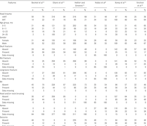
TABLE 2 Univariate Summaries According to Study
Features Bechtel et al12 Ettaro et al13 Hettler and
Greenes14
Hobbs et al9 Kemp et al15 Vinchon
et al16
n % n % n % n % n % n %
Head trauma
nAHT 50 78 316 84 216 69 31 48 67 40 25 36
AHT 14 22 61 16 95 31 34 52 100 60 44 64
Age group, mo
0–5 31 48 121 32 202 65 34 52 51 31 45 65
6–11 20 31 77 20 68 22 18 28 33 20 14 20
12–23 10 16 79 21 41 13 4 6 53 32 10 14
24–35 3 5 100 27 0 0 9 14 30 18 0 0
Gender
Female 31 48 155 41 106 34 29 45 58 35 23 33
Male 33 52 222 59 205 66 36 55 109 65 46 67
Skull fracture
Absent 28 44 154 41 149 48 0 0 142 85 57 83
Present 36 56 220 58 162 52 0 0 25 15 12 17
Data missing 0 0 3 1 0 0 65 100 0 0 0 0
Rib fracture
Absent 16 25 358 95 306 98 0 0 141 84 52 75
Present 3 5 14 4 5 2 0 0 26 16 17 25
Data missing 45 70 5 1 0 0 65 100 0 0 0 0
Long-bone fracture
Absent 17 27 343 91 294 95 0 0 138 83 57 83
Present 2 3 29 8 17 5 0 0 29 17 12 17
Data missing 45 70 5 1 0 0 65 100 0 0 0 0
RH
Absent 45 70 328 87 225 72 35 54 96 57 33 48
Present 16 25 44 12 86 28 30 46 56 34 36 52
Data missing 3 5 5 1 0 0 0 0 15 9 0 0
Head and/or neck bruising
Absent 30 47 358 95 0 0 0 0 107 64 35 51
Present 28 44 19 5 0 0 0 0 60 36 34 49
Data missing 6 9 0 0 311 100 65 100 0 0 0 0
Apnea
Absent 0 0 0 0 0 0 57 88 116 69 51 74
Present 0 0 0 0 0 0 8 12 36 22 18 26
Data missing 64 100 377 100 311 100 0 0 15 9 0 0
Seizures
Absent 46 72 0 0 235 76 46 71 84 50 33 48
Present 8 12 0 0 76 24 19 29 66 40 36 52
age as well did not significantly alter the probability that the child had been abused. Gender remained uninforma-tive where AHT was concerned. Skull fracture(s) were the only clinical fea-ture(s) with an OR of ⬍1 (ie, more suggestive of nAHT than AHT), although this association did not reach significance.
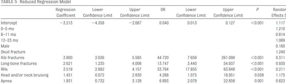
Because we found no statistical evi-dence that including these terms in a model for AHT was important, we refit-ted the model, dropping the nonsignif-icant features. The reduced, multiply-imputed multilevel analysis gave rise to Table 5, on which we base our sub-stantive conclusions.
ICI Alone
When a child younger than 3 had an ICI, with none of the clinical features noted above, the estimated probability of AHT
was ⬃4% (Table 5). Although we did account for study-specific associa-tions between AHT and age, AHT and gender, and AHT and skull fractures, we reiterate that age did not help to
determine the likelihood of abuse when all other clinical features were known.
Results When Each Feature Is Solely Present in a Child With ICI
Fractures
For rib fractures there was the stron-gest evidence of AHT, with an OR of⬃45 (Figs 1 and 2). Note, however, the wide CIs that surround this estimate, owing to the very small numbers of rib frac-tures present in both etiology groups.
Long-bone fractures are similarly in-dicative of AHT (OR: 13.75), slightly less strongly than rib fractures. Because
the prevalence of long-bone fractures was marginally higher than that of rib fractures, the CIs surrounding this es-timate are narrower.
RH, Head and/or Neck Bruising, Apnea, and Seizures
RHs were 1 of the more frequently recorded items, and had an esti-mated OR of ⬃35. Therefore, whereas a child with an ICI alone had a probability of AHT of 4%, this rose to 58% if ICI and only RHs were pres-ent. Head and/or neck bruising had a weaker, marginally significant rela-tionship with AHT. A combination of ICI and bruising to the head and/or neck increased the probability of AHT from 4% to 15%. It is worth empha-sizing, however, that this left 85% of children with this pattern of injury who did not have AHT.
TABLE 4 Full Regression Model of Demographic and Clinical Features Expressed as ORs With 95% CIs,PValues, and Estimates of the SDs of the Random Effects Regression Coefficient Lower Confidence Limit Upper Confidence Limit OR Lower Confidence Limit Upper Confidence Limit P Random Effects SD Intercept ⫺4.002 ⫺5.505 ⫺2.500 0.018 0.004 0.082 ⬍0.001 1.238
0–5 mo 1.138 ⫺0.339 2.615 3.120 0.712 13.667 0.068 1.371
6–11 mo 1.284 0.277 2.291 3.612 1.320 9.884 — ⬍0.001
12–23 mo 0.267 ⫺1.093 1.627 1.306 0.335 5.088 — 0.910
Male 0.149 ⫺0.435 0.732 1.160 0.647 2.079 0.616 0.194
Skull fractures ⫺0.749 ⫺1.928 0.430 0.473 0.145 1.537 0.213 1.142 Rib fractures 3.755 1.963 5.548 42.746 7.119 256.668 ⬍0.001 0.446 Long-bone fractures 2.635 1.179 4.092 13.949 3.250 59.865 ⬍0.001 0.971
RHs 3.450 2.824 4.076 31.497 16.840 58.912 ⬍0.001 0.159
Head and/or neck bruising 1.513 0.160 2.865 4.539 1.174 17.546 0.027 1.115
Apnea 1.938 0.742 3.135 6.948 2.100 22.985 0.001 0.617
Seizures 1.624 0.675 2.574 5.075 1.963 13.117 0.001 0.762
TABLE 5 Reduced Regression Model
Regression Coefficient Lower Confidence Limit Upper Confidence Limit OR Lower Confidence Limit Upper Confidence Limit P Random Effects SD Intercept ⫺3.213 ⫺4.358 ⫺2.067 0.040 0.013 0.127 ⬍0.001 1.117
0–5 mo 1.210
6–11 mo 0.814
12–23 mo 1.069
Male 0.160
Skull fracture 1.240
Rib fractures 3.800 2.036 5.565 44.720 7.659 261.098 ⬍0.001 0.311 Long-bone fractures 2.621 1.235 4.006 13.747 3.440 54.937 ⬍0.001 0.930
RHs 3.519 2.882 4.157 33.764 17.855 63.848 ⬍0.001 0.211
Head and/or neck bruising 1.451 0.072 2.830 4.268 1.075 16.951 0.038 1.175
Apnea 1.931 0.732 3.129 6.893 2.079 22.858 0.001 0.623
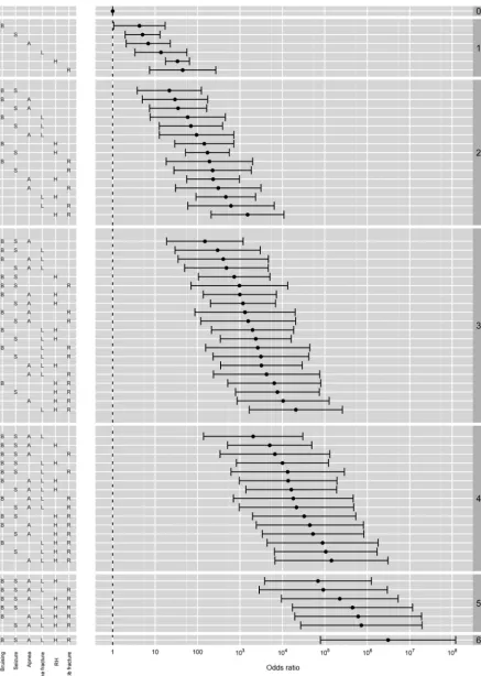
FIGURE 1
FIGURE 2
Either apnea or seizures alone, in as-sociation with ICI, significantly in-creased the likelihood of AHT, both at a level comparable to that of head and/or neck bruising alone (an OR of 5). Apnea was the slightly stronger of the 2, although this finding is subject to a degree of uncertainty because of the large amount of missing data (re-corded in only 301 of 1053 cases). It is hoped that this feature will be more commonly recorded in future studies because of increasing recognition of its association with AHT.
Results When Multiple Features Are Present
A unique advantage of our study is the ability to consider the presence or ab-sence of multiple clinical features in combination. Thus far, we have used the data to focus on situations where we knew that only a single clinical fea-ture was present; however, we now turn to the more frequent scenario where more than 1 feature may be present (Figs 1 and 2). It is immediately apparent that when only 1 or 2 fea-tures were present, the ability to dis-tinguish AHT from nAHT depends heav-ily on the specific feature(s) in question. For instance, if a child with ICI had head and/or neck bruising in combination with apnea, the estimated probability of AHT was 54% (OR: 29); however, if the child had apnea com-bined with RHs, this estimated proba-bility rose to 90%, and the OR was al-most 10 times higher. Likewise, if the child presented with apnea and sei-zures but no other features, the esti-mated probability of AHT was only 58% (OR: 35); however, if a child had sei-zures and rib fractures, the probability of AHT rose to 90%, and the OR was 227.
Once 3 or more of the significant fea-tures were present, ORs were ⬎100, and PPVs for AHT were uniformly above 85%, irrespective of the specific fea-tures. In Fig 2 we show precise
esti-mates for each of the 64 possible combinations.
Accuracy of the Model: Model Checking
In the absence of an additional, inde-pendent source of data against which to test our findings, we used cross-validation to assess the accuracy of our predictions. Details of the model checking are provided in the Appendix. We chose high cut-off limits, such that we deemed a predicted probability to be correct if the PPV for AHT or nAHT was⬎80%, and the predicted etiology was true (see Fig 2). Any PPV less than this was regarded as indeterminate. On this basis, our predicted etiology was correct 80% of the time, indeter-minate 15% of the time, and incorrect in 5% of cases. This highlights that no set of clinical features was unique to AHT or nAHT, and such features must be considered in the context of all other medical and social aspects of the case in question.
DISCUSSION
Our analysis of more than 1000 infants and young children with ICI of con-firmed etiology represents the largest published analysis of combined clini-cal features to estimate the probability of AHT. We have shown that in a child younger than 3 with an ICI and 1 or 2 of the key clinical features, the probabil-ity of AHT varied depending on the number and specific features present, with RHs and rib fractures being the most discriminating. Three or more of the key features were highly predictive of AHT. This analysis offers the potential to underpin a clinical opinion with a valid scientific estimate of probability.
In the study we draw on the raw data from the most recent large-scale com-parative epidemiologic publications, and we are indebted to the authors of the primary studies included. This is the first detailed multivariate analysis
to be produced and, in common with all studies in this difficult field, there are limitations; however, we believe that this work makes a timely and valu-able contribution to the clinical field, in particular at a time when the validity of diagnosing AHT from combinations of clinical features is being questioned.3
Although information on the presence of apnea was missing in a large num-ber of cases, the majority of these (377⫹311) came from 2 large studies that had otherwise very complete re-cording of features. Where data (eg, apnea) was missing, we addressed this by using similar individuals within the data set to impute missing data, an approach that is statistically valid pro-vided the data are missing at ran-dom.18In statistical terms, clinical
de-cisions about what investigations to perform define the mechanism leading to missing data. Because such deci-sions are usually taken sequentially and on the basis of the results of the previous investigations (that is, the ob-served data), the “missing at random” assumption is reasonable in this con-text. All of the features, other than ap-nea, were investigated for in the ma-jority of the studies.
reveals a greater understanding of the individual diagnostic indicators of AHT. However, the features included in our analysis are those that all frontline cli-nicians are likely to identify before re-ferring the child into a specialist cen-ter for more detailed assessment, and, as such, the results of this study could be used to support such additional de-tailed assessments.
This model has been developed from recent high quality, large scale com-parative studies currently available from the international scientific litera-ture. However, we believe that it has the potential for future development, into a sophisticated tool to aid the clin-ical decision process when assessing a young child with unexplained head trauma. A large-scale prospective study is urgently needed to collect a more extensive standardized clinical data set from a cross-section of young children with ICI and apply a multivar-iate analysis. A proposed study of this nature would need to be conducted on an international basis to collect suffi-cient case numbers over a reasonable time scale and to incorporate more de-tail about the nature of the currently recognized clinical indicators such as seizure type and duration, the precise location and pattern of RHs,19,20and
de-tailed neuroradiological features of ICI found in each group.21Other features
may also be contributory eg, history on presentation,14 vomiting,22
co-existent injury,1,23conscious level on
arrival,16and burns1; with increasing
features included in the analysis, larger numbers will be required spe-cifically to ensure that enough cases with each possible combination (fea-tures present or absent) are in-cluded to enable valid statistical analysis of every scenario.
Diagnostic studies in this field are open to criticism of circularity be-cause of their dependence on a con-stellation of clinical features, as
op-posed to a single gold-standard diagnostic test, which does not exist. Ultimately, in any individual case, a child either has, or has not, suffered AHT and, consequently, a diagnosis of AHT either is, or is not, correct. How-ever, except in cases of independently witnessed injury, a diagnosis must rest on a probabilistic assessment of how likely it is that AHT took place. It is not possible to restrict research in this field to independently witnessed abuse, which represents a tiny propor-tion of cases. We have attempted to minimize the risk of circularity by only analyzing cases where abuse was con-firmed by a comprehensive evaluation of all the medical and social features, after a multidisciplinary assessment of the full case details and, in many cases, by “finding of fact” in care or criminal legal proceedings or perpe-trator admissions. Likewise, nAHT cases were only included when the eti-ology was confirmed. To adequately power studies of what is, in statistical terms, a small population, some au-thors frequently combine presumptive and ‘suspected’ abuse into 1 cate-gory.13 Others10 add indeterminate
cases to the noninflicted cases or com-bine suspected abuse with confirmed abuse. We have rigidly excluded such cases from our analysis and the ex-tremely large data set and internal cross validation offers reassurance that our estimates are valid. In addi-tion, the strength of this work lies in a regression analysis of 6 significant variables in 1053 children, generating 64 possible combinations, which rep-resents a wider range of possible com-binations than could be achieved from any single study. The findings from this comparative study mitigate against the circularity argument to some ex-tent. We have demonstrated that there is a difference between the predictive probability of different combinations of features, strongly influenced by the specific features in question. However,
even for strongly influential features, such as RHs, not all cases were be-cause of AHT, as shown by a PPV of 58% for those children with an ICI and RHs and no other clinical features. These data includes the most challenging clinical scenario, namely the likelihood of abuse when a child has ICI but none of the other distinguishing clinical fea-tures. For a child younger than 3 with an ICI alone, the estimated probability that the brain injury is an AHT is 4% in this data set.
Although the use of this analysis to de-termine the probability of AHT in a given case will never replace the diag-nostic skills of the clinician, it has the potential to contribute to decision-making. This could assist frontline pro-fessionals when deciding whether to refer a child for specialist clinical and multiagency investigation of possible AHT and contribute to decision-making at various points along the referral and assessment process. It could also assist clinicians offering medical testi-mony in civil or criminal proceedings, in demonstrating why certain combi-nations of features are more or less predictive of an abusive etiology. Con-trary to the view expressed by Tuerkheimer3 that “the scientific
un-derpinnings of SBS have crumbled over the past decade,” this large-scale analysis confirms the association of AHT with specific combinations of clin-ical features and, furthermore, it has enormous potential as a prototype for a larger-scale prospective study to in-clude more sophisticated details of clinical indicators, which would be valuable to clinicians and other profes-sionals working in this field.
ACKNOWLEDGMENTS
Data were provided by Dr Kemp; K. Bech-tel, Yale University (New Haven, CT); L. Et-taro, University of Pittsburgh
(Pitts-burgh, PA); J. Hettler, Baystate Medical Center (Springfield, MA); C. Hobbs, St James’s University Hospital (Leeds, West
Yorkshire, United Kingdom); and M. Vin-chon, Centre Hospitalier Régional Univer-sitaire (Lille, France).
REFERENCES
1. Keenan HT, Runyan DK, Marshall SW, Nocera MA, Merten DF. A population-based compar-ison of clinical and outcome characteristics of young children with serious inflicted and noninflicted traumatic brain injury. Pediat-rics. 2004;114(3):633– 639
2. Oral R, Yagmur F, Nashelsky M, Turkmen M, Kirby P. Fatal abusive head trauma cases: consequence of medical staff missing milder forms of physical abuse.Pediatr Emerg Care. 2008;24(12):816 – 821 3. Tuerkheimer D. The Next Innocence Project:
shaken baby syndrome and the criminal courts. Wash Univ Law Rev. 2009;87(1):1–58. Available at: http://lawreview.wustl.edu/inprint/87/1/ dtuerkheimer.pdf. Accessed April 1, 2011 4. Tuerkheimer D. Anatomy of a misdiagnosis.
The New York Times. September 20, 2010. Available at: www.nytimes.com/2010/09/ 21/opinion/21tuerkheimer.html. Accessed April 1, 2011
5. Barr M, Leventhal JM. A spotlight on shaken baby syndrome.The New York Times. Sep-tember 24, 2010. Available at: www.nytimes. com/2010/09/25/opinion/l25baby.html. Ac-cessed April 1, 2011
6. Kennedy H. Sudden unexpected death in infancy: a multi-agency protocol for care and investigation. The Royal College of Pa-thologists and The Royal College of Paediat-rics and Child Health; September 2004. Available at: www.rcpath.org/index. asp?pageID⫽455
7. Ewing-Cobbs L, Kramer L, Prasad M, et al. Neuroimaging, physical, and developmental findings after inflicted and noninflicted
traumatic brain injury in young children. Pediatrics. 1998;102(2 pt 1):300 –307 8. Wells RG, Vetter C, Laud P. Intracranial
hem-orrhage in children younger than 3 years: prediction of intent.Arch Pediatr Adolesc Med. 2002;156(3):252–257
9. Hobbs C, Childs AM, Wynne J, Livingston J, Seal A. Subdural haematoma and effusion in infancy: an epidemiological study.Arch Dis Child. 2005;90(9):952–955
10. Hoskote A, Richards P, Anslow P, McShane T. Subdural haematoma and non-accidental head injury in children.Childs Nerv Syst. 2002;18(6 –7):311–317
11. Maguire S, Pickerd N, Farewell D, Mann M, Tempest V, Kemp AM. Which clinical fea-tures distinguish inflicted from non-inflicted brain injury? A systematic review. Arch Dis Child. 2009;94(11):860 – 867 12. Bechtel K, Stoessel K, Leventhal JM, et al.
Characteristics that distinguish accidental from abusive injury in hospitalized young children with head trauma. Pediatrics. 2004;114(1):165–168
13. Ettaro L, Berger RP, Songer T. Abusive head trauma in young children: characteristics and medical charges in a hospitalized pop-ulation. Child Abuse Negl. 2004;28(10): 1099 –111
14. Hettler J, Greenes DS. Can the initial history predict whether a child with a head injury has been abused?Pediatrics. 2003;111(3): 602– 607
15. Kemp AM, Stoodley N, Cobley C, Coles L, Kemp KW. Apnoea and brain swelling in
non-accidental head injury.Arch Dis Child. 2003; 88(6):472– 476
16. Vinchon M, Defoort-Dhellemmes S, Desur-mont M, Dhellemmes P. Accidental and non-accidental head injuries in infants: a pro-s p e c t i v e pro-s t u d y . J N e u r o s u r g. 2 0 0 5 ; 102(suppl 4):380 –384
17. Christian CW, Block R, Committee on Child Abuse and Neglect, American Academy of Pediatrics. Abusive head trauma in infants and children. Pediatrics. 2009;123(5): 1409 –1411
18. Little RJA, Rubin DB.Statistical Analysis With Missing Data. 2nd ed. Hoboken, NJ: John Wiley & Sons Inc; 2002
19. Duhaime AC, Christian C, Armonda R, Hunter J, Hertle R. Disappearing subdural hemato-mas in children.Pediatr Neurosurg. 1996; 25(3):116 –122
20. Pierre-Kahn V, Roche O, Dureau P, et al. Oph-thalmologic findings in suspected child abuse victims with subdural hematomas. Ophthalmology. 2003;110(9):1718 –1723 21. Morad Y, Kim YM, Mian M, Huyer D, Capra L,
Levin AV. Nonophthalmologist accuracy in diagnosing retinal hemorrhages in the shaken baby syndrome.J Pediatr. 2003; 142(4):431– 434
22. Tzioumi D, Oates RK. Subdural hematomas in children under 2 years: accidental or in-flicted? A 10-year experience.Child Abuse Negl. 1998;22(11):1105–1112
APPENDIX: DETAILS OF STATISTICAL METHODS
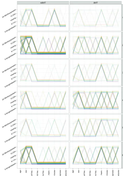
We began our analysis by visualizing the raw data with a parallel-coordinates plot (Fig A1). Each child is represented in this plot by a connected line that joins the recorded values of the variables of interest. Variables are transformed to share a common scale, and their values can be read off the vertical axes. The plot is then divided by injury etiology and by the source study, and lines are jittered and col-ored to alleviate problems with over-plotting. Densely colored areas of the plots represent more children than the light, sparse areas. For example, the predominant pattern of features seen in the AHT group of children in the Vinchon et al1study is the youngest age
group, boys, without skull, rib, or long-bone fractures, with retinal haemor-rhages, no information on bruising and apnea but with seizures.
Inspection of the raw data informed our hotdeck multiple-imputation strat-egy. The Kemp et al2 study had
com-plete information on all children and, thus, required no imputation. Hobbs et al3had only a small amount of missing
data, and (where at least 5 matches could be found) this was imputed from either itself or from the Kemp et al study.2Ettaro et al4had a small amount
of missing fracture data, and neither apnea nor seizures were recorded for these children. The fracture data we imputed from within the same study matched on all available features. This being done, we then found children in the Vinchon et al study1who matched
the Ettaro et al sample4on all features
common to both studies, using this to impute information on seizures. A sim-ilar strategy was used to bring infor-mation on bruising from the Ettaro et al study4into the Vinchon et al1cases.
At this stage, both Ettaro et al4and
Vin-chon et al1 had complete information
except for apnea (always excepting
any instances in which insufficient matching children could be found).
After a within-Bechtel et al5imputation
to fill in the sporadically missing items, data from (the imputed versions of) the Ettaro et al4and Vinchon et al1
stud-ies were used to update the Bechtel et al5 data. At this stage Bechtel et al,5
Ettaro et al,4and Vinchon et al1were all
used to form a sampling frame from which to impute into Hettler and Greenes,6 who chose not to record
many items but, importantly, recorded information on the presence or ab-sence of apnea. At this point Hettler and Greenes,6Hobbs et al,3and Kemp
et al2had (in principle) complete
infor-mation, whereas Bechtel et al,5Ettaro
et al,4 and Vinchon et al1 lacked data
only on apnea. By sampling informa-tion on apnea from similar children in the former set into the latter, we com-pleted the hotdeck stage of imputation.
In practice, there were several in-stances in this process where fewer than 5 matching children could be found. The choice of a minimum of 5 matched individuals is somewhat arbi-trary, but it is reasonably likely that 4 or fewer children may have exactly the same observed information on the missing item(s), especially if the prev-alence of the relevant feature is partic-ularly low or particpartic-ularly high. Thus, imputing from such a sample could er-roneously suggest that the missing item(s) were, in fact, known. To cir-cumvent this problem, the final stage of our imputation was to sample from the margin of the feature concerned conditional on the etiology of the neu-rologic injury.
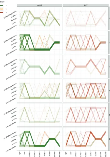
Figure A2 displays the 10 imputed data sets, made slightly transparent so that similarities and differences between imputations may be seen. Note that re-sults of the 2 subdural-only studies (Hobbs et al3and Kemp et al2) do not
show the propensity for skull frac-tures in the nAHT group seen (or
im-puted) in the other 4 studies. This may be because of a number of organic causes of subdural hemorrhage in this group, whereas the nAHT groups in the other studies were trauma-related. An interesting feature of the Hettler and Greenes6 imputations is that rib and
long-bone fractures are never imputed to be present, in contrast with skull fractures and bruising. Another obvi-ous difference between the 2 etiology groups is the number of isolated fea-tures in the nAHT groups as opposed to the high prevalence of multiple re-corded features in the AHT groups.
Following the modelling described in the article, we gave detailed consider-ation to the predictive performance of our chosen model. Figure A3 shows the raw data again but this time colored according to the predicted probability of AHT (based on the final, reduced model). Because there was substantial missing information, these predic-tions were averaged across the 10 im-puted data sets. A green color sug-gests AHT with a probability less than 20%; orange denotes ambiguous cases with predicted probabilities of AHT that are between 20% and 80%; and a red color corresponds to high probabili-ties (⬎80%) of AHT.
We see that our modelling is reason-ably successful at discriminating be-tween the 2 groups of interest. The pre-dominance of green and orange in the left-hand plots and the frequency of red lines in the right-hand plots sug-gest that, on the whole, the model is fitting quite well. However, of (at least) equal importance is the degree to which the model can suggest incorrect etiologies; it is possible to have a rib fracture in the nAHT group; conversely, some children with no obvious injury (particularly evident in the Hobbs et al3
study) can have AHT.
within-FIGURE A1
FIGURE A2
FIGURE A3
case data. To address this concern, we conducted a cross-validation investi-gation to attempt to quantify our ability to predict AHT. We used fivefold cross-validation, whereby the data is
parti-tioned (at random) into 5 subsets. Each of these 5 subsets is used in turn as the test data, and the reduced model is refitted by using the other 4 subsets as the training data.
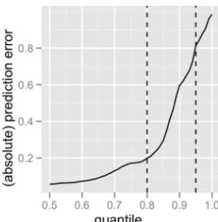
Results were consistent across the 5 cross-validations. Graphing the quan-tiles of absolute prediction error (Fig A4), we found that in the best 80% of cases the predictions are confident (positive predictive values either ⬎80% or ⬍20%) and correctly so. However, in the worst 5% of the cases, the predictions are confident (using the same definition) but incorrect. Im-portant, however, is that most of these confident-but-wrong predictions in-correctly suggest nAHT, which high-lights the need for any assessment of likelihood of abuse to be based on a full multidisciplinary assessment rather than relying on clinical features alone.
1. Vinchon M, Defoort-Dhellemmes S, Desur-mont M, Dhellemmes P. Accidental and non-accidental head injuries in infants: a pro-spective study. J Neurosurg. 2005;102(4 suppl):380 –384
2. Kemp AM, Stoodley N, Cobley C, Coles L, Kemp KW. Apnoea and brain swelling in non-accidental head injury.Arch Dis Child. 2003; 88(6):472– 476
3. Hobbs C, Childs AM, Wynne J, Livingston J, Seal A. Subdural haematoma and effusion in infancy: an epidemiological study.Arch Dis Child. 2005;90(9):952–955
4. Ettaro L, Berger RP, Songer T. Abusive head trauma in young children: characteristics and medical charges in a hospitalized popu-lation. Child Abuse Negl. 2004;28(10): 1099 –1111
5. Bechtel K, Stoessel K, Leventhal JM, et al. Characteristics that distinguish accidental from abusive injury in hospitalized young children with head trauma.Pediatrics. 2004; 114(1):165–168
6. Hettler J, Greenes DS. Can the initial history predict whether a child with a head injury has been abused? Pediatrics. 2003;111(3): 602– 607
FIGURE A4
DOI: 10.1542/peds.2010-2949 originally published online August 15, 2011;
2011;128;e550
Pediatrics
Farewell
Services
Updated Information &
http://pediatrics.aappublications.org/content/128/3/e550 including high resolution figures, can be found at:
References
http://pediatrics.aappublications.org/content/128/3/e550#BIBL This article cites 29 articles, 17 of which you can access for free at:
Subspecialty Collections
ub
http://www.aappublications.org/cgi/collection/child_abuse_neglect_s
Child Abuse and Neglect
following collection(s):
This article, along with others on similar topics, appears in the
Permissions & Licensing
http://www.aappublications.org/site/misc/Permissions.xhtml in its entirety can be found online at:
Information about reproducing this article in parts (figures, tables) or
Reprints
DOI: 10.1542/peds.2010-2949 originally published online August 15, 2011;
2011;128;e550
Pediatrics
Farewell
Sabine Ann Maguire, Alison Mary Kemp, Rebecca Caroline Lumb and Daniel Mark
Estimating the Probability of Abusive Head Trauma: A Pooled Analysis
http://pediatrics.aappublications.org/content/128/3/e550
located on the World Wide Web at:
The online version of this article, along with updated information and services, is
by the American Academy of Pediatrics. All rights reserved. Print ISSN: 1073-0397.