REPORT
De Novo Truncating Mutations in the Last
and Penultimate Exons of
PPM1D
Cause
an Intellectual Disability Syndrome
Sandra Jansen,1 Sinje Geuer,1 Rolph Pfundt,1 Rachel Brough,2 Priyanka Ghongane,2
Johanna C. Herkert,3 Elysa J. Marco,4 Marjolein H. Willemsen,1 Tjitske Kleefstra,1 Mark Hannibal,5 Joseph T. Shieh,6 Sally Ann Lynch,7,8 Frances Flinter,9 David R. FitzPatrick,10 Alice Gardham,11 Birgitta Bernhard,11 Nicola Ragge,12,13 Ruth Newbury-Ecob,14 Raphael Bernier,15 Malin Kvarnung,16 E.A. Helena Magnusson,17 Marja W. Wessels,18 Marjon A. van Slegtenhorst,18 Kristin G. Monaghan,19 Petra de Vries,1 Joris A. Veltman,1,20 Deciphering Developmental Disorders Study, Christopher J. Lord,2 Lisenka E.L.M. Vissers,1 and Bert B.A. de Vries1,*
Intellectual disability (ID) is a highly heterogeneous disorder involving at least 600 genes, yet a genetic diagnosis remains elusive in ~35%–40% of individuals with moderate to severe ID. Recent meta-analyses statistically analyzing de novo mutations in>7,000 indi-viduals with neurodevelopmental disorders highlighted mutations inPPM1Das a possible cause of ID. PPM1D is a type 2C phosphatase that functions as a negative regulator of cellular stress-response pathways by mediating a feedback loop of p38-p53 signaling, thereby contributing to growth inhibition and suppression of stress-induced apoptosis. We identified 14 individuals with mild to severe ID and/or developmental delay and de novo truncatingPPM1Dmutations. Additionally, deep phenotyping revealed overlapping behav-ioral problems (ASD, ADHD, and anxiety disorders), hypotonia, broad-based gait, facial dysmorphisms, and periods of fever and vom-iting.PPM1Dis expressed during fetal brain development and in the adult brain. All mutations were located in the last or penultimate exon, suggesting escape from nonsense-mediated mRNA decay. BothPPM1Dexpression analysis and cDNA sequencing in EBV LCLs of individuals support the presence of a stable truncated transcript, consistent with this hypothesis. Exposure of cells derived from indi-viduals withPPM1Dtruncating mutations to ionizing radiation resulted in normal p53 activation, suggesting that p53 signaling is unaffected. However, a cell-growth disadvantage was observed, suggesting a possible effect on the stress-response pathway. Thus, we show that de novo truncatingPPM1Dmutations in the last and penultimate exons cause syndromic ID, which provides additional insight into the role of cell-cycle checkpoint genes in neurodevelopmental disorders.
Next-generation sequencing (NGS) techniques have accel-erated the discovery of genes associated with intellectual disability (ID).1–7Mutations in more than 600 autosomal and X-linked genes have been implicated,8 but many more are likely to be elucidated. Recently, two separate meta-analyses used the de novo mutations identified in>7,000 individuals affected by a neurodevelopmental disorder to identify mutations that might also cause ID.9,10 In both meta-analyses, PPM1D (protein phos-phatase, Mg2þ/Mn2þ-dependent 1D [MIM: 605100]),
encoding a negative regulator of cellular stress-response pathways, had significantly more damaging de novo muta-tions than expected given the cohort size. However, the clinical characteristics of these individuals were not pro-vided in detail, and insights on the pathophysiological mechanism remained unidentified. Including seven indi-viduals identified in the meta-analyses, we collected a total of 14 unrelated individuals with mild to severe ID and/or developmental delay (DD) through international collabo-ration with colleagues and data-sharing resources such as
1Department of Human Genetics, Donders Centre for Neuroscience, Radboud University Medical Center, PO Box 9101, 6500 HB Nijmegen, the
Netherlands;2Cancer Research UK Gene Function Laboratory and Breast Cancer Now Research Centre, Institute of Cancer Research, London SW3 6JB,
UK;3Department of Genetics, University Medical Center Groningen, University of Groningen, PO Box 30.001, 9700 RB Groningen, the Netherlands; 4
Departments of Neurology, Pediatrics, and Psychiatry, University of California, San Francisco, 675 Nelson Rising Lane, Suite 405, San Francisco, CA 94143, USA;5Division of Pediatric Genetics, Metabolism & Genomic Medicine, University of Michigan Medical School, D5257 Medical Professional
Build-ing, 1500 East Medical Center Drive, Ann Arbor, MI 48109-5718, USA;6Division of Medical Genetics, Department of Pediatrics, UCSF Benioff Children’s
Hospital, Institute for Human Genetics, University of California, San Francisco, San Francisco, CA 94143-0793, USA;7Clinical Genetics, Children’s
Univer-sity Hospital, Temple Street, Dublin 1, Ireland;8Academic Centre on Rare Diseases, School of Medicine and Medical Sciences, University College Dublin,
Dublin 1, Ireland;9Department of Clinical Genetics, Guy’s and St. Thomas’ NHS Foundation Trust, Great Maze Pond, London SE1 9RT, UK;10Medical
Research Council Human Genetics Unit, Institute of Genetics and Molecular Medicine, University of Edinburgh, Western General Hospital, Crewe Road South, Edinburgh EH4 2XU, UK;11North West Thames Regional Genetic Service (Kennedy Galton Centre), North West London Hospitals, Watford
Road, London HA1 3UJ, UK;12Faculty of Health and Life Sciences, Oxford Brookes University, Gipsy Lane, Oxford OX3 0BP, UK;13West Midlands Regional
Clinical Genetics Service and Birmingham Health Partners, Birmingham Women’s Hospital NHS Foundation Trust, Birmingham B15 2TG, UK;14 Depart-ment of Clinical Genetics, University Hospitals Bristol NHS Foundation Trust, St. Michael’s Hospital, Southwell Street, Bristol BS2 8EG, UK;15Center on
Human Development and Disability, University of Washington, PO Box 357920, Seattle, WA 98195-7920, USA;16Department of Clinical Genetics,
Karo-linska University Hospital Solna, KaroKaro-linska Institutet, 171 77 Stockholm, Sweden;17Department of Medicine and Neurology, Habilitation Organization,
Region Ska˚ne, 291 89 Kristianstad, Sweden;18Department of Clinical Genetics, Erasmus Medical Center, PO Box 2040, 3000 CA Rotterdam, the
Netherlands;19GeneDx, Gaithersburg, MD 20877, USA;20Department of Clinical Genetics, Maastricht University Medical Centre, Universiteitssingel
50, 9229 ER Maastricht, the Netherlands *Correspondence:[email protected] http://dx.doi.org/10.1016/j.ajhg.2017.02.005.
650 The American Journal of Human Genetics100, 650–658, April 6, 2017
GeneMatcher.9–12One individual was previously described as part of the Simons Simplex Collection cohort.13Herein, we report on an ID syndrome caused by de novo germline mutations in the last and penultimate exons ofPPM1D, as well as the implication of such mutations in the role of
PPM1Din stress responses.
We collected clinical data by inviting individuals back to the clinic for re-evaluation and deep phenotyping. Detailed clinical information of the 14 individuals (2–21 years old) is described in the Supplemental Note and summarized in Table 1. All but one individual had mild to severe ID (93%), and 11 individuals (79%) had behavioral problems, such as anxiety disorders, attention deficit hyperactivity disorder (ADHD), obsessive behavior, sensory integration problems, and autism spectrum disor-der (ASD). The individual with a normal IQ of 96 did, how-ever, need extra tutoring at school and showed an anxiety disorder and attention problems. Seven individuals were hypersensitive for sounds. Hypotonia was a common feature in the individuals for whom this information was available (10/14 [71%]), and several individuals had a broad-based gait (5/10 [50%]). Brain MRI was performed for nine individuals (64%) without any substantial find-ings, except for moderate cortical and cerebellar atrophy and abnormal vascular structures in individual 13, who was also diagnosed with Potocki-Shaffer syndrome. Eight individuals (62%) had short stature, but weight and head circumference were variable. Feeding difficulties were a common feature (10/14 [71%]), and remarkably, eight in-dividuals (62%) had periods of illness with fever and/or vomiting. In addition, nine individuals had a high pain threshold (90%). One individual had problems emerging from anesthesia. Vision problems, such as myopia, hyper-metropia, and strabismus, were seen in nine individuals (64%). There was no apparent shared or consistent facial gestalt despite the presence of overlapping facial features, including a broad forehead, low-set posteriorly rotated ears, upturned nose, and broad mouth with thin upper lip (Figure 1A). Ten individuals (91%) had small hands often with brachydactyly, seven individuals had small feet, and six individuals had hypoplastic toenails. To further delineate the clinical spectrum associated with de novo mutations in PPM1D, we established a website to collect detailed clinical information of additional indi-viduals to be identified over the coming year (see Web Resources).
Whole-exome sequencing was performed in all individ-uals as previously described,9,10and all were identified to have a deleterious PPM1D mutation. This study was approved by the institutional review board Commissie Mensgebonden Onderzoek Regio Arnhem-Nijmegen NL36191.091.11 and received UK research ethics commit-tee (REC) approval (10/H0305/83 granted by the Cam-bridge South REC and GEN/284/12 granted by the Republic of Ireland REC). Written informed consent was obtained from all individuals. Subsequent confirmation by Sanger sequencing and investigation of parental DNA
samples of 13 individuals indicated thatPPM1Dmutations had occurred de novo. Interestingly, all 14 ID-associated
PPM1Dmutations are located in the last and penultimate exons (Figure 1B) and are predicted to result in a premature stop codon in exon 6 or in the last 55 nucleotides of exon 5 (Figure 1C). The truncated mRNA is therefore presumed to escape nonsense-mediated decay (NMD) and result in a truncated PPM1D still containing its functional protein phosphatase Mg2þ/Mn2þ-dependent (PPM)-type phospha-tase domain but lacking its nuclear localization signal (NLS). To analyze PPM1D mRNA expression on cDNA derived from lymphoblastoid cell lines (LCLs) of individ-uals with a mutation inPPM1D, we obtained LCLs from human blood by immortalization via Epstein-Barr virus (EBV) transformation according to standard procedures.
PPM1D mRNA expression analysis using two different sets of primer pairs showed no significant difference in mRNA levels between control lines and cDNA derived from LCLs of individuals 2 and 3, indeed confirming that the truncated mRNA was not subjected to NMD (Figure 2B). Subsequent Sanger sequencing, performed in four individ-uals (1–3 and 7) with a primer set targeting the mutated area (PPM1D_1 and PPM1D_3), confirmed the presence of truncated PPM1D transcripts. We showed that the de novo mutations inPPM1Dlead to stable transcription of truncated mRNA with normal expression levels. Because the truncated protein lacks its NLS, it might no longer reach the nucleus to exert its function. Immunohisto-chemical staining of PPM1D showed cytosolic and nuclear staining in LCLs in a control line (Figure S1). Similar stain-ing in EBV-transformed LCLs from an individual with a heterozygous de novo mutation in PPM1D showed a similar localization (Figure S1). However, the latter can be explained by the presence of the wild-type allele, which still results in a fully functional PPM1D. Notably, given the specific need of PPM1D in cellular stress, it is possible that localization of the mutant PPM1D is mostly affected during this state. Further quantification of PPM1D in the different cell compartments and under different phys-iological scenarios could help to improve our understand-ing of the biological mechanism underlyunderstand-ing PPM1D
pathology.
Importantly, de novo mutations in PPM1D have not been observed in over 2,000 control trios.1,15–18 Interroga-tion of large databases, such as the Exome AggregaInterroga-tion Consortium (ExAC) Browser, shows thatPPM1Dis under constraint for missense mutations (Zscore 3.13). Interest-ingly, however, PPM1D seems to be tolerant of loss-of-function mutations (with pLI ¼ 0.00).19Together, these scores could indicate that the pathophysiological mecha-nism underlying ID-associatedPPM1Dmutations is more complex and that a mechanism involving a C-terminally truncated protein is more disruptive than complete loss of it, similar to what has been identified for DVL1
and DVL3 frameshift mutations causing Robinow syn-drome.20–22 This highlights the importance of not disre-garding these genes without consideration, given that
Table 1. Main Clinical Features of Affected Individuals
Individual
Total
1 2 3 4 5 6 7 8 9 10 11 12 13 14
General Information
Age 14 y 5 y 5 y 2 y 5 y 10 y 9 y, 9 m 21 y 16 y 6 y 18 y 15 y 7 y 7 y 2–21 y
Gender F M F M M M M M F F M F F F 7 M, 6 F
Mutation c.1221T>A (p.Cys407*) c.1216del (p.Thr406 Profs*3) c.1260þ 1dup (p.Ser421 Thrfs*12)
c.1210C>T (p.Gln404*)
c.1269_ 1270dup (p.Glu424 Glyfs*8)
c.1339G>T (p.Glu447*) c.1188_ 1191del (p.Asp397 Alafs*11) c.1250dup (p.Pro418 Thrfs*16) c.1270dup (p.Glu424 Glyfs*10) c.1270dup (p.Glu424 Glyfs*10)
c.1281G>A (p.Trp427*)
c.1281G>A (p.Trp427*)
c.1654C>T (p.Arg552*) c.1404_ 1411del (p.Lys469 Argfs*4) NA
Inheritance de novo de novo de novo de novo de novo de novo de novo de novo de novo de novo de novo de novo NKa
de novo NA
Growth
Birthweight (g) 2,780 4,000 2,655 3,742 3,527 3,180 2,200 2,211 3,061 2,730 3,200 3,429 NK 3,140 NA
Height (SD) 2.7 1.5 2.8 0 NK 3 <3 <2.5 2.3 2.5 2.7 þ0.41 2.6 2 NA
Weight (SD) þ2.3 þ0.5 1.9 0 NK þ2 þ0.5 <2.5 2.2 2 1.8 >þ2.5 4.9 0 NA
Head circumference (SD)
þ0.7 0.5 1.5 0.5 0.5 1 1.8 <2.5 3.1 2.5 2.5 þ3.49 NK 1.5 NA
Neurological
ID (severity) þ(mild to moderate)
þ(mild to moderate)
þ(mild) þ þ b þ
(mild to moderate)
þ(mild to moderate)
þ(severe) þ(moderate)þ(moderate)þ(severe) þ(severe) þ(mild) 13/14 (93%)
Hypotonia þ þ þ þ þ þ þ þ þ þ 10/14 (71%)
Broad-based gaitþ NK þ þ þ þ NK NK NK 5/10 (50%)
Sensitivity to sounds
NK NK þ þ NK NK þ þ þ þ NK NK NK þ 7/7 (100%)
Behavioral features ADHD, ODD, anxiety disorder sensory integration problems short attention, panic attacks sensory integration problems, hyperarousal, short attention anxiety disorder, attention difficulties, biting sensory integration problems, anxiety, short attention
anxiety ASD ASD,
attention problems, oppositional, aggression
ASD ASD 11/14 (79%)
Facial
Broad forehead þ þ NK þ þ þ þ NK þ þ 8/12 (67%)
Low-set, posteriorly rotated ears
þ þ þ þ(right) NK NK þ þ þ NK NK þ(only
posteriorly rotated)
8/10 (80%)
Upturned nose
þ þ NK þ þ NK þ 5/12 (42%)
Thin upper lip þ þ þ NK þ þ þ þ þ NK þ þ 10/12 83%
Broad mouth þ þ þ NK þ þ þ NK 6/12 50%
(Continued on next page)
Table 1. Continued
Individual
Total
1 2 3 4 5 6 7 8 9 10 11 12 13 14
Gastrointestinal
Feeding difficulty
þ(neonatal)þ(neonatal) þ(neonatal) þ þ þ þ þ þ þ(neonatal) 10/14 (71%)
GER and/or vomiting
þ þ þ þ NK þ þ þ(infancy) þ þ þ 10/13 (77%)
Constipation þ þ þ þ þ(infancy) þ NK þ þ 8/13 (55%)
Skeletal
Small hands NK þ þ NK þ þ þ þ þ þ NK þ þ 10/11 (91%)
Small feet NK NK þ NK NK þ þ þ NK þ NK þ þ 7/8 (88%)
Hyperlordosis þ þ NK þ þ þ NK þ NK NK þ 7/10 (70%)
Other
Periodic
illnessc þ þ þ þ þ þ þ þ NK 8/13 (62%)
High pain threshold
NK þ þ þ NK þ þ þ þ þ þ NK NK 9/10 (90%)
Congenital abnormalities
bicuspid aortic valve
retractile testes, small genital
small VSD and small ODA
bilateral
cryptor-chidism
laryngo-malacia
bilateral
parietal foramina, exostoses, diaphragmatic hernia, volvulus intestined
6/14 (43%)
Vision problems
myopia, nystagmus, amblyopia
hyper-metropia, cilinder, strabismus
hyper-metropia
myopia, strabismus, astigmatism, CVI
hyper-metropia, strabismus, astigmatism, nystagmus
myopia, strabismus
hyper-metropia, strabismus
hyper-metropia, strabismus
strabismus,
nystagmus, iridocyclitis, retinal detachment
9/14 (64%)
Hypoplastic nails
þ(toenails) þ(toenails) þ(toenails) þ(toenails) þ(fifth toenails)
þ NK NK 6/12 (50%)
Recurrent infections
NK NK NK þ NK þ NK þ þ þ 5/9 (56%)
Abbreviations are as follows:þ, present;, absent; y, years; m, months; M, male; F, female; NK, not known; ID, intellectual disability; ADHD, attention deficit hyperactivity disorder; ODD, oppositional defiant disorder; ASD, autism spectrum disorder; GER, gastro esophageal reflux; VSD, ventricle septum defect; ODA, open ductus arteriosus; and CVI, cerebral visual impairment.
aParental DNA not available.
bIndividual did have learning difficulties.
cIncluding cyclic vomiting.
dIndividual 13 also had a confirmed diagnosis of Potocki-Shaffer syndrome.
The
American
Journal
of
Human
Genetics
100
,
650–658,
April
6,
2017
the disease-causing mutations could have other patho-physiological mechanisms not directly inferable from such metrics in the ExAC Browser.
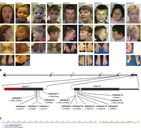
PPM1Dhas previously been shown to be expressed in both mouse and human brain,23,24 but a second hint toward a role forPPM1D in the occurrence of ID would Figure 1. Photographs of Nine Individuals with a Truncating Mutation inPPM1D, De Novo Mutations inPPM1D, and Predicted Consequences at the Protein Level
(A) Shared facial features including a broad forehead, upturned nose, broad mouth with thin upper lip, and low-set posteriorly rotated ears. Extremities show small hands and feet with brachydactyly and hypoplastic toenails. Individual 13 also had a confirmed diagnosis of Potocki-Shaffer syndrome. Parents provided informed consent for the publication of these photographs.
(B) Schematic representation of the coding sequence ofPPM1D(GenBank: NM_003620.3), including zoomed-in exons 5 and 6. All de novoPPM1Dmutations identified are depicted according to their location in the coding sequence. Protein domain structures encoded by exons 5 and 6 are highlighted in color: red for the PPM-type phosphatase domain (in exon 5) and green for the nuclear localization signal (in exon 6).
(C) Predicted protein sequences in individuals 1–14. The last part of the translated sequence of exon 5 is in blue, and the amino acids encoded by the first part of exon 6 are in orange. For individuals 1–14, the predicted mutant amino acids are depicted in red. Abbrevi-ations are as follows: WT, wild-type; and aa, amino acid.
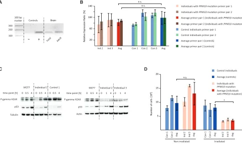
be its expression in the developing brain. We therefore investigatedPPM1Dexpression in cDNA libraries obtained from fetal and adult brain by using semiquantitative PCR. Human cDNA libraries from different human tissues were purchased from Stratagene. Expression ofPPM1D in em-bryonic and adult brain and EBV-LCLs (positive control) was investigated with PPM1D-transcript-specific semi-quantitative PCR using the primer set PPM1D_3. We
de-tected PPM1D expression in both fetal and adult brain (Figure 2A). In addition, our analysis showed wider fetal (developmental), wherebyPPM1Dexpression was detected in fetal liver and skeletal muscle, but not in their adult counterparts (data not shown). The expression ofPPM1D
in the fetal brain suggests a role during fetal brain develop-ment and thus potentially in developing normal cogni-tion. Although we were not able to further narrow down
A
C D
B
Figure 2. Functional Effects ofPPM1DMutations at the RNA and Protein Levels and Downstream Effects on p53 Activation and Cell Growth
(A) Semiquantitative PCR using primers PPM1D_3 (forward: 50-AACCTGACTGACAGCCCTTC-30; reverse: 50-ACCAGGGCAGGTA TATGGTC-30) on tissue-specific cDNA libraries showsPPM1Dexpression in fetal and adult brain. EBV-LCL cDNA is the positive control (þ), and ddH2O is the negative control ().
(B) Expression levels ofPPM1Dwere quantified by qPCR using cDNA obtained from EBV-LCLs derived from individuals with a mutation inPPM1D(individuals 2 and 3). Experiments were performed in triplicate with two sets of primer pairs: PPM1D_1 (forward: 50-TGC TTGTGAATCGAGCATTG-30; reverse: 50-CCCTGATTGTCCACTTCTGG-30) and PPM1D_2 (forward: 50-AAGTCGAAGTAGTGGTGCT CAG-30; reverse: 50-TCTTCTGGCCCCTAAGTCTG-30). Analysis was performed with SDS software according to standard procedures, and GUSB expression was used as a calibrator (forward: 50-AGAGTGGTGCTGAGGATTGG-30; reverse: 50-CCCTCATGCTCTAGC GTGTC-30).14There was no significant change inPPM1DmRNA expression between affected individuals and control lines.
Abbrevia-tions are as follows: n.s., not significant; Ind, individual; Con, control individual; and Avg, average. Results are presented as the average5SD.
(C) Radiation-induced activation of p53 was investigated in fibroblasts derived from individual 1, EBV-LCLs derived from individuals 2 and 3, healthy control cells, and the cancer cell line with active PPM1D (MCF7 as the positive control). Cells were exposed to gamma irradiation (5 Gy) from an X-ray source. Whole-cell lysates were generated from cells 30–60 min and 4 hr after irradiation and subjected to protein electrophoresis. Immunoblotting of electrophoresed lysates was performed with antibodies specific to p53 (9282S), phospho-HistonegH2AX (ser139) (9718S), and actin (I-19), orb-tubulin (D-10). Anti-rabbit, anti-mouse, and anti-rabbit secondary antibodies were incubated with the blot (1:5,000) for 1 hr at room temperature, and then exposure using enhanced chemiluminescent detection followed. Western blot analysis showed no difference between case and control cell lines but did show increased p53 activation in the PPM1Dmutant breast cancer cell line MCF7.
(D) Growth behavior of EBV-LCLs derived from individuals with a mutation inPPM1D(individuals 2 and 3) was compared with that of age- and sex-matched control EBV-LCLs. Cells were irradiated (UV light: 60 J/m2) and cultured in a concentration of 33105cells/mL.
Cell numbers were counted in triplicate after 48 hr, and the experiment was repeated three times. 48 hr after irradiation, the number of EBV-LCLs derived from individuals with a mutation inPPM1D(n¼2) was significantly lower than that of control cells (n¼2), whereas the growth of untreated cells was unaffected. Abbreviations are as follows: *, p<0.05; n.s., no significance; Ind, individual; Con, control individual; and Avg, average. Results are presented as the average5SD.
the expression to detailed brain regions, the highestPpm1d
expression in mice has been reported in the cerebellum,23 which is the center for coordination. Several of our affected individuals had a broad-based gait, a possible sign of cere-bellar disturbance, which might therefore be associated with the mutation in PPM1D. However, the individuals did not display other symptoms of coordination defects, and in the individuals who had received brain MRI, no structural abnormalities of the cerebellum could be identified.
PPM1D has also been reported to be an important regu-lator of global heterochromatin silencing and thus critical in maintaining genome integrity.25The latter was exam-ined in germ cells ofPpm1d-deficient mice, which showed enlarged heterochromatin centers with enriched immuno-fluorescent staining for H3K9me3 and HP1g, both markers for transcriptional repression.25 When PPM1D dysfunc-tion indeed alters gene expression, this might have an effect on fetal (brain) development. Moreover,Ppm1d -defi-cient mice show an increase in anxiety and depression-like behavior,26suggesting a potential protective function of PPM1D in mood stabilization.26Interestingly, four of the individuals with aPPM1Dmutation showed anxiety.
PPM1D(also known asWip1) is, like other genes encod-ing PPM and PP2C phosphatases, a regulator of stress response.27In particular, PPM1D regulates the DNA dam-age response (DDR) pathway by inhibiting p53 and other tumor suppressors (p38, ATM, Chk1, and Chk2) through dephosphorylation of these proteins.27Previously substan-tial amounts of work have gone into the role ofPPM1Din tumorigenesis, given that acquiredPPM1Dmutations have been identified in individuals with breast, ovarian, colon, and lung cancer and are postulated to exert their effect through gain of function.27–31 However, these mosaic mutations in lymphocytes were shown to occur only in individuals who had undergone chemotherapy and were shown to be absent in DNA isolated from the germline prior to chemotherapy.27–31 The gain-of-function effect was shown by overexpression of cancer-associated
PPM1D mutations in tumor cells, which suppressed ionizing radiation (IR)-induced molecular responses.28In normal conditions, exposure to IR causes upregulation of p53 levels and phosphorylation of the histone H2AX (gH2AX), events that are normally prevented by PPM1D activity. In tumor cells with overexpression of cancer-asso-ciated PPM1D mutations, p53 and gH2AX upregulation after IR exposure is impaired, suggesting that in tumor cells, truncatingPPM1Dmutations are hyperactive.28We tested the upregulation of p53 andgH2AX in MCF7 cells, a breast cancer cell line serving as a positive control, which showed the expected upregulation of p53 andgH2AX, sug-gesting that the IR exposure was successful (Figure 2C). Compared with healthy control cells, fibroblasts from indi-vidual 1 and EBV-LCLs derived from indiindi-viduals 2 and 3 showed normal p53 andgH2AX responses (Figure 2C). In conclusion, these data indicate that ID-associatedPPM1D
mutations do not cause p53 depletion, which suggests a
pathophysiological mechanism different from the ac-quiredPPM1Dcancer-associated mutations.
As a regulator of the DDR, PPM1D plays an important role in cell-cycle control by positively upregulating G1-to-S phase progression.32 We hypothesized that the ID-associated mutations in PPM1D would lose this positive upregulation and thereby cause cells to stall in the G1-to-S phase, leading to reduced cell proliferation. We therefore next tested whether EBV-LCLs derived from indi-viduals 2 and 3 showed growth abnormalities in compari-son with age- and sex-matched control EBV-LCLs. For this, cells were exposed to IR and analyzed for growth character-istics (Figure 2D). Indeed, cells derived from individuals showed 50% less growth than control lines, whereas the growth of untreated cells was unaffected (Figure 2D), showing that heterozygous PPM1D truncation leads to growth disadvantage after radiation. Hence, if the effect of the truncating mutations in our cases is a gain of func-tion, this does not seem to affect the role of PPM1D on p53. However, a cell-growth disadvantage was observed after IR, suggesting that another function related to PPM1D cell-cycle checkpoints might be compromised.
Several genes are known to have somatic mutations that lead to cancer but germline mutations that cause an ID phenotype. Examples include genes encoding compo-nents of the RAS-MAPK pathway, SETBP1 (SET binding protein 1 [MIM: 611060]), and CTNNB1 (catenin beta 1 [MIM: 116806]).33–36Also, some of these germline muta-tions give rise to a higher cancer risk, whereas others do not.33–36Germline PPM1Dmutations in individuals with cancer have to our knowledge not yet been reported. Although it is known that germline mutations in some genes, for instance NF1 (neurofibromin 1 [MIM: 613113]), cause ID and a higher risk of (benign) tumors,37none of the individuals studied here (2–21 years old) have developed cancer. Thus, we cannot exclude nor confirm the possibility that thePPM1Dmutations in the individuals with ID predispose to cancer.
In conclusion, de novo truncating germline mutations in the last and penultimate exons of PPM1D lead to an ID syndrome with behavioral problems, hypotonia, broad-based gait, periods of fever and vomiting, high pain threshold, short stature, small hands and feet, and overlapping facial dysmorphisms. Exposure of affected cells to IR resulted in normal p53 activation, suggesting that p53 signaling is not affected by the truncated protein. Nonetheless, a cell-growth disadvantage after IR was observed. The significant enrichment of de novo muta-tions in individuals with ID, the expression in the devel-oping and mature brain, and this clinical ID syndrome underscore the role ofPPM1Din neurodevelopment.
Supplemental Data
Supplemental Data include a Supplemental Note and one figure and can be found with this article online athttp://dx.doi.org/10. 1016/j.ajhg.2017.02.005.
Conflicts of Interest
K.G.M. is an employee of GeneDx, Inc.
Acknowledgments
We thank the individuals and their parents for participating in this study. We thank Caroline Wright for her help in contacting refer-ring clinicians from the Decipherefer-ring Developmental Disorders (DDD) study, Megan Cho for her help in contacting referring clini-cians from GeneDx, Jessica Radley for clinical support, and Ms. Saskia van der Velde-Visser for culturing cells. This work was financially supported by grants from the Netherlands Organisa-tion for Health Research and Development (917-86-319 to B.B.A.d.V., 912-12-109 to B.B.A.d.V. and J.A.V., 907-00-365 to T.K., and 918-15-667 to J.A.V.) and the European Research Council (starting grant DENOVO 281964 to J.A.V.). D.R.F. is funded by a Medical Research Council University Unit grant to the University of Edinburgh. The DDD study presents independent research commissioned by the Health Innovation Challenge Fund (grant HICF-1009-003), a parallel funding partnership among the Well-come Trust, Department of Health, and WellWell-come Trust Sanger Institute (grant WT098051). The views expressed in this publica-tion are those of the authors and not necessarily those of the Well-come Trust or the Department of Health. The research team acknowledges the support of the National Institute for Health Research through the Comprehensive Clinical Research Network.
Received: October 10, 2016 Accepted: February 3, 2017 Published: March 23, 2017
Web Resources
Allen Brain Atlas,http://human.brain-map.org Allen Mouse Brain Atlas,http://mouse.brain-map.org DECIPHER,https://decipher.sanger.ac.uk/
ExAC Browser,http://exac.broadinstitute.org/ GeneMatcher,https://genematcher.org/ OMIM,http://www.omim.org/
OurPPM1Dwebsite,http://www.ppm1dgene.com RefSeq,https://www.ncbi.nlm.nih.gov/refseq/
References
1. Rauch, A., Wieczorek, D., Graf, E., Wieland, T., Endele, S., Schwarzmayr, T., Albrecht, B., Bartholdi, D., Beygo, J., Di Donato, N., et al. (2012). Range of genetic mutations associ-ated with severe non-syndromic sporadic intellectual disability: an exome sequencing study. Lancet 380, 1674– 1682.
2. Vissers, L.E., de Ligt, J., Gilissen, C., Janssen, I., Steehouwer, M., de Vries, P., van Lier, B., Arts, P., Wieskamp, N., del Rosario, M., et al. (2010). A de novo paradigm for mental retardation. Nat. Genet.42, 1109–1112.
3. de Ligt, J., Willemsen, M.H., van Bon, B.W., Kleefstra, T., Yn-tema, H.G., Kroes, T., Vulto-van Silfhout, A.T., Koolen, D.A., de Vries, P., Gilissen, C., et al. (2012). Diagnostic exome sequencing in persons with severe intellectual disability. N. Engl. J. Med.367, 1921–1929.
4. Gilissen, C., Hehir-Kwa, J.Y., Thung, D.T., van de Vorst, M., van Bon, B.W., Willemsen, M.H., Kwint, M., Janssen, I.M.,
Hoischen, A., Schenck, A., et al. (2014). Genome sequencing identifies major causes of severe intellectual disability. Nature
511, 344–347.
5. Grozeva, D., Carss, K., Spasic-Boskovic, O., Tejada, M.I., Gecz, J., Shaw, M., Corbett, M., Haan, E., Thompson, E., Friend, K., et al.; Italian X-linked Mental Retardation Project; UK10K Consortium; and GOLD Consortium (2015). Targeted Next-Generation Sequencing Analysis of 1,000 Individuals with Intellectual Disability. Hum. Mutat.36, 1197–1204.
6. Deciphering Developmental Disorders Study (2015). Large-scale discovery of novel genetic causes of developmental disor-ders. Nature519, 223–228.
7. Hamdan, F.F., Srour, M., Capo-Chichi, J.M., Daoud, H., Nassif, C., Patry, L., Massicotte, C., Ambalavanan, A., Spiegelman, D., Diallo, O., et al. (2014). De novo mutations in moderate or severe intellectual disability. PLoS Genet.10, e1004772. 8. Vissers, L.E., Gilissen, C., and Veltman, J.A. (2016). Genetic
studies in intellectual disability and related disorders. Nat. Rev. Genet.17, 9–18.
9. Lelieveld, S.H., Reijnders, M.R., Pfundt, R., Yntema, H.G., Kamsteeg, E.J., de Vries, P., de Vries, B.B., Willemsen, M.H., Kleefstra, T., Lo¨hner, K., et al. (2016). Meta-analysis of 2,104 trios provides support for 10 new genes for intellectual disability. Nat. Neurosci.19, 1194–1196.
10. McRae, J.F., Clayton, S., Fitzgerald, T.W., Kaplanis, J., Prig-more, E., Rajan, D., Sifrim, A., Aitken, S., Akawi, N., Alvi, M., et al. (2016). Prevalence, phenotype and architecture of devel-opmental disorders caused by de novo mutation. bioRxiv. https://doi.org/10.1101/049056.
11. Sobreira, N., Schiettecatte, F., Boehm, C., Valle, D., and Ha-mosh, A. (2015). New tools for Mendelian disease gene identi-fication: PhenoDB variant analysis module; and GeneMatcher, a web-based tool for linking investigators with an interest in the same gene. Hum. Mutat.36, 425–431.
12. Sobreira, N., Schiettecatte, F., Valle, D., and Hamosh, A. (2015). GeneMatcher: a matching tool for connecting investi-gators with an interest in the same gene. Hum. Mutat.36, 928–930.
13. Sanders, S.J., Murtha, M.T., Gupta, A.R., Murdoch, J.D., Raube-son, M.J., Willsey, A.J., Ercan-Sencicek, A.G., DiLullo, N.M., Parikshak, N.N., Stein, J.L., et al. (2012). De novo mutations revealed by whole-exome sequencing are strongly associated with autism. Nature485, 237–241.
14. Mukhopadhyay, A., Nikopoulos, K., Maugeri, A., de Brouwer, A.P., van Nouhuys, C.E., Boon, C.J., Perveen, R., Zegers, H.A., Wittebol-Post, D., van den Biesen, P.R., et al. (2006). Erosive vitreoretinopathy and wagner disease are caused by intronic mutations in CSPG2/Versican that result in an imbal-ance of splice variants. Invest. Ophthalmol. Vis. Sci.47, 3565– 3572.
15. Iossifov, I., O’Roak, B.J., Sanders, S.J., Ronemus, M., Krumm, N., Levy, D., Stessman, H.A., Witherspoon, K.T., Vives, L., Pat-terson, K.E., et al. (2014). The contribution of de novo coding mutations to autism spectrum disorder. Nature515, 216–221. 16. Genome of the Netherlands Consortium (2014). Whole-genome sequence variation, population structure and demo-graphic history of the Dutch population. Nat. Genet. 46, 818–825.
17. Gulsuner, S., Walsh, T., Watts, A.C., Lee, M.K., Thornton, A.M., Casadei, S., Rippey, C., Shahin, H., Nimgaonkar, V.L., Go, R.C., et al.; Consortium on the Genetics of Schizophrenia (COGS); and PAARTNERS Study Group (2013). Spatial and
temporal mapping of de novo mutations in schizophrenia to a fetal prefrontal cortical network. Cell154, 518–529.
18. Xu, B., Ionita-Laza, I., Roos, J.L., Boone, B., Woodrick, S., Sun, Y., Levy, S., Gogos, J.A., and Karayiorgou, M. (2012). De novo gene mutations highlight patterns of genetic and neural complexity in schizophrenia. Nat. Genet.44, 1365–1369. 19. Lek, M., Karczewski, K.J., Minikel, E.V., Samocha, K.E., Banks,
E., Fennell, T., O’Donnell-Luria, A.H., Ware, J.S., Hill, A.J., Cummings, B.B., et al.; Exome Aggregation Consortium (2016). Analysis of protein-coding genetic variation in 60,706 humans. Nature536, 285–291.
20. White, J., Mazzeu, J.F., Hoischen, A., Jhangiani, S.N., Gambin, T., Alcino, M.C., Penney, S., Saraiva, J.M., Hove, H., Skovby, F., et al.; Baylor-Hopkins Center for Mendelian Genomics (2015). DVL1 frameshift mutations clustering in the penultimate exon cause autosomal-dominant Robinow syndrome. Am. J. Hum. Genet.96, 612–622.
21. White, J.J., Mazzeu, J.F., Hoischen, A., Bayram, Y., Withers, M., Gezdirici, A., Kimonis, V., Steehouwer, M., Jhangiani, S.N., Muzny, D.M., et al.; Baylor-Hopkins Center for Mendelian Genomics (2016). DVL3 Alleles Resulting in a -1 Frameshift of the Last Exon Mediate Autosomal-Dominant Robinow Syn-drome. Am. J. Hum. Genet.98, 553–561.
22. Bunn, K.J., Daniel, P., Ro¨sken, H.S., O’Neill, A.C., Cameron-Christie, S.R., Morgan, T., Brunner, H.G., Lai, A., Kunst, H.P., Markie, D.M., and Robertson, S.P. (2015). Mutations in DVL1 cause an osteosclerotic form of Robinow syndrome. Am. J. Hum. Genet.96, 623–630.
23. Lein, E.S., Hawrylycz, M.J., Ao, N., Ayres, M., Bensinger, A., Bernard, A., Boe, A.F., Boguski, M.S., Brockway, K.S., Byrnes, E.J., et al. (2007). Genome-wide atlas of gene expression in the adult mouse brain. Nature445, 168–176.
24. Hawrylycz, M.J., Lein, E.S., Guillozet-Bongaarts, A.L., Shen, E.H., Ng, L., Miller, J.A., van de Lagemaat, L.N., Smith, K.A., Ebbert, A., Riley, Z.L., et al. (2012). An anatomically compre-hensive atlas of the adult human brain transcriptome. Nature
489, 391–399.
25. Filipponi, D., Muller, J., Emelyanov, A., and Bulavin, D.V. (2013). Wip1 controls global heterochromatin silencing via ATM/BRCA1-dependent DNA methylation. Cancer Cell24, 528–541.
26. Ruan, C.S., Zhou, F.H., He, Z.Y., Wang, S.F., Yang, C.R., Shen, Y.J., Guo, Y., Zhao, H.B., Chen, L., Liu, D., et al. (2015). Mice deficient for wild-type p53-induced phosphatase 1 display elevated anxiety- and depression-like behaviors. Neuroscience
293, 12–22.
27. Lu, X., Nguyen, T.A., Moon, S.H., Darlington, Y., Sommer, M., and Donehower, L.A. (2008). The type 2C phosphatase Wip1:
an oncogenic regulator of tumor suppressor and DNA damage response pathways. Cancer Metastasis Rev.27, 123–135. 28. Ruark, E., Snape, K., Humburg, P., Loveday, C., Bajrami, I.,
Brough, R., Rodrigues, D.N., Renwick, A., Seal, S., Ramsay, E., et al.; Breast and Ovarian Cancer Susceptibility Collaboration; and Wellcome Trust Case Control Consortium (2013). Mosaic PPM1D mutations are associated with predisposition to breast and ovarian cancer. Nature493, 406–410.
29. Pharoah, P.D., Song, H., Dicks, E., Intermaggio, M.P., Harring-ton, P., Baynes, C., Alsop, K., Bogdanova, N., Cicek, M.S., Cun-ningham, J.M., et al.; Australian Ovarian Cancer Study Group; and Ovarian Cancer Association Consortium (2016). PPM1D Mosaic Truncating Variants in Ovarian Cancer Cases May Be Treatment-Related Somatic Mutations. J. Natl. Cancer Inst. 108.http://dx.doi.org/10.1093/jnci/djv347.
30. Swisher, E.M., Harrell, M.I., Norquist, B.M., Walsh, T., Brady, M., Lee, M., Hershberg, R., Kalli, K.R., Lankes, H., Konnick, E.Q., et al. (2016). Somatic Mosaic Mutations in PPM1D and TP53 in the Blood of Women With Ovarian Carcinoma. JAMA Oncol.2, 370–372.
31. Zajkowicz, A., Butkiewicz, D., Drosik, A., Giglok, M., Suwinski, R., and Rusin, M. (2015). Truncating mutations of PPM1D are found in blood DNA samples of lung cancer patients. Br. J. Cancer112, 1114–1120.
32. Kleiblova, P., Shaltiel, I.A., Benada, J.,Sevcı´k, J., Pecha´ckova´, S., Pohlreich, P., Voest, E.E., Dundr, P., Bartek, J., Kleibl, Z., et al. (2013). Gain-of-function mutations of PPM1D/Wip1 impair the p53-dependent G1 checkpoint. J. Cell Biol.201, 511–521.
33. Niemeyer, C.M. (2014). RAS diseases in children. Haematolog-ica99, 1653–1662.
34. Hoischen, A., van Bon, B.W., Gilissen, C., Arts, P., van Lier, B., Steehouwer, M., de Vries, P., de Reuver, R., Wieskamp, N., Mortier, G., et al. (2010). De novo mutations of SETBP1 cause Schinzel-Giedion syndrome. Nat. Genet.42, 483–485. 35. Makishima, H., Yoshida, K., Nguyen, N., Przychodzen, B.,
San-ada, M., Okuno, Y., Ng, K.P., Gudmundsson, K.O., Vishwa-karma, B.A., Jerez, A., et al. (2013). Somatic SETBP1 mutations in myeloid malignancies. Nat. Genet.45, 942–946.
36. Kuechler, A., Willemsen, M.H., Albrecht, B., Bacino, C.A., Bar-tholomew, D.W., van Bokhoven, H., van den Boogaard, M.J., Bramswig, N., Bu¨ttner, C., Cremer, K., et al. (2015). De novo mutations in beta-catenin (CTNNB1) appear to be a frequent cause of intellectual disability: expanding the mutational and clinical spectrum. Hum. Genet.134, 97–109.
37. Ferner, R.E., and Gutmann, D.H. (2013). Neurofibromatosis type 1 (NF1): diagnosis and management. Handb. Clin. Neurol.115, 939–955.