International Journal of Pharmaceutical Research & Allied Sciences, 2016, 5(2):135-143
Research Article
CODEN(USA) : IJPRPM
ISSN : 2277-3657
Reduction of High Glucose-Induced Nitric Oxide Synthase Expression in
Human Vascular Endothelial Cells by Ascorbic Acid and
α
-Tocopherol
Mohammad Ali Ghaffari
1, Mohammad Aberumand
2, Hoshang Roshanmehr
2and Seyed Ahmad Hosseini
*3
1Cellular and Molecular Research Center, Ahvaz Jundishapur, University of Medical Sciences, Ahvaz, Iran
2Department of Biochemistry, School of Medical, Ahvaz Jundishapur University of Medical Sciences, Ahvaz, Iran, 3Nutrition and Metabolic Diseases Research Center, Ahvaz Jundishapur, University of Medical Sciences, Ahvaz,
Iran
*Email: [email protected]
_____________________________________________________________________________________________
ABSTRACT
Hyperglycemia is generally regarded as one of the major causes of vascular complications in diabetic patients. It has been shown that high-glucose concentration increases nitric oxide synthase (NOS) expression and NO generation in cultured human aortic endothelial cells. The epidemiological studies have demonstrated that there is an inverse relation between vitamin use and cardiovascular disease in diabetes patients. This study tested whether α-tocopherol and ascorbic acid can alter high glucose-induced expression of NOS in vascular endothelial cells. Human umbilical vascular endothelial cells (HUVECs) were treated with α-tocopherol and ascorbic acid, and stimulated by high glucose. Expression of endothelial nitric oxide synthase (eNOS) and inducible nitric oxide synthase (iNOS) was measured by real-time PCR (RT-PCR). Ascorbic acid (100 µM) and α-tocopherol (200 µM) significantly inhibited glucose (25 mM)-induced expression of eNOS mRNA and iNOS mRNA. Ascorbic acid and α -tocopherol are also able to reduce concentration of NOS in HUVECs. Additionally, there is a significant positive relation between expression of eNOS and iNOS mRNA with concentration of NOS. These results suggest that ascorbic acid and α-tocopherol can inhibit high glucose-induced production of NOS proteins by downregulation of its gene expression. Therefore, important role of ascorbic acid and α-tocopherol in the treatment of vascular dysfunction associated with diabetes disease may be via this molecular mechanism. Of course, further studies on regulation of these vitamins to signaling pathways are necessary.
Key words: Glucose, HUVECs, nitric oxide synthase, α-tocopherol, ascorbic acid
_____________________________________________________________________________________________
INTRODUCTION
It has been reported that high extra-cellular glucose induces reactive oxygen species (ROS) production (2). Several studies have demonstrated that nitric oxide (NO) is abnormal in patients with type 2 diabetes (3). A multitude of experimental arguments have led to the concept that NO is not only involved in the control of vasomotor tone but also in vascular homeostasis function. NO is an endothelium-derived relaxing factor that produced through the conversion of arginin to citrulline by endothelial nitric oxide synthase (eNOS) or inducible nitric oxide synthase (iNOS) in vascular endothelial cells (4). The promoter region of eNOS gene contains tentative regulatory sequences, including cAMP, phorbol esters, and sterol-responsive elements (5). It has been shown that high-glucose concentration increases eNOS expression and NO generation in cultured human aortic endothelial cells (6). Increasing of NO production rates, often coupled with accelerated NO removal through poorly understood pathways, leads to impaired NO signaling and secondary generation of toxic NO-derived species (7). The reaction of NO with superoxide anion (O2
-), yielding peroxynitrite (ONOO-), accounts for a major part of the accelerated NO removal (7). Thus this reaction can have been an important role in enhanced rates of NO consumption as endothelium-derived relaxing factor.
In vitro studies have shown that various antioxidants, including vitamin E (α-tocopherol) and vitamin C (ascorbic acid), can prevent hyperglycemia-induced endothelial injury of dysfunction (8),(9). In addition, the epidemiological studies have demonstrated acute administration of vitamins improves endothelium-dependent vasodilatation (9), and there is an inverse relation between vitamin E and cardiovascular disease in diabetes patients (10).
Although a lot of studies have been done for protective role of antioxidants, especially vitamins, in the pathophysiology of diabetes and cardiovascular disease (9), (10), (11), (12), (13), (14), (15). but the literature data concerning the molecular mechanism of α-tocopherol and ascorbic acid effect on the production of NO are limited. We have previously shown that α-tocopherol and ascorbic acid are able to decrease LDL glycation as dose dependent in a high-glucose in vitro model (16). Therefore, our objective in this study is to evaluate of the effect of
α-tocopherol and ascorbic acid on concentration of NOS, and eNOS and iNOS expression in high glucose-exposed human umbilical vein endothelial cells (HUVECs).
MATERIALS AND METHODS
Materials
Human umbilical vascular endothelial cells (HUVECs) were obtained from National Cell Bank Iran (NCBI), Pasteur Institute of Iran (Tehran, Iran). They were cultured in Dulbecco’s modified Eagle’s medium (DMEM) containing 0.1 mg/ml heparin (Gibco BRL), 100 U/ml penicillin, 100 µg/ml streptomycin, and 15% fetal bovine serum (Gibco BRL). The cultures were maintained at 37oC in an atmosphere of 95% air and 5% CO2. Medium was refreshed every
three days. HUVECs of passages 3-5 were used for experiments. α-tocopherol, and ascorbic acid were purchased from Sigma (St. Louis, Mo, U.S.A), Dimethyl sulfoxide (DMSO) were obtained from Merck (Darmstadt, Germany).
Methods
Cell treatment and cell viability. In this study, HUVECs were treated with a medium containing 5.5 mM glucose
(control) or 25 mM glucose (high glucose) for 24 hours (17), (18) in the presence or absence 50, 100, or 200 µM of ascorbic acid or α-tocopherol. The incubation medium was then removed for analysis on NOS expression and NOS concentration, as described below. Cells viability were measured by adding 15 µl of 2 mg/ml MTT (3-(4,5-dimethyl-thiazol-2-yl)-2,5-diphenyltetrazolium) to cells in 96-well plates and incubating for 24 hour at 37oC. Then the medium was removed, 250 µl DMSO was added to each well and the cells were incubated at 37oC for 4 hours. Absorption at 550 nm was read using a microplate reader (19).
Quantitative real-time PCR (RT-PCR) analysis. Total RNA was purified from HUVECs using the Qiagen RNA
dilutions of a cDNA sample was stablished and then used to calculate relative levels of each gene [(20). All RT-PCR assays were performed in triplicate.
Assay of nitric oxide synthase (NOS) concentration. The concentration of NOS in cell culture was measured by
enzyme-linked immunosorbent assay with commercial ELISA kits from Abnova Corporation (Taiwan). The cells that were used to measurement of NOS concentration were washed twice with Dulbecco's PBS, and cells were lysed with 400 µl of lysis buffer (25 mM Tris/HCl (pH 7.6), 150 mM NaCl, 1 mg/l aprotinin, 10 mg/l leupeptin, 1 mM EDTA, 50 mM NaF, and 1% Triton-X-100). All samples were assayed in triplicate and expressed as pg/ml.
Statistical analysis
All data are shown as mean ± SD. Treatment effects were analyzed using one-way analysis of variance (ANOVA). The level of significance was p < 0.05.
RESULTS
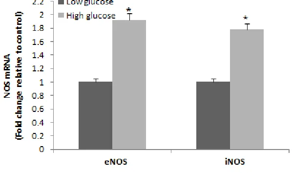
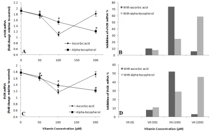
To verify inducement of high glucose on nitric oxide synthase (NOS), HUVECs were stimulated by 25 mM glucose concentration for 24 hours. High glucose increased the expression of eNOS mRNA and iNOS mRNA in comparison with low glucose (5.5 mM) approximately 92% and 78%, respectively (Figure 1). For determination of the best concentration of vitamins, we assayed the effect different concentrations (50 -200 µM) of ascorbic acid and α -tocopherol on glucose (25 mM)-induced eNOS and iNOS expression for 24 hours. Ascorbic acid inhibited glucose-induced eNOS and iNOS mRNA expression in a significant dose-dependent manner (p < 0.05) when the concentration was limited between 50 and 100 µM, whereas α-tocopherol showed a significant inhibition of glucose-induced eNOS and iNOS mRNA expression as dose dependent in concentrations of 50 to 200 µM (Figure 2A,C). According to this study 100 µM concentration of ascorbic acid is able to reduce eNOS and iNOS mRNA expression approximately 74% and 52%, respectively, but 200 µM concentration of α-tocopherol is able to reduce eNOS and iNOS mRNA expression approximately 59% and 49%, respectively (Figure 2B,D).
Figure 1. The effect of 5.5 mM glucose concentration, as basal concentration, (black bar) and 25 mM glucose concentration (grey bar) on eNOS and iNOS expression assessed by RT-PCR in HUVECs after 24 hours incubation (37oC, 5% CO
2)
Data are represented as the mean±SD of triplicate determination. *p < 0.05 compared with control (black bar).
Figure 2. The effect of 50, 100, and 200 µM concentrations of ascorbic acid (♦) and α-tocopherol (■) on eNOS (A), and iNOS (C) expression in presence high glucose concentration (25 mM); The comparison of inhibition percent of eNOS (B), and iNOS (D) expression
in presence 25 mM glucose concentration and different concentrations (50-200 µM) of ascorbic acid and α-tocopherol. HUVECs
treatment with vitamins at 37oC with air of 5% CO
2 for 24 hours
Data are represented as the mean±SD of triplicate determination. *p < 0.05 compared with control (in the absence of vitamins).
Figure 3. Effect of low glucose (5.5 mM), as basal concentration, and high glucose (25 mM) on concentration of NOS in HUVECs after 24 hours incubation (37oC, 5% CO
2)
Data are represented as the mean±SD of triplicate determination. *p < 0.05 compared with control (in the presence of low glucose).
Figure 4. (A) The effect of 50, 100, and 200 µM concentrations of ascorbic acid (♦) and α-tocopherol (■) on HUVECs nitric oxide synthase (NOS) concentration in presence high glucose concentration (25 mM); (B) The comparison of inhibition percent of NOS protein concentration in presence high level of glucose (25 mM) and different concentrations (50-200 µM) of ascorbic acid and α-tocopherol.
HUVECs treatment with vitamins at 37oC with air of 5% CO
2 for 24 hours
Data are represented as the mean±SD of triplicate determination. *p < 0.05 compared with control (in the absence of vitamins)
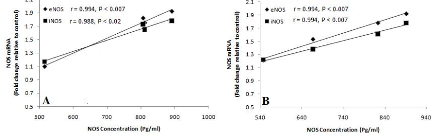
Figure 5. Correlation between expression and concentration of eNOS (♦) and iNOS (■) in presence of high glucose (25 mM) and different concentrations (50 – 200 µM) of ascorbic acid (A) and α-tocopherol (B) in HUVECs
DISCUSSION
Dysfunction of endothelial cells are early manifestation of atherosclerosis that the most important result it is cardiovascular disease (21). Diabetes mellitus is an important risk factor for cardiovascular disease (2),(22). Pathways of nitric oxide (NO) signaling and reactions of lipid oxidation are important in both the maintenance of vascular homeostasis and the progression of vascular disease. Studies biochemical and cell biology have demonestrated that NO with oxidizing lipids could lead to either vascular protection or vascular injury (23), (24). The effect of NO is dependent to amounts it, if NO generated at low levels by nitric oxide synthase (NOS) can terminate propagating lipid radicals, reaction that would be protective. However, if NO generated at elevated levels, it can be converted to prooxidant species that can potentiate injury to vascular cells (24).
lipid oxidation and endothelial cells dysfunction (29), (30). Peroxynitrite is unique as a lipid oxidant, because it mediates oxidation of unsaturated fatty acids in the absence of transition metal catalysts (29). This NO-derived reactive species is more than two orders of magnitude potent than hydrogen peroxide in catalyzing lipid oxidation in
vitro (31). Nitrogen oxide will both oxidize and nitrate unsaturated lipids (30), (32). These reactions result in
formation of a complex mixture of products including nitrated lipid derivatives and alkylnitrites. The oxidation of nitrite will yield nitryl chloride, which initiates lipid oxidation and can yield on LDL particle similar to that found in foam cells and so may be operative in atherogenesis (33), (34).
Vaziri et al. (35) demonstrated that antioxidant therapy ameliorated hypertension in rats, as well as, prospective studies have been shown cardiovascular benefits from antioxidant therapy (8), (9), (10). In the present study, ascorbic acid and α-tocopherol were used as protective agents against high glucose-induced NOS expression. It has been shown in several studies that antioxidants reduced vascular oxidative stress (36), (37). Additionally, ascorbic acid increased vasodilatation of forearm resistance arteries in humans with hypercholesterolemia (38), long-term smokers (39), essential hypertension (40), and coronary artery disease (41). Although numerous studies have shown antioxidants play a key role in oxidative stress inhibition. However, the literature concerning the effect vitamins on the expression and concentration of NOS in presence of high glucose is limited. In this investigation, we indicated that treatment of HUVECs with ascorbic acid and α-tocopherol decreased high glucose-induced eNOS and iNOS expression.
A dose-dependent manner was observed when the concentration of ascorbic acid and/or α-tocopherol was limited to the range of 50 to 100 µM or 50 to 200 µM, respectively. According to this study, optimum concentration for ascorbic acid and α-tocopherol obtained 100 µM and 200 µM, respectively. In presence of ascorbic acid (100 µM) high glucose-induced eNOS and iNOS expression were inhibited by 74% (1.92-fold decreased to 1.1-fold) and 52% (1.78-fold decreased to 1.17-fold), respectively. α-tocopherol (200 µM) also inhibited the expression of eNOS and iNOS in presence of high glucose by 59% (1.92-fold decreased to 1.21-fold) and 49% (1.78-fold decreased to 1.22-fold), respectively. According to these results, we suggest that 100 µM ascorbic acid and 200 µM α-tocopherol could restore eNOS and iNOS expression to levels observed in 5.5 mM glucose.
Our results are in agreement with the finding of several studies. For instance, Kang et al. (42) demonstrated that α -lipoic acid is able to decrease eNOS expression in bladder of rats with streptozotocin-induced diabetic. Similarly, Askwith et al.(43) also reported that in vitro taurine treatment reduces expression of NOS in high glucose-exposed human Schwann cells. In addition, there are other numerous studies that have been showed antioxidants, for example, melatonin, vitamin C (44), hesperidin (45), and vitamin E (46) could decrease expression of NOS isoforms
in vitro and/or in vivo.
The mechanism of ascorbic acid and α-tocopherol on NOS downregulation is still unknown. However, according to study of Askwith et al. (43) the effect of antioxidants on NOS expression may be mediated by an antioxidant action, as well as, they also showed which this effect could be mediated by carbonyl scavenging or perhaps by restoring Ca+2 signaling (43).
In the present study, we also indicated that high glucose (25 mM) increase concentration of NOS protein in HUVECs by 86%, and found that ascorbic acid and α-tocopherol could reduce high glucose-induced NOS concentration in a significant and dose-dependent manner. The effective concentration of ascorbic acid and α -tocopherol that obtained in the present experimental study was 100 µM and 200 µM, respectively. These results confirmed our previous data in this study.
Finally, this investigation showed a positive and significant correlation between NOS protein content with NOS mRNA expression in HUVECs at present high glucose and ascorbic acid and α-tocopherol. According to these results, we suggest that ascorbic acid and α-tocopherol could decrease concentration of high glucose-induced NOS protein in HUVECs via NOS gene downregulation. Therefore, this is may be one of molecular mechanisms that ascorbic acid and α-tocopherol could prevent the increase in atherosclerosis induced by diabetes in human.
Acknowledgements
This work was supported by a research grand from the Cellular and Molecular Research Center of Jundishapur University of Medical Sciences, Ahvaz, Iran (Project No. CMRC-59).
REFERENCES
[1] Contreras F, Rivera M, Vasquez J, De la Parte M, Velasco M. Diabetes and hypertension physiopathology
and therapeutics. Journal of human hypertension. 2000;14:S26-S31.
[2] Libby P, Ridker PM, Maseri A. Inflammation and atherosclerosis. Circulation. 2002;105(9):1135-43.
[3] Williams SB, Cusco JA, Roddy M-A, Johnstone MT, Creager MA. Impaired nitric oxide-mediated
vasodilation in patients with non-insulin-dependent diabetes mellitus. Journal of the American College of
Cardiology. 1996;27(3):567-74.
[4] Lamas S, Marsden PA, Li GK, Tempst P, Michel T. Endothelial nitric oxide synthase: molecular cloning
and characterization of a distinct constitutive enzyme isoform. Proceedings of the National Academy of
Sciences. 1992;89(14):6348-52.
[5] Marsden PA, Heng H, Scherer S, Stewart R, Hall A, Shi X, et al. Structure and chromosomal localization
of the human constitutive endothelial nitric oxide synthase gene. Journal of biological chemistry. 1993;268(23):17478-88.
[6] Cosentino F, Hishikawa K, Katusic ZS, Lüscher TF. High glucose increases nitric oxide synthase
expression and superoxide anion generation in human aortic endothelial cells. Circulation. 1997;96(1):25-8. [7] Di Wang H, Hope S, Du Y, Quinn MT, Cayatte A, Pagano PJ, et al. Paracrine role of adventitial superoxide anion in mediating spontaneous tone of the isolated rat aorta in angiotensin II-induced hypertension. Hypertension. 1999;33(5):1225-32.
[8] Du X, Stockklauser-Färber K, Rösen P. Generation of reactive oxygen intermediates, activation of NF-κB,
and induction of apoptosis in human endothelial cells by glucose: role of nitric oxide synthase? Free Radical
Biology and Medicine. 1999;27(7):752-63.
[9] Cummings PM, Giddens K, Nassar BA. Oral glucose loading acutely attenuates endothelium-dependent
vasodilation in healthy adults without diabetes: an effect prevented by vitamins C and E. Journal of the
American College of Cardiology. 2000;36(7):2185-91.
[10]Skyrme-Jones RAP, O’Brien RC, Berry KL, Meredith IT. Vitamin E supplementation improves
endothelial function in type I diabetes mellitus: a randomized, placebo-controlled study. Journal of the
American College of Cardiology. 2000;36(1):94-102.
[11]Golbidi S, Badran M, Laher I. Antioxidant and anti-inflammatory effects of exercise in diabetic patients.
Experimental diabetes research. 2011;2012.
[12]Montonen J, Knekt P, Järvinen R, Reunanen A. Dietary antioxidant intake and risk of type 2 diabetes.
Diabetes Care. 2004;27(2):362-6.
[13]Devasagayam T, Tilak J, Boloor K, Sane KS, Ghaskadbi SS, Lele R. Free radicals and antioxidants in human health: current status and future prospects. Japi. 2004;52(794804):4.
[14]Laight D, Carrier M, Änggård E. Antioxidants, diabetes and endothelial dysfunction. Cardiovascular
research. 2000;47(3):457-64.
[15]Ruhe RC, McDonald RB. Use of antioxidant nutrients in the prevention and treatment of type 2 diabetes.
Journal of the American College of Nutrition. 2001;20(sup5):363S-9S.
[16]Ghaffari MA, Mojab S. In Vitro Effect of?-Tocopherol, Ascorbic Acid and Lycopene on Low Density
Lipoprotein Glycation. Iranian Journal of Pharmaceutical Research. 2010:265-71.
[17]Ho FM, Lin WW, Chen BC, Chao CM, Yang C-R, Lin LY, et al. High glucose-induced apoptosis in
human vascular endothelial cells is mediated through NF-κB and c-Jun NH 2-terminal kinase pathway and prevented by PI3K/Akt/eNOS pathway. Cellular signalling. 2006;18(3):391-9.
[18]Ho FM, Liu SH, Liau CS, Huang J, Lin-Shiau SY. High glucose–induced apoptosis in human endothelial
cells is mediated by sequential activations of c-Jun NH2-terminal kinase and caspase-3. Circulation. 2000;101(22):2618-24.
[19]Chen Y-J, Hsu K-W, Chen Y-L. Acute glucose overload potentiates nitric oxide production in
lipopolysaccharide-stimulated macrophages: the role of purinergic receptor activation. Cell biology
international. 2006;30(10):817-22.
[20]Turpaev K, Bouton C, Diet A, Glatigny A, Drapier J-C. Analysis of differentially expressed genes in nitric
oxide-exposed human monocytic cells. Free Radical Biology and Medicine. 2005;38(10):1392-400.
[22]Haffner SM, Lehto S, Rönnemaa T, Pyörälä K, Laakso M. Mortality from coronary heart disease in subjects with type 2 diabetes and in nondiabetic subjects with and without prior myocardial infarction. New
England journal of medicine. 1998;339(4):229-34.
[23]Dusting GJ, Macdonald PS. Endogenous nitric oxide in cardiovascular disease and transplantation. Annals
of medicine. 1995;27(3):395-406.
[24]Eiserich JP, Patel RP, O’Donnell VB. Pathophysiology of nitric oxide and related species: free radical reactions and modification of biomolecules. Molecular aspects of medicine. 1998;19(4):221-357.
[25]Ding Q, Hayashi T, Packiasamy AJ, Miyazaki A, Fukatsu A, Shiraishi H, et al. The effect of high glucose
on NO and O 2− through endothelial GTPCH1 and NADPH oxidase. Life sciences. 2004;75(26):3185-94.
[26]Zhu M, Chen J, Tan Z, Wang J. Propofol Protects Against High Glucose–Induced Endothelial Dysfunction
in Human Umbilical Vein Endothelial Cells. Anesthesia & Analgesia. 2012;114(2):303-9.
[27]Srinivasan S, Hatley M, Bolick D, Palmer L, Edelstein D, Brownlee M, et al. Hyperglycaemia-induced superoxide production decreases eNOS expression via AP-1 activation in aortic endothelial cells. Diabetologia. 2004;47(10):1727-34.
[28]Tesfamariam B, Brown ML, Cohen RA. Elevated glucose impairs endothelium-dependent relaxation by
activating protein kinase C. Journal of Clinical Investigation. 1991;87(5):1643.
[29]Radi R, Beckman JS, Bush KM, Freeman BA. Peroxynitrite-induced membrane lipid peroxidation: the
cytotoxic potential of superoxide and nitric oxide. Archives of biochemistry and biophysics. 1991;288(2):481-7.
[30]Gallon AA, Pryor WA. The reaction of low levels of nitrogen dioxide with methyl linoleate in the presence
and absence of oxygen. Lipids. 1994;29(3):171-6.
[31]Patel R, Diczfalusy U, Dzeletovic S, Wilson M, Darley-Usmar V. Formation of oxysterols during
oxidation of low density lipoprotein by peroxynitrite, myoglobin, and copper. Journal of lipid research. 1996;37(11):2361-71.
[32]O'Donnell VB, Eiserich JP, Chumley PH, Jablonsky MJ, Krishna NR, Kirk M, et al. Nitration of
unsaturated fatty acids by nitric oxide-derived reactive nitrogen species peroxynitrite, nitrous acid, nitrogen dioxide, and nitronium ion. Chemical research in toxicology. 1999;12(1):83-92.
[33]Eiserich JP, Cross CE, Jones AD, Halliwell B, van der Vliet A. Formation of nitrating and chlorinating species by reaction of nitrite with hypochlorous acid a novel mechanism for nitric oxide-mediated protein modification. Journal of Biological Chemistry. 1996;271(32):19199-208.
[34]Hazell L, Arnold L, Flowers D, Waeg G, Malle E, Stocker R. Presence of hypochlorite-modified proteins
in human atherosclerotic lesions. Journal of Clinical Investigation. 1996;97(6):1535.
[35]Vaziri ND, Ni Z, Oveisi F, Trnavsky-Hobbs DL. Effect of antioxidant therapy on blood pressure and NO
synthase expression in hypertensive rats. Hypertension. 2000;36(6):957-64.
[36]Bauersachs J, Fleming I, Fraccarollo D, Busse R, Ertl G. Prevention of endothelial dysfunction in heart failure by vitamin E. Cardiovascular research. 2001;51(2):344-50.
[37]Chen X, Touyz RM, Park JB, Schiffrin EL. Antioxidant effects of vitamins C and E are associated with altered activation of vascular NADPH oxidase and superoxide dismutase in stroke-prone SHR. Hypertension. 2001;38(3):606-11.
[38]Ting HH, Timimi FK, Haley EA, Roddy M-A, Ganz P, Creager MA. Vitamin C improves
endothelium-dependent vasodilation in forearm resistance vessels of humans with hypercholesterolemia. Circulation. 1997;95(12):2617-22.
[39]Heitzer T, Mu T. Antioxidant vitamin C improves endothelial dysfunction in chronic smokers. Circulation.
1996;94(1):6-9.
[40]Taddei S, Virdis A, Ghiadoni L, Magagna A, Salvetti A. Vitamin C improves endothelium-dependent
vasodilation by restoring nitric oxide activity in essential hypertension. Circulation. 1998;97(22):2222-9. [41]Gokce N, Keaney JF, Frei B, Holbrook M, Olesiak M, Zachariah BJ, et al. Long-term ascorbic acid administration reverses endothelial vasomotor dysfunction in patients with coronary artery disease.
Circulation. 1999;99(25):3234-40.
[42]Kang DI, Kim SH, Lee SD, Kwak HS, Choi SH, Kim DR, et al. Effects of Alpha-lipoic acid on nitric oxide synthase expression and ultrastructural changes in the bladder of rats with streptozotocin-induced diabetes. Korean Journal of Urology. 2007;48(2):212-8.
[43]Askwith T, Zeng W, Eggo MC, Stevens MJ. Taurine reduces nitrosative stress and nitric oxide synthase expression in high glucose-exposed human Schwann cells. Experimental neurology. 2012;233(1):154-62.
[44]Sönmez M, Narin F, Akkuş D, Özdamar S. Effect of melatonin and vitamin C on expression of endothelial
[45]Xiaoting L, Xiangyun Z, Shumei L, Minghua D, Liang X. Effect of hesperidin on expression of inducible nitric oxide synthase in cultured rabbit retinal pigment epithelial cells. Retinal Degenerative Diseases: Springer; 2010. p. 193-201.
[46]Calvisi DF, Ladu S, Hironaka K, Factor VM, Thorgeirsson SS. Vitamin E down-modulates iNOS and
NADPH oxidase in c-Myc/TGF-α transgenic mouse model of liver cancer. Journal of hepatology.