RESEARCH
Dimerized translationally controlled
tumor protein increases interleukin-8
expression through MAPK and NF-κB pathways
in a human bronchial epithelial cell line
Heewon Lee and Kyunglim Lee
*Abstract
Background: Histamine releasing factor (HRF) is a unique cytokine known to regulate a variety of immune cells in late allergic reactions. In the previous study, we revealed that the biologically active form of HRF is the dimerized translationally controlled tumor protein (dTCTP) for the first time, and confirmed the secretion of IL-8 cytokine by dTCTP in human bronchial epithelial cells. However, the signaling pathway by which dTCTP promotes the secretion of IL-8 is not known.
Results: When the cells were stimulated with dTCTP, the canonical NF-κB pathway and ERK, JNK and p38 MAPK become activated. dTCTP promoted transcription of IL-8, which involved NF-κB and AP-1 transcription factors. NF-κB was found to be essential for the transcriptional activation of IL-8, while AP-1 was partially responsible for the tran-scriptional activation by dTCTP. p38 MAPK was found to be involved in post-trantran-scriptional regulation of dTCTP by stabilizing IL-8 mRNA.
Conclusions: This study demonstrated that dTCTP induces IL-8 secretion in BEAS-2B cells through transcriptional and post-transcriptional regulation of MAPK and NF-κB pathways. This study provides insight into the mechanism by which dTCTP induces inflammation.
Keywords: BEAS-2B, Dimerized TCTP, Interleukin-8, MAPK, NF-κB
© The Author(s) 2018. This article is distributed under the terms of the Creative Commons Attribution 4.0 International License (http://creativecommons.org/licenses/by/4.0/), which permits unrestricted use, distribution, and reproduction in any medium, provided you give appropriate credit to the original author(s) and the source, provide a link to the Creative Commons license, and indicate if changes were made. The Creative Commons Public Domain Dedication waiver (http://creativecommons.org/ publicdomain/zero/1.0/) applies to the data made available in this article, unless otherwise stated.
Background
Airway epithelium is not only the target of the inflamma-tory response, but it is also an important effector tissue that controls secondary mediators. It is the first tissue to encounter inhaled allergens and contributes to endog-enous defense by regulating mucus secretion and ciliary movement. Airway epithelial cells are known to secrete various cytokines and chemokines under external stim-uli. Interleukin-8 (IL-8) previously known as neutro-phil chemotactic factor is one of them. IL-8 secretion is increased by oxidant stress, which thereby cause the recruitment of inflammatory cells and induces a further
increase in oxidant stress mediators, making it a key parameter in localized inflammation [1].
Histamine releasing factor (HRF) was first described as a substance capable of histamine release from basophils present in the secretion of activated mononuclear cells [2]. Its clinical importance has been emphasized since it was found in the biological fluids of allergic patients. In particular, the fact that it was found in the sera of asth-matic patients and bronchoalveolar lavage fluids of airway inflamed mice suggests its role in airway inflammatory diseases. In a previous report, amino-terminal sequenc-ing led to the identification of Translationally controlled tumor protein (TCTP) as the same as HRF [3]. TCTP, a highly conserved and ubiquitously expressed protein in all eukaryotic cells, has been reported to be involved in a variety of cellular processes such as cell growth and
Open Access
*Correspondence: [email protected]
development [4]. However, since not all TCTP secreted by cells exhibited histamine releasing activity, the rela-tionship between HRF and TCTP remained unclear.
Our previous studies demonstrated that the biologi-cally active form of TCTP is its dimeric form of TCTP (dTCTP) and that dimerization is essential for generat-ing its cytokine-like activity of TCTP [5]. The BEAS-2B cell line derived from human normal bronchial epithe-lium was the first non-immune cell of HRF target cells reported. When oxidative stress was applied to BEAS-2B cells, HRF was secreted and extracellular HRF promoted the release of inflammatory cytokines such as IL-8 and GM-CSF [6], suggesting the presence of dTCTP-specific receptors on the BEAS-2B cell line.
In this study, we investigated the IL-8 secretion sign-aling pathways by dTCTP in BEAS-2B cells and dem-onstrated that in BEAS-2B cells, dTCTP induces IL-8, an inflammatory cytokine, through MAPK and NF-κB signaling.
Methods
Cell culture
Human bronchial epithelial cells, BEAS-2B, were pur-chased from the American Type Culture Collection (ATCC, CRL-9609) and cultured in bronchial epithelial cell growth medium (BEGM, Lonza) at 37 °C and 5% CO2. Airway Epithelial Cell Growth Medium (Promocell,
C-21060) was used in small interfering RNA (siRNA) transfection experiments.
Reagents and antibodies
The sources of reagents and antibodies used in this study are as follows: IPTG was from Duchefa (Haarlem, The Netherlands); Ampicillin sodium salt and chlorampheni-col were from USB (OH, USA); Penicillin–Streptomycin was from Thermo Fisher Scientific (UT, USA). MAPK Inhibitors PD98059, SP600125, SB203580 and BAY11-7082 were from Calbiochem (CA, USA). The oligonu-cleotides used in these experiments were synthesized by Bioneer (Seoul, Korea). Antibodies against phospho-p44/p42 MAPK (Thr202/Tyr204), phospho-p44/p42 MAPK, phospho-SAPK/JNK (Thr183/Tyr185), SAPK/JNK, phospho-p38 MAPK (Thr180/Tyr182), p38 MAPK, phos-pho-MAPKAPK-2 (Thr222), MAPKAPK-2, phospho-IκBα (Ser32/36), phospho-IκBα, Lamin A/C and β-actin were from Cell Signaling Technology (MA, USA); GAPDH antibody was from AbFrontier (Seoul, Korea); NF-κB (p65) anti-body was from Enzo Life Sciences (NY, USA).
Recombinant protein production
We prepared the recombinant proteins as previously described [5]. Briefly, pRSET A/Del-N11 TCTP was transformed to E. coli strain BL21(DE3)pLysS and
cells were grown at 37 °C and 220 rpm in Luria–Ber-tani medium containing the 100 μg/ml ampicillin and 34 μg/ml chloramphenicol. The pre-culture medium was diluted 1:100 with 400 ml and cultured until OD600
reached 0.6–0.8 (Hitachi, U-3000). After IPTG was added to a final concentration of 0.4 mM, the culture was incu-bated at 37 °C and 200 rpm for 2 h 30 min. Cells were harvested by centrifugation at 7140×g (Sorvall, SLA-3000 rotor) for 10 min at 4 °C and stored at − 70 °C or used directly. Cell pellets were resuspend in ice-cold equilibration buffer (50 mM sodium phosphate, 300 mM NaCl, 10 mM imidazole; pH 7.4) with 1 mM PMSF and disrupted by sonication (Kyung Ill, KTA-400). Sonication was performed three times on ice for 30 s at 1/10 of max-imal amplitude followed by centrifugation for 40 min at 12,000×g (Sorvall, SS34-rotor).
The supernatants containing soluble proteins were purified with HisPur™ Cobalt Resin (Thermo, 89965) according to the manufacturer’s instruction. Subse-quently, the proteins were eluted and the mixtures desalted using a PD-10 desalting column (GE Healthcare, 17-0851-01) and loaded onto HiTrap Q HP column (GE Healthcare, 17-1153-01) which was equilibrated with buffer A (20 mM Tris, 1 mM EDTA, 50 mM NaCl; pH 7.4). Proteins were separated by AKTA FPLC systems (GE Healthcare) and eluted with buffer B (20 mM Tris, 1 mM EDTA, 1 M NaCl; pH 7.4) with a constant flow rate of 1 ml/min. The eluted fractions were separated by SDS-PAGE and the samples containing dTCTP were collected and desalted with PBS. Purified dTCTP was concentrated using Vivaspin 500 (Sartorius, VS0122) and stored at − 70 °C until use.
Measurement of IL‑8
BEAS-2B cells were seeded at 4000 cells per well in 48-well plates (Nunc). When the cells became 60% con-fluent, they were washed twice with 1% penicillin–strep-tomycin/BEBM, and treated with or without inhibitor for 30 min at indicated concentrations followed by 10 μg/ml dTCTP. After 16–20 h, IL-8 in the media was measured with Legend MAX™ Human IL-8 ELISA Kit (BioLegnd, 431508) according to the manufacturer’s protocol.
Immunofluorescence confocal microscopy
1 h at room temperature. Rabbit NF-κB (p65) anti-body (Enzo, ALX-210-574) was diluted 1:300 with block-ing buffer and treated to the cells overnight at 4 °C. The stained cells were washed with PBS and probed with Alexa Fluor 488-conjugated goat anti-rabbit IgG (Invitro-gen, A11008) diluted 1:1000 in blocking buffer for 30 min at room temperature in a shaded chamber. After wash-ing with PBS, cells were counterstained with DAPI uswash-ing ProLong™ Gold Antifade Mountant (Invitrogen, P36931). Confocal microscopy was performed using a Carl Zeiss Laser Scanning Systems LSM 510.
Immunoblotting
BEAS-2B cells were washed with PBS 3 times and har-vested by scraping with ice-cold lysis buffer (50 mM Tris–HCl; pH 7.4, 150 mM NaCl, 1 mM EDTA, 0.25% deoxycholate, 1% Triton X-100) containing protease inhibitor cocktail (Roche, 11 836 170 001) and phos-phatase inhibitor cocktail (Sigma Aldrich, P5726 and P0044). Cells were vortexed thoroughly for 30 s and set on ice for every 10 min. After 30 min, lysates were cen-trifuged at 12,000×g for 20 min at 4 °C, and the super-natants were collected. Protein contents were quantified by Bradford assay (Bio-Rad, CA, USA) and 5–15 μg of each sample were separated by SDS-PAGE, transferred to PVDF membranes and probed with primary antibodies followed by incubation with HRP-conjugated goat anti-mouse/rabbit IgG antibody (Bio-rad, CA, USA). Proteins of interest were visualized with chemiluminescent sensi-tive plus HRP substrate (Surmodics, LERI-0110-2C) and detected with LAS-3000 (Fujifilm, Tokyo, Japan).
Reverse transcription‑PCR
Total RNA was isolated from cells using RNeasy Mini Kit (Qiagen, 74104) as described by the manufacturer. The isolated RNA had an A260/A280 ratio of 2.0–2.1 and 1 μg of
each RNA samples were reverse transcribed into cDNA using High capacity cDNA Reverse transcription Kit (Applied Biosystems, 4368814). 1 μl of the resulting cDNA, 10 pmol of each forward and reverse primer, and the DNA polymerase mixture provided in AccuPower PCR Pre-Mix (Bioneer, K-2016) in 20 μl reaction volume were amplified using PCR. The primers were synthesized by Genotech Co. Ltd. (Korea) and the sequences were: IL-8,
5′-CATGACTTCCA AGCTGGCCGTG-3′ (forward) and
5′-TCACTGATTCTTGGATACCACA GAG-3′ (reverse)
and β-actin, 5′-CAGCTCGTAGCTCTTCTCCA-3′
(forward) and 5′-CAGCTCGTAGCTCTT CTCCA-3′
(reverse). The reaction mixtures were pre-denatured by incubating at 94 °C for 5 min followed by 27 cycles (for IL-8) or 30 cycles (for β-actin) of 94 °C for 30 s, 55 °C for 30 s and 72 °C for 30 s, and for final extension, they were
incubated at 72 °C for 10 min. 8 μl of the amplified prod-ucts were resolved on a 1% agarose gel containing SYBR™ Safe DNA gel stain (Invitrogen). Images were taken using E-Graph AE-9000 (Atto, Japan).
Real‑time quantitative PCR
Changes in IL-8 transcripts were confirmed by real-time PCR in cells treated with various doses of dTCTP. Lev-els of IL-8 mRNA was analyzed using TaqMan Gene Expression Assays (Applied Biosystems, 4331182) (Assay IDs: IL-8, Hs00174103_m1; GAPDH, Hs99999905_m1) according to the manufacturer’s protocol. 2 μl of cDNA was used for 20 μl reaction, and each samples were run in triplicates. Reaction mixtures were amplified with an initial denature step at 95 °C for 10 min, followed by 40 cycles of 95 °C for 15 s and 60 °C for 1 min using Applied Biosystems 7300 Real-Time PCR System (Applied Bio-systems, ABI 7300). The expression of IL-8 mRNA was normalized to GAPDH mRNA. The efficiencies of the reactions were calculated by a fivefold serial dilution and estimated to be 90–95%.
Luciferase assay
BEAS-2B cells were plated at 6 × 10⁴ cells/well into 96
Electrophoretic mobility gel shift assay (EMSA)
BEAS-2B cells grown in 100 mm culture dish (Nunc) were serum starved for 2 h and stimulated with either PBS or dTCTP. The nuclear extracts were prepared using NE-PER™ Nuclear and Cytoplasmic Extraction Reagents (Thermo, 78833) according to the manufacturer’s man-ual. The protein contents of nuclear extracts were quan-tified, aliquoted, and used directly or stored at − 70 °C until use.
The sequences of the sense-strand oligonucleotides used in these experiments were as follows: NF-κB,
5′-AGTTGAGGGGACTTTCCCAGGC-3′, AP-1,
5′-CGCTTGATGAGT CAGCCGGAA-3′ (Promega
Cor-poration, USA), mutant NF-κB, 5′-AGTTGAGGTAA
CTTTCCCAGGC-3′, and mutant AP-1, 5′
-CGCTTGA-TATGTCAGCCGGAA-3′. The complementary pairs of
oligonucleotides were annealed using a heating block. The pair of nucleotides were mixed at a molar ratio 1:1, incubated at 95 °C for 5 min, and allowed to stand over-night at room temperature.
EMSA was performed as previously described with some modification. Briefly, 6% polyacrylamide gel was pre-run for 40 min in 0.5× TBE buffer. During this time, binding reactions were performed by adding 5 μg of nuclear extracts (as 2–4 μl of NE-PER) in 5× bind-ing buffer (20% Glycerol, 5 mM MgCl2, 2.5 mM EDTA,
2.5 mM DTT, 250 mM NaCl, 50 mM Tris, 2.5 mg/ml poly dI·dC). Then 3′ end-biotinylated double stranded DNA was added at a final concentration of 1 pmol and incu-bated at room temperature for 20 min. The specificity of the binding reaction was confirmed by pre-incubating the nuclear extracts with tenfold molar excess of unlabeled DNA. The reaction mixtures were electrophoresed and transferred to nylon membrane (Roche, 11 209 299 001) at 4 °C. The membrane was cross-linked for 10 min using E-Graph AE-9000 (Atto Incorporation, Japan) equipped with 312 nm UV transilluminator. The biotin-labeled DNA on the membrane was detected using LightShift Chemiluminescent EMSA Kit (Thermo, 20148) accord-ing to the instructions.
Small interfering RNA (siRNA)
BEAS-2B cells were seeded in 12 well plates (160,000 cells/well) and incubated for 24–36 h in growth media. At 60–65% confluency, cells were transfected with AccuTarget™ FAM labeled for Negative Control (Bioneer, SN-1021) or siRNA targeting p65 mRNA (Bioneer,
1128171) using Lipofectamine® RNAiMAX Reagent
(Invitrogen) at a final concentration of 20 nM in Opti-MEM® (Thermo). After 6 h, media were changed with growth media and incubated for another 18 h. 24 h post-transfection, the transfection efficiency was confirmed by fluorescence microscopy (data not shown) and the cells
were treated with or without 10 μg/ml of dTCTP for 16–20 h. The resulting supernatants were harvested and analyzed for IL-8 contents.
Statistical analysis
Data are presented as means ± standard errors. Data were analyzed with GraphPad Prisms 5 software (Graph-Pad Software Inc., CA, USA). Statistical significance was determined using Student’s two-tailed unpaired t test for comparisons between two groups. For more than 3 groups, one-way ANOVA analysis was performed.
Results
dTCTP‑induced IL‑8 secretion is transcriptionally regulated
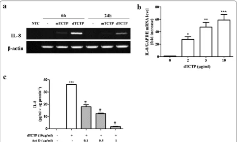
Previously, Kim et al. showed that recombinant dTCTP, but not monomer TCTP (mTCTP), induced IL-8 secre-tion in BEAS-2B cells [5]. We analyzed IL-8 mRNA by RT-PCR in BEAS-2B cells which were treated with either mTCTP or dTCTP for 6 or 24 h. As shown in Fig. 1a, IL-8 mRNA was significantly induced in response to dTCTP treatment for 6 h. After 24 h, IL-8 mRNA decreased. However, when stimulated with mTCTP, BEAS-2B cells did not generate IL-8 mRNA at both time points. Changes in IL-8 transcripts were confirmed by real-time PCR in cells treated with various doses of dTCTP. Figure 1b shows that the IL-8 transcripts increased in a dose-dependent manner. During 6 h incubation with 2, 5 or 10 μg/ml of dTCTP, IL-8 transcripts were increased to mean ± SD of 27.7 ± 7.4, 47.4 ± 10.8 or 58.9 ± 15.5-fold respectively, indicating that dTCTP had specific action on IL-8 gene expression. Next, we investigated whether IL-8 induction by dTCTP is affected by actino-mycin D (Fig. 1c). Cells stimulated with dTCTP for 6 h were treated with actinomycin D for 1 h and the resulting IL-8 protein was measured. When treated with actino-mycin D at a low dose of 0.1 μg/ml, IL-8 protein expres-sion was reduced to 49.7% of the control. In addition, the amount of IL-8 protein decreased in a actinomycin D concentration-dependent manner. These results suggest that dTCTP induces IL-8 by promoting the transcription of IL-8 gene.
dTCTP activates transcription factor NF‑κB and AP‑1 in BEAS‑2B cells
mechanism mediating IL-8 release from activated T lym-phocytes [10] and from cystic fibrosis bronchial epithelial cell lines [11]. Although not widely studied, it has been also suggested that cyclic AMP-responsive element bind-ing protein (CREB) transcription factor may be involved in the regulation of the CXC chemokine [12]. Using in silico analysis, Bezzerri et al. [13] identified four tran-scription factor binding sites in the proximal region of the IL-8 promoter in human bronchial epithelial IB3-1 cell. The consensus sequences for NF-κB (− 80/− 72 bp), NF-IL6 (− 93/− 84 bp), AP-1 (− 126/− 120 bp), and CREB (− 171/− 164 bp) were mapped.
However, among those transcription factors implicated in IL-8 gene expression, NF-κB and AP-1 are most well-known key regulators for IL-8 gene expression in airway epithelial cells (reviewed in [14]). And specific elements involved in the regulation of NF-κB and AP-1 pathways have been reported to be different depending on the cell type and stimulus.
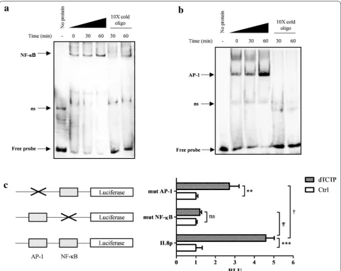
To investigate whether NF-κB and AP-1 transcription factors are involved in dTCTP-induced signaling path-way, nuclear extracts from BEAS-2B cells stimulated with dTCTP for 0, 30 and 60 min were analyzed by EMSA using probes for NF-κB (Fig. 2a) and AP-1 (Fig. 2b). When the transcription factor complexes with the cor-responding sequence, the band shifts due to the increase in the molecular weight. In both cases, the free probes were reduced by the addition of nuclear extracts and the DNA–protein complex increased most at 60 min after dTCTP treatment. This suggests that the nuclear extracts contain proteins that can bind the NF-κB and AP-1 con-sensus sequences. The DNA binding specificity of the protein was confirmed by the adding a tenfold excess of cold unlabeled oligonucleotide to the mixture of DNA and nuclear extracts. The cold probe competes with the labeled DNA probe to bind to the proteins, so that the intensity of the shifted band is reduced or eliminated. The binding specificity was also confirmed using a tenfold
molar excess of unlabeled binding site mutant oligonu-cleotide that did not affect the formation of protein-DNA complexes (see Additional file 1: Figure S1).
These results demonstrated that dTCTP induces DNA binding of NF-κB and AP-1 transcription factors. Hoff-mann et al. [14] revealed that the binding of p65 NF-κB to the endogenous IL-8 promoter contributed to the ini-tiation of the transcription process by recruiting RNA
polymerase II within 30 min upon IL-1 stimulation. The specific mechanism of transcriptional regulation of the IL-8 gene by the AP-1 dimer is not known at the atomic level, but is believed to contribute to an environment suitable for transcription initiation by complexing with other proteins. Therefore, EMSA results suggested that dTCTP can initiate IL-8 transcription via NF-κB and AP-1.
NF‑κB and AP‑1 are required for IL‑8 induction by dTCTP
To verify whether two candidates, NF-κB and AP-1, have a role in dTCTP-induced IL-8 expression, we used lucif-erase reporter gene designed to be driven by IL-8 pro-moter (Fig. 2c). Specifically, IL-8 promoter which was ligated into pGL3-Basic vector, designated as pGLIL8p, was transiently transfected to BEAS-2B cells for 24 h, and stimulated with dTCTP for 18 h. As a surrogate marker for IL-8 promoter activity, luminescence from lysed cells was measured. We compared luciferase activity of pGLIL8p transfected cells with that of pIL8pmutAP-1 and pIL8pmutNF-κB transfected cells, which have a mutation in binding sites for AP-1 and NF-κB, respec-tively. In cells introduced with pGLIL8p, dTCTP elevated luciferase activity to 4.6 fold over non-stimulated con-dition. However, defect in κB binding sites significantly impaired luciferase expression driven by IL-8 promoter, implying that IL-8 induction is predominantly mediated by NF-κB transcription factor. Without NF-κB, dTCTP did not enhance IL-8 transcription. However, cells trans-fected with pIL8mutAP-1 showed reduced promoter activity by 59.3%, but the reduction was not as signifi-cant as that in mutation with NF-κB. In the absence of activation process through AP-1, dTCTP enhanced IL-8 transcription 2.7-fold. These results show that both NF-κB and AP-1 are involved in the transcription of IL-8 by dTCTP. However, although NF-κB is essential for the activation of IL-8 transcription, AP-1 appears to contrib-ute to maximal gene expression.
dTCTP activates NF‑κB pathway and inhibition of NF‑κB abrogates IL‑8 induction by dTCTP
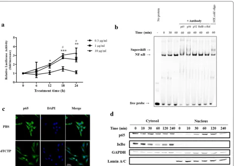
NF-κB represents the nuclear factor-κB and is a tran-scription factor that regulates gene expression. It is involved in immune and inflammatory mechanisms and regulates cell proliferation, differentiation, death, and tumor formation. It is also known to play an important role in internal signaling in the development of vertebrate animals. As NF-κB seems to play an important player in IL-8 induction by dTCTP, we investigated whether dTCTP activates NF-κB pathway. We first tested the ability of dTCTP to induce luciferase expression using the luciferase reporter plasmid under the control of the NF-κB response element. Figure 3a demonstrates that dTCTP induces NF-κB transcriptional activity. Specifi-cally, the luciferase activity of cells treated with 10 μg/ml of dTCTP for 24 h was increased to 3.3 times that of the control.
The NF-κB protein family includes NF-κB1, NF-κB2, Relish, RelA (p65) RelB, c-Rel, and the Drosophila pro-teins Dorsal and Dif, which shares the Rel homology domain (RHD). It acts as homodimer or heterodimer on
the kb site to regulate transcriptional activity, and the specificity of the response to external stimuli depends on which pair of dimer is formed. To characterize which subunit comprises NF-κB dimer in dTCTP-induced sign-aling, a supershift assay was performed using antibod-ies specific for the Rel and NF-κB family members. In Fig. 3b, incubation with p65 or p50 antibodies caused a retarded mobility in DNA–protein complex, suggesting that nuclear extracts from dTCTP stimulated cells con-tain both p65 and p50 subunits. The result is consistent with the western blotting analysis in Fig. 3c to confirm the translocation of p65 subunit from cytosol to the nucleus. In the experiment, BEAS-2B cells were treated with dTCTP for indicated times and the nuclear com-partment was fractionated and blotted for p65 and IκBα expression in each compartment. In the nucleus fraction, p65 was clearly introduced within 30 min of stimulation and degraded after 240 min of stimulation. IκBα begins to be proteolyzed at 30 min and restored to its basal level after 120 min. Mechanistically, IκBα degradation precedes p65 translocation, the time laps between p65 and IκBα are comprehensible. GAPDH and Lamin A/C expression were measured to ensure equal loading and homogeneity of each fraction. In Fig. 3d, localization of NF-κB p65 in cells was visualized using immunofluores-cence staining. These data demonstrated that canoni-cal NF-κB pathway was activated in response to dTCTP stimulation.
MAPKs are involved in dTCTP‑induced IL‑8 expression
AP-1 is one of the early-identified mammalian transcrip-tion factors that activate cell growth, death and differen-tiation by activating target genes for a variety of external stimuli such as inflammatory cytokines, growth factors and stress. The signaling pathway to control the activity of AP-1 is typically the MAPK pathway. MAPK includes proteins of extracellular signal-regulated kinases (ERKs), c-Jun N-terminal kinases (JNKs) and p38 mitogen-acti-vated protein kinases (p38 MAPK), which share many properties such as activation by phosphorylation of ser-ine and threonser-ine residues, three-step activation pathway and similar substrate recognition sites.
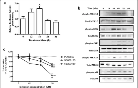
We first measured the AP-1 transcriptional activity against dTCTP using the inducible AP-1 luciferase vec-tor. Twenty-four hours after transfection of the reporter vector into BEAS-2B cells, dTCTP was treated for the indicated time and luciferase activity was measured. As a result, transcription activity by dTCTP increased up to 1.7-fold (Fig. 5a). We next examined the phospho-rylation of MAPKK and MAPK by dTCTP treatment in order to identify the upper MAPK pathway that activates AP-1. In Fig. 5b, MEK1/2 phosphorylation was observed 10 min after treatment with dTCTP, and ERK signal also increased from 30 min, and then decreased after 120 min. In the case of JNK, slight phosphorylation was
observed at 60–120 min. MKK3/6 and p38 MAPK were significantly phosphorylated from 30 min and gradually decreased. We used specific inhibitors of MAPK to inves-tigate the extent to which each MAPK contributes to IL-8 production by dTCTP (Fig. 5c). Prior to this experi-ment, we confirmed that the use of 10 μM of each inhibi-tor did not affect cell viability or the activity of two other MAPKs (data not shown). Cells treated with inhibitors for 30 min were stimulated with dTCTP and the amount of IL-8 produced after 16 h was measured. All three MAPK inhibitors showed a dose-dependent decrease in IL-8 secretion, among which the p38 inhibitor SB203580 showed the most dramatic reduction. At the highest con-centration of 10 μM, IL-8 production was reduced to 69.1% for PD98059, 60.6% for SP600125, and 23.9% for SB203580 compared to the control. From these results, it can be seen that the three MAPKs, particularly the p38 pathway, are involved in the signaling pathway of dTCTP.
These experiments confirmed that p38 MAPK and NF-κB are the major contributors to IL-8 expression by dTCTP. p38 MAPK signaling has been reported to be NF-κB-dependent or independent, depending on
the type or duration of stimulation and the cell type examined.
p38 MAPK is independent of NF‑κB activation and increases the stability of IL‑8 mRNA
We then investigated whether dTCTP-induced NF-κB activation is affected by the p38 MAPK inhibitor. After incubation for 30 min with SB203580, cells were stimu-lated with dTCTP for 1 h and the changes in IκBα phos-phorylation were detected by immunoblotting (Fig. 6a). The results showed that inhibition of p38 MAPK did not affect IκBα phosphorylation. Next, we performed EMSA to determine whether p38 MAPK affects the DNA bind-ing activity of NF-κB. Figure 6b shows, increased DNA– protein complexes in samples treated with dTCTP did not change upon treatment with SB203580.
One of the unique functions of p38 MAPK is its role in regulating mRNA stability of proinflammatory cytokines. Most inflammatory cytokine mRNA derived from normal cells has a short half-life due to AU-rich elements (AREs) of 3′ untranslated region. It is well known that IL-8 mRNA, which contains ARE, induces chronic inflamma-tion by inducing phosphorylainflamma-tion of ARE binding protein
tristetraprolin (TPP) in response to various stimuli due to activation of p38 MAPK, thereby preventing mRNA from decaying. We therefore investigated whether dTCTP-activated p38 MAPK potentiates IL-8 expression by modulating mRNA stability (Fig. 6c). Cells treated or not treated with inhibitor were stimulated with dTCTP for a period of time and incubated with actinomycin D and the residual mRNA was measured over time. In the dTCTP-treated group, the half-life of IL-8 mRNA was estimated as 4.5 h. However, when the activity of p38 MAPK was inhibited, half-life of IL-8 mRNA decreased by 65.5% to 1.5 h. Thus, in our system using dTCTP, we conclude that p38 MAPK potentiates IL-8 gene expression by stabiliz-ing IL-8 mRNA without affectstabiliz-ing the NF-κB pathway.
Discussion
In early studies using basophils isolated from allergic donors, HRF was thought to work in an IgE-dependent manner because HRF provoked histamine release only in
some basophils [15, 16]. These differences led to the clas-sification of IgE by IgE+/IgE−. However, in subsequent studies, it was reported that HRF enhanced IgE anti-body-induced histamine release in basophils regardless of type of cell surface IgE, and that the IgE antibody and HRF did not bind directly [17, 18].
Allergic response through antigen-IgE-FcεRI axis is well-known. However, it has been shown that without multivalent hapten, certain populations of monomeric IgE can also activate FcεRI aggregation and deliver the subse-quent signal transduction events [19]. IgE molecules have a broad spectrum of mast cell activation potential [20], and the IgEs categorized as highly cytokinergic tend to bind better to autoantigen such as HRF. This means that the results can vary widely depending on the IgE molecules used in the experiment. In this context, we can compre-hensively understand the conflicting results of the relation-ship between HRF and IgE. As the new function of IgE was established, Kawakami et al. reassessed the relationship
between IgE and HRF and suggested that IgE and IgG are HRF receptors that have been sought for a long time [21]. Using a large panel of IgE and IgG, they demonstrated that HRF binds to the Fab sites of a subset of IgE and IgG. Peptides that interfere with HRF-Ig binding have been shown to inhibit mast cell activation and suppress passive cutaneous anaphylaxis and mast cell dependent airway inflammation. Furthermore, they revealed that pulmonary inflammation induced by nasal administration of HRF in naïve mice occurs via B cells, mast cells and FcεRIα and Fcγ using genetically deficient mice.
Although the primary involvement of IgE and Fc epsi-lon receptor in the inflammatory response by HRF has
been clearly demonstrated by the ‘knock out’ system, we cannot dispense with the possibility of another HRF-specific receptor other than IgE for the following rea-sons. First of all, in HRF-responsive basophils in which HRF-mediated direct histamine release was found, phos-phorylation of intercellular signaling molecules did not accompany FcεRIγ phosphorylation [22]. Next, HRF and anti-IgE showed different results in response to the treatment of a pharmacologic inhibitor, rottlerin, imply-ing that they induce histamine release through different signal transduction pathway [23]. Anti-IgE-induced his-tamine release was not affected by rottlerin, but HRF-mediated histamine release increased, demonstrating
that HRF signaling is similar to but not entirely equiva-lent to that of IgE. Although we cannot rule out the pos-sibility that this is due to qualitative and quantitative differences in the signals induced by Highly cytokiner-gic (HC) IgE and IgE + Ag, signals transmitted to cells are very similar except for some points because they act through essentially the same receptor [24]. Finally, HRF caused IL-8 and GM-CSF cytokine secretion in AML14-3D10 [25] and BEAS-2B [6] cell lines that do not express detectable FcεRIα (see Additional file 2: Figure S2). Functional receptors for IgE have been reported to be expressed in bronchial epithelial cells in some asth-matic patients, which implies an important message that the expression repertoire of membrane protein changes as asthmatic lesions progress [26, 27]. However, we con-firmed the absence of high-affinity IgE receptor in the BEAS-2B cell line even after stimulating the cells with dTCTP. Furthermore, the report that only dimeric TCTP binds to the cell surface of BEAS-2B suggested the pres-ence of other receptor besides FcεRI [5]. In this respect, we sought to obtain clues for the receptor by identifying signaling molecules involved in IL-8 expression by HRF in BEAS-2B cells, where the receptor is presumed to be present.
The regulation of the IL-8 gene expression has been well characterized [13, 14, 28, 29]. The promoter region of IL-8 gene is restrained by several mechanisms, so that the protein is barely detectable in normal physiology. The restrained promoter should be derepressed through following mechanisms: (1) altered function of NF-κB-repressing factor (NRF) as a coactivator which is origi-nally involved in basal silencing of IL-8 gene in resting cells, (2) replacement of octamer-1 (Oct-1) with CAAT/ enhancer-binding protein (C/EBP) which has binding site on the complementary strand of the octamer motif (5′-ATTTGCAT-3′), and (3) recruitment of CREB-bind-ing protein (CBP)/p300, followed by hyperacetylation of histones and chromatin remodeling. When the pro-moter is activated by various stimuli such as TNFα and IL-1β, IL-8 protein expression increases from tens to hundreds of fold. In previous studies on the mechanism of IL-8 expression, NF-κB and AP-1 have been shown to play pivotal roles [30–36], but the specific mechanism depends on the type of stimulus and cell used.
In our experimental conditions, dTCTP induced phosphorylation of ERK, JNK, and p38 MAPK and acti-vated canonical NF-κB pathway. We confirmed that those pathways were specific for dTCTP in the follow-ing two experiments. First, only dTCTP phosphoryl-ated ERK and IκBα and degraded IκBα, while mTCTP had no such activity (see Additional file 3: Figure S3A). In addition, dTBP2 peptide, which was found to bind to dTCTP and to inhibit its cytokine-like activity [37],
suppressed dTCTP-induced phosphorylation of ERK and IκBα (see Additional file 3: Figure S3B). Notably, the mutation in the κB site of the IL-8 promoter abrogated dTCTP-induced promoter activity, suggesting that the transcriptional activation by NF-κB was indispensable for IL-8 production (Fig. 2). However, it seemed that NF-κB alone could not develop maximal IL-8 expression by dTCTP. This was inferred from the fact that IL-8 produc-tion was induced by 5.1-fold (see Addiproduc-tional file 4: Figure S4), although there was no significant increase in NF-κB luciferase activity in Fig. 3a when the dose of dTCTP was increased from 1 to 10 μg/ml. These findings suggest the additional mechanism other than transcriptional activa-tion by NF-κB is required for the maximal IL-8 expres-sion induced by dTCTP. We demonstrated that the promoter activity decreased significantly when the AP-1 binding site was mutated, implying its role in IL-8 tran-scriptional activation (Fig. 2c). It was also confirmed that dTCTP activated MAPKs, known as the upstream mole-cule of AP-1 dimer. One of the greatest hallmarks of IL-8 is that it exists at a very low level in the resting state, but the expression level increases dramatically in response to the stimulation. This dramatic increase is known to be mainly regulated at the mRNA level, but the half-life of IL-8 mRNA is reported to be very short, about 1.5 h [38,
39]. In this respect, the threefold increase in the half-life of mRNA by p38 MAPK may have a significant impact on IL-8 production induced by dTCTP (Fig. 6c). These findings suggest that NF-κB pathway is essential, but not sufficient for IL-8 expression and that the additional tran-scriptional activation by AP-1 and post-trantran-scriptional regulation such as mRNA stabilization by p38 MAPK are required for the maximal IL-8 expression by dTCTP.
that extracellular TCTP causes the invasion and metas-tasis of tumors previously known as the function of intra-cellular TCTP, but there is no mention of whether the material used is a dimer or monomer.
These reports provide valuable information. However, in the interpretation of the experimental results, we must consider not only the cell type used in each experiment but also the method by which the recombinant protein was produced and its concentration used. We performed this study with the highest concentration of 10 μg/ml based on the concentration used in BEAS-2B in the pre-vious report [5, 6]. Most of the previous studies using recombinant HRF protein were carried out at concentra-tions below 10 μg/ml. Notably, HRF directly enhanced B cell proliferation at concentrations of less than 1 μg/ml, and could activate B cell growth even at 100-fold lower concentrations, especially in the presence of anti-CD40 Ab [41]. If we can rule out other experimental factors, B cells can be considered the most sensitive of the HRF target cells. Recombinant proteins were expressed in Sf9 insect cells or E. coli system, and a vector with the entire HRF sequence was used in most cases. Analysis of the purified recombinant protein on SDS-PAGE showed some degree of spontaneous dimerization of HRF. How-ever, we have established a system for producing dTCTP by inducing intermolecular disulfide bonds between exposed cysteine residue by truncating the N-termi-nal sequence to produce higher proportions of dimers. Mutants that disrupted disulfide bonds by replacing cysteine with alanine did not produce dimers and failed to induce inflammation when administered intranasally [5, 21]. However, it is not yet clear which form of dimer is found in vivo, and the answer could hold the key to HRF research.
In summary, HRF appears to function in the orches-tration of various cells at higher stages of late phase response. It is also found in normal serum, but for some reason it does not cause inflammation. But once switched on, bioactive HRF causes extensive downstream signaling as a multi-modulator of the immune system. Depending on the mode of action in each target cell, the response or subsequent signaling caused by HRF will be different. Our study demonstrated that in BEAS-2B cells, dTCTP induces IL-8, an inflammatory cytokine, through MAPK and NF-κB signaling. NF-κB activation was essential for IL-8 production by HRF, and for maximal production, AP-1 activation by MAPKs and IL-8 mRNA stabilization by p38 MAPK were also required. Identifying kinases and adapter molecules upstream of this signaling event will be an important clue for receptor classification and is currently under investigation.
Authors’ contributions
KL and HL conceived and designed the experiments. HL performed the experiments and wrote the manuscript. KL and HL analyzed the data. Both authors read and approved the final manuscript.
Acknowledgements
We thank Soon-Jung Park (Yonsei University, Seoul, Korea) for providing IL-8 promoter reporter plasmid constructs.
Competing interests
The authors declare that they have no competing interests.
Availability of data and materials
All data generated or analyzed during this study are included in this published article.
Consent for publication Not applicable.
Ethics approval and consent to participate Not applicable.
Funding
This research was supported by Basic Science Research Program through the National Research Foundation of Korea (NRF) funded by the Ministry of Science, ICT and future Planning (2017R1A2B2004023) and by a grant of the Korea Health Technology R&D Project through the Korea Health Industry Development Institute (KHIDI), funded by the Ministry of Health & Welfare, Republic of Korea (HI17C0631).
Publisher’s Note
Springer Nature remains neutral with regard to jurisdictional claims in pub-lished maps and institutional affiliations.
Additional files
Additional file 1: Figure S1. The binding reaction in EMSA assays is spe-cific for NF-κB and AP-1. Nuclear extracts were prepared from the BEAS-2B cells stimulated with or without 10 μg/ml of dTCTP for 60 min. EMSA was performed according to the Methods using biotinylated probes for (A) NF-κB and (B) AP-1. To examine the specificity of the binding reactions, tenfold molar excess of unlabeled wild-type oligo or tenfold molar excess of unlabeled mutant oligo was added to the binding mixture.
Additional file 2: Figure S2. Lack of FcεRIα expression in BEAS-2B cells. FcεRIα expression was determined by immunoblotting in BMMC and BEAS-2B cells. For BEAS-2B, 10 μg/ml of dTCTP was treated for the indi-cated times and the change in protein expression were measured. BMMC was used as a positive control for cells expressing FcεRIα.
Received: 13 September 2017 Accepted: 13 February 2018
References
1. Vlahopoulos S, Boldogh I, Casola A, Brasier AR. Nuclear factor-kappaB-dependent induction of interleukin-8 gene expression by tumor necrosis factor alpha: evidence for an antioxidant sensitive activating pathway distinct from nuclear translocation. Blood. 1999;94(6):1878–89. 2. Thueson DO, Speck LS, Lett-Brown MA, Grant JA. Histamine-releasing
activity (HRA). I. Production by mitogen- or antigen-stimulated human mononuclear cells. J Immunol. 1979;123(2):626–32.
3. MacDonald SM, Rafnar T, Langdon J, Lichtenstein LM. Molecular identification of an IgE-dependent histamine-releasing factor. Science. 1995;269(5224):688–90.
4. Bommer UA, Thiele BJ. The translationally controlled tumour protein (TCTP). Int J Biochem Cell Biol. 2004;36(3):379–85.
5. Kim M, Min HJ, Won HY, Park H, Lee JC, Park HW, Chung J, Hwang ES, Lee K. Dimerization of translationally controlled tumor protein is essential for its cytokine-like activity. PLoS ONE. 2009;4(7):e6464.
6. Yoneda K, Rokutan K, Nakamura Y, Yanagawa H, Kondo-Teshima S, Sone S. Stimulation of human bronchial epithelial cells by IgE-dependent histamine-releasing factor. Am J Physiol Lung Cell Mol Physiol. 2004;286(1):L174–81.
7. Lee HY, Hyung S, Lee NY, Yong TS, Han SH, Park SJ. Excretory-secretory products of Giardia lamblia induce interleukin-8 production in human colonic cells via activation of p38, ERK1/2, NF-kappaB and AP-1. Parasite Immunol. 2012;34(4):183–98.
8. Mukaida N, Mahe Y, Matsushima K. Cooperative interaction of nuclear factor-kappa B- and cis-regulatory enhancer binding protein-like factor binding elements in activating the interleukin-8 gene by pro-inflamma-tory cytokines. J Biol Chem. 1990;265(34):21128–33.
9. Nakamura H, Yoshimura K, Jaffe HA, Crystal RG. Interleukin-8 gene expression in human bronchial epithelial cells. J Biol Chem. 1991;266(29):19611–7.
10. Caristi S, Piraino G, Cucinotta M, Valenti A, Loddo S, Teti D. Prosta-glandin E2 induces interleukin-8 gene transcription by activating C/ EBP homologous protein in human T lymphocytes. J Biol Chem. 2005;280(15):14433–42.
11. Vij N, Amoako MO, Mazur S, Zeitlin PL. CHOP transcription factor medi-ates IL-8 signaling in cystic fibrosis bronchial epithelial cells. Am J Respir Cell Mol Biol. 2008;38(2):176–84.
12. Sun H, Chung WC, Ryu SH, Ju Z, Tran HT, Kim E, Kurie JM, Koo JS. Cyclic AMP-responsive element binding protein- and nuclear factor-kappaB-regulated CXC chemokine gene expression in lung carcinogenesis. Cancer Prev Res (Phila). 2008;1(5):316–28.
13. Bezzerri V, Borgatti M, Finotti A, Tamanini A, Gambari R, Cabrini G. Map-ping the transcriptional machinery of the IL-8 gene in human bronchial epithelial cells. J Immunol. 2011;187(11):6069–81.
14. Hoffmann E, Dittrich-Breiholz O, Holtmann H, Kracht M. Multiple control of interleukin-8 gene expression. J Leukoc Biol. 2002;72(5):847–55. 15. Sampson HA, Broadbent KR, Bernhisel-Broadbent J. Spontaneous
release of histamine from basophils and histamine-releasing factor in patients with atopic dermatitis and food hypersensitivity. N Engl J Med. 1989;321(4):228–32.
16. MacDonald SM, Lichtenstein LM, Proud D, Plaut M, Naclerio RM, MacGlashan DW, Kagey-Sobotka A. Studies of IgE-dependent histamine releasing factors: heterogeneity of IgE. J Immunol. 1987;139(2):506–12. 17. Schroeder JT, Lichtenstein LM, MacDonald SM. Recombinant
histamine-releasing factor enhances IgE-dependent IL-4 and IL-13 secretion by human basophils. J Immunol. 1997;159(1):447–52.
18. Wantke F, MacGlashan DW, Langdon JM, MacDonald SM. The human recombinant histamine releasing factor: functional evidence that it does not bind to the IgE molecule. J Allergy Clin Immunol. 1999;103(4):642–8. 19. Furuichi K, Rivera J, Isersky C. The receptor for immunoglobulin E on rat
basophilic leukemia cells: effect of ligand binding on receptor expression. Proc Natl Acad Sci USA. 1985;82(5):1522–5.
20. Kitaura J, Song J, Tsai M, Asai K, Maeda-Yamamoto M, Mocsai A, Kawakami Y, Liu FT, Lowell CA, Barisas BG, Galli SJ, Kawakami T. Evidence that IgE molecules mediate a spectrum of effects on mast cell survival and activation via aggregation of the FcepsilonRI. Proc Natl Acad Sci USA. 2003;100(22):12911–6.
21. Kashiwakura JC, Ando T, Matsumoto K, Kimura M, Kitaura J, Matho MH, Zajonc DM, Ozeki T, Ra C, MacDonald SM, Siraganian RP, Broide DH, Kawakami Y, Kawakami T. Histamine-releasing factor has a proinflam-matory role in mouse models of asthma and allergy. J Clin Invest. 2012;122(1):218–28.
22. Vonakis BM, Macglashan DW Jr, Vilarino N, Langdon JM, Scott RS, MacDonald SM. Distinct characteristics of signal transduction events by histamine-releasing factor/translationally controlled tumor protein (HRF/TCTP)-induced priming and activation of human basophils. Blood. 2008;111(4):1789–96.
23. Bheekha-Escura R, Chance SR, Langdon JM, MacGlashan DW Jr, MacDonald SM. Pharmacologic regulation of histamine release by the human recombinant histamine-releasing factor. J Allergy Clin Immunol. 1999;103(5 Pt 1):937–43.
24. Kitaura J, Eto K, Kinoshita T, Kawakami Y, Leitges M, Lowell CA, Kawakami T. Regulation of highly cytokinergic IgE-induced mast cell adhe-sion by Src, Syk, Tec, and protein kinase C family kinases. J Immunol. 2005;174(8):4495–504.
25. Bheekha-Escura R, MacGlashan DW, Langdon JM, MacDonald SM. Human recombinant histamine-releasing factor activates human eosinophils and the eosinophilic cell line, AML14-3D10. Blood. 2000;96(6):2191–8. 26. Campbell AM, Vignola AM, Chanez P, Godard P, Bousquet J. Low-affinity
receptor for IgE on human bronchial epithelial cells in asthma. Immunol-ogy. 1994;82(4):506–8.
27. Campbell AM, Vachier I, Chanez P, Vignola AM, Lebel B, Kochan J, Godard P, Bousquet J. Expression of the high-affinity receptor for IgE on bronchial epithelial cells of asthmatics. Am J Respir Cell Mol Biol. 1998;19(1):92–7. 28. Jundi K, Greene CM. Transcription of interleukin-8: how altered regulation
can affect cystic fibrosis lung disease. Biomolecules. 2015;5(3):1386–98. 29. Roebuck KA. Regulation of interleukin-8 gene expression. J Interferon
Cytokine Res. 1999;19(5):429–38.
30. Oudin S, Pugin J. Role of MAP kinase activation in interleukin-8 produc-tion by human BEAS-2B bronchial epithelial cells. Am J Respir Cell Mol Biol. 2002;27(1):107–14.
31. Schmeck B, Moog K, Zahlten J, van Laak V, N’Guessan PD, Opitz B, Rosseau S, Suttorp N, Hippenstiel S. Streptococcus pneumoniae induced c-Jun-N-terminal kinase- and AP-1 -dependent IL-8 release by lung epithelial BEAS-2B cells. Respir Res. 2006;7:98.
32. Chmura K, Bai X, Nakamura M, Kandasamy P, McGibney M, Kuronuma K, Mitsuzawa H, Voelker DR, Chan ED. Induction of IL-8 by Mycoplasma pneumoniae membrane in BEAS-2B cells. Am J Physiol Lung Cell Mol Physiol. 2008;295(1):L220–30.
33. Chen W, Monick MM, Carter AB, Hunninghake GW. Activation of ERK2 by respiratory syncytial virus in A549 cells is linked to the production of interleukin 8. Exp Lung Res. 2000;26(1):13–26.
34. Alcorn MJ, Booth JL, Coggeshall KM, Metcalf JP. Adenovirus type 7 induces interleukin-8 production via activation of extracellular regulated kinase 1/2. J Virol. 2001;75(14):6450–9.
35. Gon Y, Hashimoto S, Matsumoto K, Nakayama T, Takeshita I, Horie T. Cooling and rewarming-induced IL-8 expression in human bronchial epithelial cells through p38 MAP kinase-dependent pathway. Biochem Biophys Res Commun. 1998;249(1):156–60.
36. Matsumoto K, Hashimoto S, Gon Y, Nakayama T, Horie T. Proinflamma-tory cytokine-induced and chemical mediator-induced IL-8 expression in human bronchial epithelial cells through p38 mitogen-activated protein kinase-dependent pathway. J Allergy Clin Immunol. 1998;101(6 Pt 1):825–31.
37. Kim M, Chung J, Lee C, Jung J, Kwon Y, Lee K. A peptide binding to dimer-ized translationally controlled tumor protein modulates allergic reactions. J Mol Med (Berl). 2011;89(6):603–10.
• We accept pre-submission inquiries
• Our selector tool helps you to find the most relevant journal • We provide round the clock customer support
• Convenient online submission • Thorough peer review
• Inclusion in PubMed and all major indexing services • Maximum visibility for your research
Submit your manuscript at www.biomedcentral.com/submit
Submit your next manuscript to BioMed Central
and we will help you at every step:
39. Green J, Khabar KS, Koo BC, Williams BR, Polyak SJ. Stability of CXCL-8 and related AU-rich mRNAs in the context of hepatitis C virus replication in vitro. J Infect Dis. 2006;193(6):802–11.
40. Vonakis BM, Gibbons S Jr, Sora R, Langdon JM, MacDonald SM. Src homol-ogy 2 domain-containing inositol 5′ phosphatase is negatively associated with histamine release to human recombinant histamine-releasing factor in human basophils. J Allergy Clin Immunol. 2001;108(5):822–31.
41. Kang HS, Lee MJ, Song H, Han SH, Kim YM, Im JY, Choi I. Molecular iden-tification of IgE-dependent histamine-releasing factor as a B cell growth factor. J Immunol. 2001;166(11):6545–54.