WE
In vivo antimalarial effects of ethanol and crude
aqueous extracts of phyllantus amarus
1 1 2
1. Medical Biochemistry Unit, Department of Biochemistry, Federal University of Technology, P.M.B.1526,OwerrI Imo State, Nigeriia 2. Department of Microbiology, Federal University of Technology,
Owerri, Imo State, Nigeria
Corresponding Author e-mail : [email protected]
ABSTRACT: Phyllantus amarus was screened for in vivo antimalarial activity in albino mice. Ethanol and crude aqueous extracts were made and designated respectively as ethanol and crude aqueous extracts. Phytochemical screening of the entire plant except the roots were determined to ascertain the resident secondary metabolites which may be responsible for this antimalarial activity. The following phytochemicals were determined at varying concentrations: tannins, saponins, flavonoids, terpenoids, sterols, and alkaloids. The plant was equally screened for its mineral content, some of which were found to be abundant, such as Ca, Fe, Mn, Mg, K, Na, Cd, Pb, and Zn. Chloroquine resistant Plasmodium berghei (NK65) were injected into the animals. Forty eight hours after inducing malaria infestation, the plant extracts were administered intraperitoneally for 4
TM
used as standard antimalarials at a dose of 10 mg/Kg body wt respectively. Parasitemia was monitored microscopically in all groups for one week using thick and thin blood films obtained from the tail veins of each mouse. It was observed that the ethanol extract showed the highest antimalarial activity when compared with the aqueous extract treated group, the chloroquine treated group, the Artemeter treated group and the untreated group.The antimalarial activities of the extracts may be attributed to the phytochemicals/secondary metabolites resident in the ethnobotanical Phyllantus amarus. The plant can be used in the treatment of malarial infestation as well as the provision of essential minerals in health and nutrition.
Keywords: Antimalarials, Phyllantus amarus, albino mice, ethanol and crude aqueous extract, Plasmodium berghei
INTRODUCTION
Malaria is a mosquito borne infectious disease of humans caused by eukaryotic protists of the genus Plasmodium. It is widespread in tropical and subtropical regions of the world including much of the Sub-Saharan Africa. In Nigeria, malaria is mostly caused by Plasmodium falciparum and Plasmodium malariae. The female anopheles mosquito transmits these parasites to humans. Malaria has great morbidity and mortality than any other infectious disease of the world (WHO,2005). In humans, the parasite called sporozoites migrate to the liver, where they mature and release another form, the merozoites. These then enter the blood stream and infect the red blood cells.
Despite advances in modern medicine, malaria remains a disease difficult to eradicate, therefore a major health problem (Poinar,2005). Malaria is the most clinically investigated disease worldwide with an estimated 300 million to 500 million investigated cases annually. This according to the world health organization (WHO) results in approximately, 1.5 million to 2.7 million deaths annually. Ninety percent of the deaths occur in children under five years of age living in Sub-Saharan Africa. In Africa, some 3000 children die of malaria daily, one every second (Joy et al., 2003). In Nigeria, 60 million people experience malaria attack at least twice a year. The new data on Nigeria,further reveals that 92% of pregnant women and children under 5 years of age are very susceptible, because their resistance is low (Kahleen and Arthur,1999). Malaria infestation is commonly associated with poverty and can indeed be a cause of poverty and a major hindrance to economic development (Okwu et al., 2012).Human malaria likely originated in Africa and has co-evolved along with its hosts, mosquitoes and non-human primates. The first evidence of malaria parasites was found in mosquitoes preserved in amber from the Falacogne period, that is approximately 30 million years old (Poinar,2005) Malaria
may have become a human pathogen from the entire history of the species (Zhongbua and
Yau,1992;Hayakawa et al.,2008;WHO,2005).Humans may have originally caught Plasmodium falciparum from
⃰
Nwaoguikpe
Figure 1. Life cycle of Plasmodium Species
The gametocytic male (microgametocytes) and the female (macrogametocytes) are ingested by anopheles mosquitoes during a blood meal. The parasite multiplication in the mosquito is known as the sporogonic cycle. While in the mosquito stomach, the microgametes penetrate the macrogametes generating zygotes. The zygotes in turn become motile and elongated, and invade the mid-gut wall of the mosquito, where they develop into oocytes. . The oocytes grow, rupture and release sporozoites into a new human host, gorillas (Neils et al.,2012).About 10,000 years ago, malaria started having a major impact on human survival which coincides with the start of Agriculture (Hempelmann,2009) ; the consequence was natural selection for sickle cell disease (SCD),thalassemia, glucose 6-phosphate dehydrogenase deficiency, ovalocytosis, elliptocytosis, the loss of Garbich antigen (glycophorin C) and the duffy antigens on the erythrocytes, because, such blood disorders confer a selective advantage against malaria infection (Canil,2008). The three major types of inherited genetic resistance (Sickle cell disease, thalassemia and Glucose - 6- phosphate dehydrogenase deficiency) were present in the Mediterranean countries. Throughout human history, the most critical factors in the spread and eradication of the disease have been human behavior (shifting population centers, changing farming methods and the like) and living standards. Poverty has been and remains a reason for the persistence of the disease, while it has undergone a decline in some other locations. Climate change is likely to affect future trends in malaria transmission, but, the severity and geographical distribution of such effects is currently uncertain, though attracting scientific attention (Perham et al.,2011). In humans, malaria is caused by P.falciparum, P. malariae, P. ovale, P.vivax and P. knowlesi (8). Among these, infected P. falciparum is the most common species identified (75%) followed by P.vivax( 20%) .P. falciparum accounts for the majority of deaths (David et al.,2011) non-falciparum species have been found to be the cause of about 14% of cases of severe malaria in some human groups (David et al.,2011) P. falciparum is proportionately more common outside Africa. There has been documented human infections with several species of Plasmodia from higher apes; however, with the exception of P. knowlesi- a zoonotic species that causes malaria in macaques (Hayakawa et al.,2009).
perpetuating the malaria or plasmodium life cycle. The malaria parasite requires amino acids for the synthesis of its protein, imported from host plasma and digested from host hemoglobin. Hemoglobin is an extremely abundant protein in the erythrocytes, cytoplasm and serves as the major source of amino acids for the parasites .The food vacuole is an acidic compartment (pH 5.0-5.4) that contain protease activities. In this regard, the food vacuole resembles a lysosome, except that other acid hydrolases are absent. It is assumed that other acid hydrolases are not needed since the micro-environment of the erythrocyte is almost exclusively protein and in particular, hemoglobin.
Several distinct protease activities representing three of the four major classes of proteases have been identified. The digestion of hemoglobin probably occurs probably by a semi-ordered process involving the sequential action of different proteases (Goldberg,2012). Many plasnespsin genes have been identified in the genome of P. falciparum and four of these appear to function in the food vacuole. Plasnespsin- 1 and Plasnespsin - 2 are the best characterized and both are capable of cleaving unwanted hemoglobin between phenylalanine and leucine residues located at position 33 and 34 on the alpha(α) globin chain. These residues are located in a conserved region known as “hinge region”, which is located in a conserved domain known to be very crucial to the stability of the overall structure of hemoglobin. Cleavage at this site presumably causes the globin subunits to dissociate and partially unfold. It has been suggested that falcipain 2 (Subramananian et al,, 2009) and possibly falcipain 3(Babu and Larry,2005) were capable of digesting either native hemoglobin and therefore may also participate in the initial cleavage of hemoglobin digestion and releases heme. Free heme is toxic due to its ability to destabilize and lyze membranes, as well as inhibiting the activity of many enzymes. Three and possibly four mechanisms are involved in the detoxification of heme (a) the sequestration of the free heme into hemozoin or the natural pigment. (b) a degradation facilitated by hydrogen peroxide with the food vacuole (c) a glutathione dependent degradation which occurs in the parasite cytoplasm (d) a heme oxygenase, which has been found in P. berghei (rodent parasite) and P.knowlesi (Simian parasite), but not P. falciparum..Both the hemozoin formation pathway and the degradative pathway possibly function simultaneously with 25-50% of the free heme reconverted into hemozoin and the remainder degraded. However, some studies suggest that up to 955 molecules of free iron released during hemoglobin digestion is found in hemozoin (Babu and Larry,2005) .Over decades and centuries, man has been in search of drugs for malarial treatment .These drugs known as antimalarials range from herbal or ethnobotanical compounds such as Quinine from the bark of Cinchona tree to the synthetic antimalarials like chloroquine and the 4-aminoquinolones (Ene et al,, 2008c). As a result of drug resistance in the treatment of malaria, a new conventional approach has been at interplay to design drugs on the concept of combination therapy .The main aim being to minimize drug resistance and to potentiate the activities of antimalarials that have found use in the therapy of malaria by prompt parasitological confirmation by microscopy or other current diagnostic methods like using LDH kits (Nwaoguikpe, 2012). Quinine has a long history, stretching from Peru and the discovery of Cinchona tree, the potential uses of its bark to the current day, and a collection of derivatives that are still frequently used in the treatment of malaria.
Quinine is an alkaloid that acts as a blood schizonticide and weak gametocide against Plasmodium vivax and Plasmodium malariae. As an alkaloid, it is accumulated in the food vacuole of Plasmodium species especially P.falciparum. It acts by inhibiting the hemozoin biocrystallization, thus facilitating the aggregation of cytotoxic heme .Quinine is less toxic as a blood schizonticidal agent than chloroquine; however, it is still very effective and widely used in the treatment of acute cases of severe P. falciparum. Quinine and quinidine are the two most commonly used alkaloids related to quinine in the treatment and prevention of malaria. Quinimax is a combination of four alkaloids (quinine, quinidine, cinchoine and cinchonidine ).This combination has been more effective than quinine, supposedly due to synergism. Chloroquine was until recently, the most widely used antimalarial. It was the original prototype from which most methods of treatment are derived.. It is also the least expensive, best tested and safest of all available drugs. The emergence of drug resistant parasitic strains is rapidly decreasing the effectiveness , however, it is still the first line drug of choice in most Sub-saharan African countries. Chloroquine is a 4-aminoquinolone compound with a complicated and still unclear mechanism of action. It is believed to reach high concentrations in the vacuoles of the parasites, which is due to its alkaline nature, raises the internal pH. The most significant level of activity found is against all forms of schizonts.. Apart from chloroquine , a wide array of antimalarials synthetically produced have been used over the years for the prophylaxis and chemotherapy of malaria. These include-Amodiaquine, Pyrimethamine and Artemisinin and its derivates, Proguanil, Sulfonamide, Mefloquine ,Atovaquine, Primaquine,. Artemisinin was derived from a chinese herb (Qinghaosu) that has been used in the treatment of fever for over 1600 years, thus predating the use of Quinine in the western world. It is derived from the plant Artemisia annica and the active compound was isolated first in 1971 and named Artemisinin. It is a sesquiterpene lactone with a chemically rare peroxide bridge linkage. It is this ,that is thought to be responsible for the majority of its antimalarial actions, although the target within the parasite remains controversial.
antimalarials currently used and acts primarily on the trophozoite phase, thus preventing the progression of the disease. Semi-synthetic artemisinin derivatives such as artesunate, arthemeter are easier to use than the parent compound and are converted rapidly once in the body to the active compound dehydroartemisinin.
Malaria caused by parasites of the genus:”plasmodia” is one of the leading infectious diseases in many tropical regions including, Nigeria, where transmission occurs all year round. The high cost of malaria treatment has left the poor masses of Nigeria heavily reliant on traditional medicine practitioners and ethno-medicinal plants as remedies against fever and other symptoms of malaria. Surprisingly, a review of studies into medicinal plants used in treating malaria across all ethnic and cultural groups in Nigeria, showed that there are more than 110 plant species employed in the treatment of malaria and other fevers. The review entitled” Medicinal plants used in Nigeria was documented (Adebayo and Kareth,2010 ), such plants include: the leaves of Newboulden (Akokola) in Yoruba, Emilia chlorantha (African yellow). Other plants include: Eupartorium odorantum (Ogbogbo in Yoruba),Cajanus cajan (Pigeon pea in English), Pisidium guajava (guava),Mangifera indica (mango ), A,
Cymbopogon gigante as leaf and Uvaria Chamal bark. Another multi-herbal combination is the mixture
Alchornea cordifolia (baushe in Hausa) .Most of these antimalarial plants are used as monotherapy and only a few of these plants are taken together In combined therapy pattern. An example is the multi-herbal extract referred to as “ Agbo-Iba” , made up of Cajanus cajan (pigeon pea), Anacardium occidentale, Euphorbia lateriflora leaf, Mangifera indica leaf, and Nauclea latifolia bark, Cassia alata leafof Carica papaya leaves, Cymbopogon leaves and Neem leaves. Pharmacological studies have demonstrated under laboratory conditions, antimalarial effects of extracts from plant species used in Nigerian folkloric medicine out of 51 species tested (Adekunle,2008). Previous studies on Neem leaves attributed its antimalarial to it affecting all stages of the malaria parasite in the body unlike artemisinin and primaquine that is seen to affect the inactive stages of the malaria parasite.Phyllantus amarus Schumach and Thonn, is a plant of the family Euphorbiaceae. It is a wild herb of the Amazonian forest though widely distributed in the tropics and sub-tropics (Ajala et al., 2011;Iranloye et al.,2011) across Nigeria. It is known by several local names, but commonly called (Enyikwonwa) in Igbo language and regarded as a plant of general medicinal application. Traditional local uses in Nigeria include: the treatment of diarrhea and gastrointestinal disorders and also as a food additive for puerperal and lactating/breastfeeding mothers. It is also used in Southeastern Nigeria, to treat malaria related syndromes. It is effective against fungi, antiviral (hepatitis B). It binds hepatitis B surface antigen (HBsAg) and inhibits HIV DNA polymerase activity as well as other hepatitis viral DNA (Patel et al.,2011). The parts of the plant normally used include- the dried leaves and stem. Phyllantus amarus extract have been found to possess hepatoprotective activity (Patel et al.,2011). It has equally been found to exhibit hypoglycemia effect in diabetic induced animals and thus useful for the management of diabetes mellitus(Karuma et al., 2011;Ravikumar et al.,2011) and also possess antimicrobial properties and found to interfere with the formation of kidney stones, therefore, a useful alternative for protective treatment (Xavier et al.,2012 ). Possible antispasmolytic effects of the extracts on smooth muscles have been reported to contribute to its effect on urolithiasis. Recent reports on studies of the antibacterial, anti-inflammatory and anti-malarial activities of some Nigerian medicinal plants did not include Phyllantus amarus (Samuel et al., 2011), irrespective of the widespread use of the plant for the treatment of fevers in Southeastern, Nigeria. This study aims at unveiling the antimalarial effects of the leaves and stem extracts of the plant and the comparison of its antimalarial effect with standard antimalarials on malaria infested Albino mice.
MATERIALS AND METHODS Phytochemical analysis
Phytochemical screening of aqueous and methanol extracts of the Phyllanthus amarus was carried out using standard procedures outlined in the Analytical methods of the Association of Official Analytical Chemists (AOAC, 1990)
Preparation of Samples for Mineral Determination
The antimalarial plant “Phyllantus amarus” was collected from the herbarium of the Department of Forestry
and Wildlife of the School of Agriculture and Technology, of the Federal University of Technology, Owerri , Nigeria. The plant was authenticated by a plant taxonomist Mr Francis Iwunze, attached to the department as being of the best variety. One hundred and fifty grams (150 g) of the plant leaves were plucked from the plants
0
were blended into powder using an electric grinder. The blended sample was sieved to get fine powder. Fifty grams (50.g) of the powdered sample were soaked in 300.ml of distilled water and ethanol of analytical grade for 48 hr. Each sample was filtered using Whatman No 1 filter paper. The crude aqueous (CAE) and ethanol
0
used. The percentage yield of each extract was calculated and standardized. Two standard antimalaria drugs were used which include-Paluta Artemeter injection purchased from Eva Pharmacy PLC, PortHarcourt, prepared at a concentration of 10 mg/kg/bw and chloroquine phosphate injection was prepared at the same concentration of 10 mg/bd wt for administration.
washed under running water and dried at room temperature of 27 C until completely dried. The dried leaves
Animals
Twenty-five Swiss albino mice of both sexes were obtained from Nigerian Institute for Pharmaceutical Research and Development (NIPRD) Abuja.. The mice were acclimatized for a period of 10 days. The animals were fed standard mouse cubes and clean drinking water. The animals were caged in five (5) groups of separate cages of five animals in each.
Malaria parasite
The chloroquine resistant Plasmodium berghei (NK65) used for the study was obtained from NIPRD (Nigerian Institute for Pharmaceutical Research and Development, Idu Abuja, where five animals ( 4 males and 1 female) weighing 19 g to 22 g were selected and infested with Plasmodium berghei through passaging.
METHODS
In vivo culture of the Plasmodium berghei using Albino Mice
The infested/passaged mice were brought back to the Biochemistry Research Laboratory from Abuja. The mice were allowed to stay for 4 days for the incubation of the parasites until the parasite infestation was established. After 4 days, the passaged mice were sacrificed and their blood used to passage/infect the 25 healthy mice via the intraperitoneal route. The P .berghei infested blood cells were injected intraperitoneally into the mice from the blood diluted with phosphate buffered saline (pH 7.2), such that each 0.2 ml blood had
7
have free access to standard laboratory mice foodstuff (vital starter) and water; the animals being kept under standard laboratory conditions with frequent checks. Five different cages were made to contain five animals in each, making up the 5 groups based on their similar weights and project design.
In vivo treatment of the infested albino mice
Tests were performed using a 4 day curative standard test (David et al.,2004), employing the rodent malaria parasite P. berghei. Five mice were used for each group labeled from A to E., regarded as test/treatment groups. Forty-eight hours after infection with the malaria species, the plant extracts were administered to the experimental groups (Groups 1-3) at a dose of 10 mg/kg/bdwt/day for 4 days. The drugs were administered to the animals based on their average body weight. Chloroquine was administerd the Chloroquine standard control group at the standard dosage of 10 mg/kg/bdwt/day for 4 days. The negative control group animals were not treated. All drug administration was performed via the intraperitoneal route. The extracts were dissolved to the indicated suitable dose level in solution and suspension, the later requires dissolution in 3%/v/v Tween 80. Treatments were performed daily for 4 days starting 48 hours after infection; each animal receiving a total of 4 intraperitoneal doses of the standard drugs (David et al.,2004; WHO,2008). Smears were made, fixed with methanol, stained with Geimsa at pH 7.2 and examined under microscope using X 100 (under oil immersion) to assess the level of parasitemia. The percentage parasitemia was calculated according to the technique outlined as follows:
Percetage parasitemia = No of parasites in treated X 100/No. of parasites in control x 1
% Parasitemia= No of parasites in treated/ No of parasites in control
This is normally assumed to be,
% Parasitemia= No of parasites in treated x100 /500
Preparation of samples for mineral determination.
Two grams (2g) of each of the dried powdered samples were
weighed into separate beakers using micro-weighing balance treated with 20 ml of concentrated HNO , heated
0 0
3
to an empty beaker which served as control .The content of the beaker is allowed to cool, filtered through Whatman filter paper No. 42 into a volumetric flask and made up to 100 ml with deionized water. The flasks were then covered.
Determination of minerals.
The mineral content of the sample was analyzed by the methods of the Association of Official Analytical chemists (AOAC, 1990),using atomic absorption spectrophotometer (AAS 969 Unicam Solar 32); but the Na and K were determined by flame photometry (JENWAY PF7). All reagents and chemicals used were of analytical grade. Acid washed glasswares as well as de-ionized water were used throughout the analyses. The approximately 10 X 10 infested cells ( parasite/kg body weight). The mice (both the infected and co-infected)
3
digested samples were made up 100 ml and stored in polypropylene containers. Four working standards (1000 ppm) and a blank were prepared for each of the mineral elements, Ca, Fe, Mn, Mg, K, Na, Cd, Pb, and Zn in concentrated HNO . Absorbance values were noted for standard solutions of each element in the sample using atomic absorption spectrophotometer (AAS). Calibration curve was obtained for concentration versus absorbance and data were statistically analyzed by fitting of straight line by least square method. A blank reading was also taken and the concentrations of various elements were calculated in mgg-1. All values are means of three replicate determinations. The standard error of the mean of the mineral content determined, computed and recorded.
Staining techniques.
The staining technique is aimed at identifying chloroquine resistant malaria parasite, P. berghei . The procedure is as follows:
(a)Using sterile scissors, blood was collected from the tail vein of infested albino mice unto a glass slide (b)Placing another slide on the blood, a thin film smear was made and allowed to dry.
(c)The thin film smear was now flooded with methanol for about 2-3 minutes. This is to fix the blood parasites permanently on the slide.
(d) The film smear was flooded with Giemsa stain and allowed for about 15-20 minutes. (e) Sterile water was now used to wash the Giemsa stain and allowed to dry.
(f) Immersion oil was dropped on the slide (film) and viewed under the microscope at X100 eye piece.
Statistical analysis.
Each analysis of the parasitemic level was carried out in triplicate. Data obtained were analyzed using student’s t-test and ANOVA. Values for p≤0.01 were taken to be significant.
RESULTS
Results are presented in tables 1-5 and figures 1, 2 and 3 respectively.
Table1. Results of qualitative phytochemical screening .
3
Sample Alkaloid Flavonoid Anthraquinone Glycoside Pblobatamin Saponins Tannins Sterols Terpenoids Phyllantus
amarus
++ ++ + - + +++ +++ +++ +++
Table 2 . Yields of crude aqueous extract (CAE) and ethanol extract of Phyllantus amarus
Fraction Vol. of extract (ml) Sample weight(g) Weight of extract(g) Sample
Phyllantus amarus CAE 350.0 50.0 11.0
Phyllantus amarus EtOH 300.0 50.0 10.0
Table3. Percentage (%) body weight of parasitized animals
Sample No of animals Dosage of drug (mg/kgbw)
Initial BW of animals(g)
Final BW of % increase in animals(g) BW
EtOH extract 5.0 10.00 21.00±0.0 22.00±0.0 4.76a
CAE extract Artemeter STD(control) Chloroquine STD (control)
Untreated
5.0 10.00
5.0 10.00
10.00
5.0 10.00
5.0 10.00
21.00±0.0 21.00±0.0
21.00±0.0
21.00±0.0
21.50±0.1 2.38 23.00±0.0 9.52
19.00±0.0 - 5.26
18.50±0.0 - 7.50
a a
b
b
.
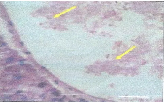
Fig. 2 Thin blood film smear showing red cells of P.berghei infected and treated with ethanol extract of Phyllantus amarus treated at day 4. Red arrows represent parasitized zones; yellow arrows show cleared zones
Fig3. Thin blood film showing red cells of Plasmodium berghei from infected mice not treated at day 4. Red arrows show parasitized zones. There are no cleared zones.
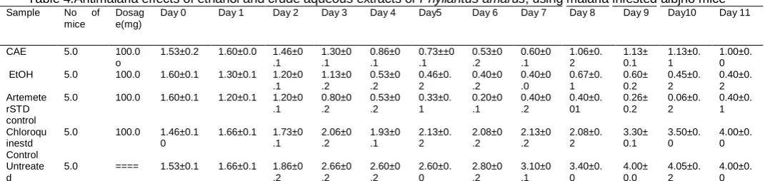
Table 4.Antimalaria effects of ethanol and crude aqueous extracts of Phyllantus amarus, using malaria infested albjno mice
Sample No of Dosag Day 0 Day 1 Day 2 mice e(mg)
Day 3 Day 4 Day5 Day 6 Day 7 Day 8 Day 9 Day10 Day 11
CAE 5.0 100.0 o
1.53±0.2 1.60±0.0 1.46±0 .1
1.30±0 0.86±0 0.73±±0 0.53±0 0.60±0 1.06±0. 1.13± 1.13±0. 1.00±0.
.1 .1 .1 .2 .1 2 0.1 1 0
EtOH Artemete rSTD control Chloroqu inestd Control Untreate d 5.0 100.0 5.0 100.0 5.0 100.0 5.0 ==== 1.60±0.1 1.60±0.1 1.46±0.1 0 1.53±0.1 1.30±0.1 1.20±0 .1 1.20±0.1 1.20±0 .1 1.66±0.1 1.73±0 .1 1.66±0.1 1.86±0 .2
1.13±0 0.53±0 0.46±0. 0.40±0 0.40±0 0.67±0. 0.60± 0.45±0. 0.40±0.
.2 .2 2 .2 .0 1 0.2 2 2
0.80±0 0.53±0 0.33±0. 0.20±0 0.40±0 0.40±0. 0.26± 0.06±0. 0.40±0.
.2 .2 1 .1 .2 01 0.2 2 1
2.06±0 1.93±0 2.13±0. 2.08±0 2.13±0 2.08±0. 3.30± 3.50±0. 4.00±0.
.2 .1 2 .2 .2 2 0.1 0 0
2.66±0 2.60±0 2.60±0. 2.80±0 3.10±0 3.40±0. 4.00± 4.05±0. 4.00±0.
.2 .2 0 .2 .1 0 0.0 2 0
Values in the table are the Mean ± SD from triplicate determinations. Values having the same superscript are significantly the same at p≤0.05 and different from others.
1
Table 5. Mineral content of the leaves of Phyllantus amarus. Values expressed as mg/g or
mgg-Mineral(mg/g) Ca K Na Mn Mg Fe Cu Zn Cd Pb
Conc(mg/g 157.42±1.42 11.34±0.10 14.33±0.2 7.92±0.2 59.62±0.15 34.55±0.15 3.08±0.11 3.30±0.22 - -Values in the table are the Mean ± STD from triplicate determinations. Cadmium and lead were not dictated in the sample.
DISCUSSION
The normal Swiss mice injected with Plasmodium berghei and not treated(Group 5) and Group 4 treated with chloroquine became weak after day 6; while the infected Group 3 treated with Artemeter (10 mg/kg bw) survived and the parasitemia cleared in day 4. However, comparing day 0 and day 11 of treatments with the ethanol extract of P. amarus , the level of parasitemia greatly reduced from (1.60±0.2 to 0.4±0.2) . The ethanol extract was more efficacious than the crude aqueous extract (CAE) in reducing the parasitemia (1.53±0.2 to 1.0±0.2) as compared to (1.53±0.1 t0 4.0±0.1) in the untreated group. Comparisons made between day 0, day 4, day 8 and day 11, imply that the parasitemic level of the infested animals treated with ethanol and crude aqueous extracts of the plant showed significant difference (p≤0.05) when compared with other treatment groups like the infested untreated group and the chloroquine treated group. There was no significant difference (p≤0.05) observed when Group 4 mice, treated with chloroquine and Group 5 (untreated animals) were compared. The difference in the efficacy of the ethanol and aqueous extracts may be due to the concentration of the active metabolites in the two extracts. Although the parasitemia was not completely cleared in the group treated with the ethanol and aqueous extracts, there was dramatic reduction as shown in table 5. The antimalarial effects of extracts from Phyllantus amarus have been documented (Ogata et al., 1992;Bagaran et al.,2011). Apart from the treatment of malaria, the plant has equally exhibited various actions in the treatment of hepatitis B and C (Akinjogunta et al.,2010;Xia et al.,2011 ) and even urolithiasis (Ajala et al.,2011)
al.,1999;Alison,2009).Apart from its role as an antimalaria plant,a lot of literature have cited and indicated enormous biochemical roles such as hepatoprotective, anticancer,anti HIV 1 and 2 (Godwin,2010;Taesokikul et al.,2012;Thakur et al.,2012).Others include dyslipidemic,antimicrobial,anti-hepatitis B and C(Appiah-Opong et al.,2011;Nayak et al.,2010;Abhyankar et al.,2010). As a result of the endemicity of malaria parasite and its ubiquitousness arising from more than 200 species of Plasmodia and of which more than 11 species use man as their secondary host; malaria remains a sickness with the highest rate of mortality and morbidity (Collins et al.,2008;Perkins and Austin,2008). We suggest that research be intensified in the discovery of phytomedicines as a panacea for the therapy of most tropical diseases such as malaria and sickle cell disease. There is no doubt that the antimalarial activity of the plant extracts(Phyllantus amarus) compared favorably with that of the most current antimalarial, Artemeter, that has overcome chloroquine resistance in chloroquine resistant Plasmodium falciparum malaria.This plant endowed with the preponderance of phytochemicals , nutrients vitamins( some antioxidants) and mineral elements, would onetheless provide radical cure for all types of benign malaria attacks and their accompanying sequale when administered at appropriate dosages..
REFERENCES
Abhyankar G, Suprasanna P. Pandey BN2010. Hairy root extract of Phyllantus amarus induces apoptic cell death. Innovative Food Science and Emerging Technologies, 11 (3):526-532
Adebayo JO, Kareth AU 2010. Potential anti-malarial from Nigeria plants, a review. Journal Ethnopharmacology, 133(3): 289-302 Adekunle MF (2008). Indigeneous uses of plant leaves to cure malaria fever at OmO,Forest reserve(OFR),
Ajala TO,Igwilo CI,Oreagba IA 2011.The antiplasmodial effect of the extracts and formulated capsules of Phyllantus amarus on Plasmodium yoelli infection in mice. Asian Pacific Journal of Tropical Medicine, 4(4):283-287
Akinjogunta OJ,Eghafona NO, Enabulele IO(2010) Antibacterial activity of ethanolic extracts of Phyllantus amarus against extended spectrum of beta(β) lactamase producing Escherichia coli isolated from stool samples of HIV sero-positive patients with or without diarrhea. African Journal of Pharmacy and Pharmacology,4 (6); 402-407
Alison AC 2009. Genetic control of resistance to human malaria . Current Opinion in Immunology, 21(5):499-505 AOAC 1990.Official methods of Analysis of the Association of Official Analytical Chemists.
Appiah-Opong R,Nyarko AK, Dodoo D 2011.Antiplasmodial activity of extract of Tridax procambens and Phyllantus amarus in in vitro
Plasmodium falciparum culture system. Ghana Medical Journal, 45(4):143-150
Asfaw D , Abebe D and Urga K 1999.Traditional medicine in Ethiopia. Perspectives and Development Efforts .Journal Ethiopian M edical Practice, 1(2): 114-117
Babu LT, Larry AW 2005 .Targeting the hemozion synthesis pathway for antimalarial drug recovery: Technologies for In vitro β-Hematin formation assay.Combination Chemistry and High Throughput Screening. Proceedings Natl, Acad. Sci, 8:63-79
Bagaran A, Rahuman AA, Kamaraji C, Kaushik NK, Mohana Krishman D, Sahal D 2011. Antispasmodial activity of botanical extracts against Plasmodium falciparum. Parasitol Res, 108(5):1099-1109
Canali S. 2008.Researches on thalassemia and Malaria in Italy and the origins of the Haldane hypothesis. Med. Secoli, 20(3):827-846 Carter R, Mendis KN 2002 .Evolutionary and Historical Aspects of the burden of Malaria. Clinical Microbiology Rev. 15(4): 864 - 941
Collins WE, Sullivan JS, Nacc D,Williams A, Barnwell JW 2008 .Observations on the sporozoite transmission of plasmodium vi vax to monkeys.Journal Parasitol. , 94(1):287-288
Cox F 2002. History of Human Parasitology. Clinical Microbiology Review, 15(4):595-612
David AF, Philip JR, Simon RC, Reto B, Solomon N 2004.Antimalarial drug discovery; efficiency models for compound sc reening. Nat. Review, 3: 509-520
Dobson MJ 1994.Malaria in England. A geographical and historical perspective .Parasitology , 36(1-2):35-60
Ene AC, Amah DA, Kwanashie HD, Agomo PU and Atawodi SE 2008a. Preliminary in vivo anti-malarial screening of petroleum ether, chloroform and methanol extracts of fifteen(15) plants grown in Nigeria. Journal Pharmacological Toxicology,3 (4): 254 - 260 Ene AC, Atawodi SE, Ameh DA, Kwanashie HO, Agomo PU 2008b Experimental Induction of chloroquine resistance in Plasmodium
berghei (NK65). Trends in Med. Research,3 (1):16-20
Ene AC,Atawodi SE, Ameh DA, Kwaneshie HO, Agomuo ,PU 2008c Locally used plants for malaria therapy among Hausa, Yoruba and Ibo communities in Maiduguri, North eastern Nigeria.
Godwin A 2010. Effectiveness of some medicinal plants decoction in the treatment of Malaria in Nigeria. Annals of Biological Research, 1(2):230-237
Goldberg M 2012.Survival of malaria parasites in the human host. Howard Hughes Medical Institute,USA
Hayakawa T, Arisue N, Udono T 2009. Identification of Plasmodium malariae, a human malaria parasite in imported Chimpanzees. PLoS ONE 4(10):e7412
HayakawaT,Culleton R, Otai H, Horri T, Tanabe 2008.Big bang in the evolution of extant malaria parasites. Mol. Biol. Evol.,25 (10): 2233-2239
Hempelmann E, Tesarowiez I, Oleksya BJ 2009.“ Kuregefasste Geschichte der Malaria-Chemotherapre Von Zwiebelbiszum Artemisinin” Pharm Unsever Zeit , 38(6): 500-507
Iranloye BO, Owoyele VB, Kelani OR 2011. Analgesic activity of aqueous leaf extracts of Phyllantus amarus. African Journal of Medicine and Medical Sciences, 40(1): 47-50
Joy DA,Feng X ,Mu J, Furaya T, Chotivanich K, Kretti AU,Ho M, Wang A., White NJ, Suh E, Beerti P, Su Xa 2000 “Early origin and recent expansion o Plasmodium falciparum” Science,300(5617): 318-321
rd
Publishers,USA PP 730-732
Karuma R, Bharathi VG, Reddy SS 2011 Protective effects of Phyllantus amarus aqueous extract against renal oxidative stress in Streptozotocin-induced diabetic rats. Indian Journal of Pharmacology, 43(4):414-418
Nayak PS, Upadhyay A, Dirivedi SK 2010.Quantitative determination of Phyllanthrin in Phyllantus amarus by high performance thin layer chromatography. Plantas Medicinales Aromaticas, 9(5):353
Neils O Verhulst, Renate C Smallegange, Willem Takken 2012. Mosquitoes as potential bridge vectors of malaria parasites from non-human primates to non-humans. Frontiers of Physiology,3(197)1-3
Njunda AL , Assob NJC, Nsagha SD, Kamga FHL,Mokenyu MD, Kwenti T 2013 Comparison of capillary and venous blood using bood fi lm microscopy in the detection of malaria parasites. A hospital based study. Scientific Journal of Microbiology,2(5):89-94
Nwaoguikpe RN 2012 . Functions of Dehydrogenases in Health and Disease. In Dehydrogenases -INTECH-Open mind and Open Science (chapter 7).Rose Angela Canuto (Ed.) ISBN-978-953-307-019-3 PP 165-180
Ogata T, Higuchi H, Mochida S. 1992“HIV-1 reverse transcriptase inhibitor from Phyllantus niruri” AIDS Research and Human Retroviruses, 8(11):1937-1994
Ogun State), Nigeria. Ethiopean Journal of Environmental Studies and Management, 1(1) 321
Okwu OO, Sanyaolu LO, Olakunbo AF 2012.Malaria and working performance in a Nigerian Uiversity. Research Journal of Biology,02 (05):151-156
Patel JR, Tripathi P,Sharma V 2011. Phyllantus amarus, ethnomedicinal uses, phytochemistry and pharmacology,a review. Journal Ethnopharmacology,138 (2): 286-313
Patel JR,Tripathi P. Sharma V , Chauhen NS, Dixit VK 2011. Phyllantus amarus, ethomedicinal uses, phytochemistry and pharmac ology, a review. Journal of Ethnopharmacology, 135(2): 286-313
Perham PE, Christiansen-Jucht , Pople D and Michael E 2011. Understanding and Modeling the impact of climate change on infectious diseases. In Tech- Progress and Future challenges in climate change. Blanco J and Khevadmand H (eds) ISBN 979-9533072776:43-46
Perkins SL. Austin C 2008. Four new species of Plasmodium from New Guinea Lizards: integrating morphology and molecules. Journal Parasitology, 95(2):1-6
Poinar G. 2005 . Plasmodium dominicann (Plasmodiidac hemospororida) from tertiary Dominican Amber. System Parasitology, 6(1): 47-52 RavikumarYS, Ray U, Nandhitha M 2011 Inhibition of hepatitis C virus replication by herbal extracts of Phyllantus amarus, as potent natural
source. Virus Research, 158(1-2):89-97
Samuel TA,Okonessien ED, Akande IS 2011. Phytochemical screening and the effects of aqueous extr acts of Phyllantus amarus leaves on lipid profile and cardiac muscle cyclic guanosine monophosphate of male guinea pigs. Planta Medica, 77(12): 1324 - 1325 Subrmananian S, Hardt M, Choe Y, Niles RK, Johansen EB,Leqac J, Gut J, Kerr ID,Craik CS, Rosenthal PJ 2009. Hemoglobin cleava ge
site-specificity of the Plasmodium cysteine proteases falcipain - 2 and falcipain-3 digests hemoglobin to provide free amino acids for parasite protein synthesis. PLoS ONE, 4(4):e5156
Taesokikul T,Nakajima M,Tassanepyaku IW 2012 .Effects of Phyllantus amarus on the Pharmacokinetics of Midazolam and Cytochrome P450 activities in rats . Xenobiotica, 42 (7):641-648
Thakur JS, Agarwal RK,Kharya MD 2012. Immobilization mediated enhancement of Phyllantrin and Hypophyllantrin from Phyllantus amarus. Chinese Journal of Natural Medicines,10 (3): 207-212
WHO 2005.World Medical Report. Roll Back Malaria (WHO-UNICEF) WHO/HTM/MAL/2005’1102
Xavier JR,Gnanam R, Muragan MP 2010.Clonal propagation of Phyllantus amarus. A hepatoprotector. Pharmacocognosy Magazine, 8(29):78-82.
Xia Y,Lao JP, Gluud C 2011.Phyllantus species for chronic hepatitis B virus infection. Cochrane Database Syst. Res. 4CD0089 60.