-'.'~
ID
Venous wall function in the pathogenesis of
..
vancose vems
G. Heather Clarke, PhD, S. N. Vasdekis, MD, J. T. Hobbs, FRCS, and A. N. Nicolaides, MS, London, U.K., and ,i\.felbourne, Australia
the cause
oi uaricose ueins, citing
wunrespect to these three factors. Duplex scanning techniques were used to assess the venous valves, and simultaneous measurements of calj volume (strain-gauge plethysmography} and venous pressure made dunng venous occlusion plethysmography were used to determine the elasticity of the venous wall and the Tale of arterial inftow. Fifty-one controllegs and36 legs with superficial venous insufficiency were examined. Risk factors were used tq divide the controllegs into two groups: low risk or normal
(23 legs) and high risk (28Iegs). The results obtained in the high-risk limbs demonstrated a significantly reduced vein wall elasticity (p < 0.001) and increased arterial inftow (p < 0.005) compared with the normallimbs, with no corresponding in crease in the incidence of valvular incompetence. I}""", .."""It" rl"n..I.. ".,""",,1
tJ,nl-the role o the vein wall an t e Tale o arterIa In ow.
From the lrvine Laboratory for Cardiovascular lnvestigation and Research, Academic Surgical Unit, St Mary's HosPital Medical School, London, U.K.
V ARICOSE VEINS ARE ASSOCIA TED, by definition, with incompetent malfunctioning val ves and dilatation and weakening of the vein walls. It is not clear, however,
whether the valvular incompetence
occurs first and
produces the vein wall dilatation or vice versa. Three theories have evolved to explain the cause of varicose veins, each attributing their development to an inherent primary factor.1 The three factors proposed are a weakness of the venous valves,l, 2 a weakness of the vein wall,I,3 and multiple arteriovenous communica-tions.l, 4 The association between varicose veins and malfunctioning venous valves or dilated vein walls is self-evident. This is not so with multiple arteriovenous anastomoses. The latter was proposed initially on the basis of serial arteriography4 demonstrating an
abnor-SupfIOrted bythe Cardiovascular Disease Educational and Research Trust, the Greek State Scholarship Foundation, and the Scholl Foundation.
Accepted for publication Jan. 21, 1991.
Reprint requests: Heather Clarke, Department of Medical
Radiations Science, Royal M~lboume Institute of
Technol-ogy, Melboume, Victoria 3001, Australia. 11/56/28745
mally rapid movement of blood from the arterial to the venous system in limbs with varicose veins and periop-erative visualization of direct arteriovenous communi-cations with an operating microscope. Subsequent non-invasive investigations that used plethysmographic tech-niques indicated that the maximum arterial inflow to the limb was significantly higher in limbs ,vith varicose veins.5-7 The exact nature of the communications has not been established, however, and there is no evidence to refute the suggestion that they might be damaged and dilated capillary vessels.8
The relative etiologic roles of the three factors proposed have not been resolved by previous investiga-tion. This omission may largely be attributed to the ab-sence of any established method of assessing venous wall function and the problems of identifying patients in the early stages of development of varicose veins suitable for such etiologic studies. The development of a method for determining the elasticity of the venous wall in vivo has helped to overcome these difficulties.9, 10
The aim of this study was to i.nvestigate normallimbs, high-risk limbs, and limbs with established varicose veins by evaluating the venous valve function, the vein
Volume 777
Number 4 V enous ~all jUnction and varicose veins 403
100
a
J: E E--~
~~
~ ~~
..
no 90%(RP-AVP)
.l RT90-RP AVP1
Time
-o
Fig. 1. Schematic drawing of a typical ambulatory venous pressure recording shows the resting pressure (RP) the measured ambulatory venous pressure (AVP), and 90% refilling time (RTgo).
Methods of investigation. Four investigative proce-dures were performed in this study: ambulatory venous pressure measurements,10,11 duplex Doppler scan-ning,10, 12, 13 assessment ofvenous elasticity,9,10 and measurements of maximum arterial inflow.5-7, 10 The ambulatory venous pressure measurements were per-formed to assist in selecting the high-risk limbs. The etiologic studies ,vere based on the other three methods of investigation. Duplex Doppler scanning ,vas used to assess the valve function, the measurement of venous elasticity to assess venous wall function, and the mea-surements of maximum arterial inflow to assess arterial inflow.
Ambulatory venous pressure measurements.
Am-bulatory venous pressure was measured directly by
cannulating a vein on the dorsum of the foot. The pa-tient was asked to stand, holding a frame for support and allowing the baseline resting pressure to be established. The patient was then asked to perform 10 tiptoe move-ments at arate of one per second to empty the veins and then to remain still while the veins refilled. The two
measurements
taken from these recordings were the
ambulatory venous pressure, defined as the pressure immediately after exercise, and the R T 90, which is the time taken for the pressure to effect a 90% recovery of the preexercise level (Fig. 1).
Duplex Doppler scanning.10,12 Duplex scanning was used toassess valvular efficiency in the lower-limb veins of the subjects studied, with a 7.5 MHz imaging probe coupled with a 5 MHz Doppler crystal (DRF 300; Diasonics Incorporated, Milpitas, Calif.). The probe was placed ayer the vessel of interest, and the Doppler sample volume was positioned within the vein so that the angle of insonation was approximately 60 wall function, and the rate of arterial inftow, in an
at-tempt to determine the cause of varicose veins.
MATERIAL AND METHODS
Subjects. Twenty-three volunteers (32Iimbs) and 36 patients (55 limbs) referred from the vein clinic were investigated. (Care ,vas taken to exclude subjects who were taking vasoactive medication or wearing elastic support stockings.) The 87 limbs studied were com-prised of 51 controllimbs and 36 limbs with superficial venous incompetence and normal deep veins. The con-trol limbs included both volunteer limbs (n = 32) and the apparently normal contralateral limbs of patients with unilateral venous disease (n = 19). These control limbs were classified into two groups of normal and high-risk limbs according to the following criteria: (1) family history of varicose veins, (2) occupation involv-ing standinvolv-ing, (3) history of symptoms or signs associated with varicose veins, (4) presence of reftux detected with Doppler ultrasonography, and (5) abnormal ambula-tory venous pressure recordings: ambulaambula-tory venous pressure greater than 40 mm Hg and 90% refilling time (R T 90) less than 18 seconds.
If two or more of these criteria were present, the limb was classified as being at high risk of developing vari-cose veins (28 limbs). Otherwise they were classified as normal (23 limbs).
The material used in this investigation therefore is comprised of subjects in whom only one limb was stud-ied and subjects in whom both limbs were studstud-ied and classified either in the same group or in different groups. The distribution of patients was allowed for in the sta-tistical analysis of the measurements obtained by
404 Clarke el al. Surgery APril 1992 17cm Thlgh Cutt Inflaled lo 80 mmHg
2
1~__~;:;;--~
/
'
"'-~-Fig. 2. Simultaneous measurement of pressure and volume with proximal occlusion.
degrees. Manualcompression of the limb was applied distally, producing an outAow ofblood. The presence of retrograde Aow on release of compression indicated val-vular incompetence.
This method was used to determine the function of the valves in the deep (femoral, popliteal, gastrocnemius, posterior tibial, anterior tibial, and peroneal) and superficial (long saphenous, short saphenous, and Gia-comini) veins of the lower limbs. (The Giacomini vein ascends from the proximal part of the short saphenous vein deep in the posterior aspect of the thigh, parallel to the skin, and terminates as the posteromedial tributary of the long saphenous vein.) Variations from the stan-dard anatomy of the lower-limb venous system are fre-quent, and the investigations of the valve function were modified to the requirements of the individuallimb, ex-amining each vein at multilevels to ensure that there was no localized incompetence.
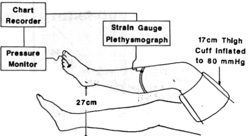
Measurement oí venous elasticity.9, 10 The elastic-ity of the lo,ver-limb venous system was assessed by standard strain-gauge plethysmography and direct mea-surements of venous pressure (Fig. 2). The patient was placed supine with one leg elevated 27 cm at the heel to allow the veins to empty. The knee was Aexed and ro-tated externally, and the thigh was supported to ensure that the leg ,vas relaxed. Strain-gauge plethysmography was performed with an electrically calibrated mercury-in-silicone rubber strain gauge placed around the calf at the maximum circumference, alÍowing percentage vol-ume change to be determined. Measurements of venous pressure were obtained by inserting a 21-gauge butter-Ay needle in a vein on the dorsum of the foot. The strain-gauge plethysmograph and pressure monitor were connected to a t,vo-channel chart recorder, allow-ing simultaneous measurements of pressure and volume to be made.
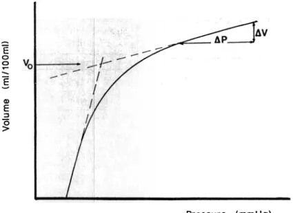
The pressure-volume relationship of the lo\ver-limb venous system was then obtained by placing a 17 cm \vide cuff around the thigh and inflating it to 80 mm Hg to occlude venous outflow. The subsequent changes in pressure and volume were recorded simultaneously and corresponding readings of pressure and volume were taken from these recordings at 15-second intervals and plotted on a graph (Fig. 3).
The change in pressure (~P) and the corresponding change in volume (~V) were measured in the linear, high-pressure part of the pressure-volume curve (Fig. 3). This linear region of the curve and the initial slope \vere extrapolated, as shown in Fig. 3, to obtain Vo, the theoretic value of the initial volume pertaining to the changes in volume at high pressures. The elasti.c mod-ulus (K) was calculated by substituting ~P, ~ V, and Vo, as shown in equation 1. This was converted to SI units of Nm-2 with a standard conversion factor from milli-meters of mercury (132.9): K = ~P/~V /Vo (1).
This method of calculating K is inherently variable beca use of the difficulty in determining the extent of the linear region of the curve and selecting the gradient. To standardize this maximum and minimum gradients \vere obtained from each graph, and the mean value was used to calculate K.
Measurement oí maximum arteria! inflow.5-7,10 The maximum arterial inflow was measured from the plethysmographic recording obtained in measuring the venous elasticity. The maximum arterial inflo\v was determined from the initial slope of the recording, as il-lustrated in Fig. 4, and expressed in units of percentage volume change per minute.
The gradient of the initial slope of the volume curve was not always clear, because In some recordings there was a very sharp change in volume initially, and the subsequentchanges in volume were more typical. This
E
o o.
-E
~ Q) E ;:, "O>
Pressure (mmHg)Fig. 3. Schematic drawing shows the calculation of the elastic modulus from the pressure volume relationship.
/
Plateau
/
E
o o...
-E
-~ E ~g
/
1/ MAl I/mln) 11 2% JJ~m~
Time.
I I Cuff InflatlonFig. 4. Schematic drawing shows the calculation of the maximum arterial inflo\v (MAl) from the strain-gauge plethysmographic recording.
initial artifactual change in volume may be attributed to one of two causes: patient movement or rapid distal movement of the blood in the veins beneath the occlud-ing cuff. The maximum arterial inflow in these limbs was measured by determining the initial gradient of the curve relating to the subsequent changes in volume.
were detected (Table 1). It should algo be noted that the re flux found in the four high-risk limbs was consistently evident in only one limb; in the other three limbs it was detected on one visit but not on a subsequent visito This
phenomenon has been described as intermittent reflux. These findings were statistically compared with a x2test (Table 11).
Measurements oí K. The measurements of K ob-tained in the three groups of normal, high-risk, and primary varicose veins are shown in Fig. 5 on a logarithmic scale. It is clearly evident that K is higher in the normal group than in either the high-risk group or the group of limbs with primary varicose veins, and
RESULTS
Duplex Doppler Lindings. The presence of valvular incompetence in the superficial veins only was estab-lished in all the limbs with primary varicose veins and in four of the risk limbs; in the remaining 24 high-risk limbs and all of the normallimbs no sites of reflux
406 Clarke et al. Surgery
April. 1992
Table 11. Comparison bet"leen the three groups of limbs studied of the statistical significance (p value) obtained with the three methods of assessment used: val ve function (reflux), K, and arterial inflow Table l. Summary of results of etiologic
investigations, val ve function (re(fux), K, and arterial inftow
N
HRG
SVI
o
o
36 100 4 14 Reflux K Arteria! inflow13:6
2.0-90.6
1.10.5-2.8
N, Normal; HRG, high risk; SVI, superficial vein incompetence.Reflux No of limbs
0;.
K (104 Nm-2) Mean Range j: 2 SD MAl (mljdljmin) Median Range No. of limbs0.4-2.8
2.10.4-5.0
233.1
0.6-5.9
28
3.2
1.0-5.9 36N, Normallimbs; HRG, rugh-risk limbs; SVI, Jimbs with superficial vein in-competen~; MAl, maximum arterial inflow.
these same measurements in the patients with estab-lished varicose veins, there is no corresponding similar-ity in the incidence of valvular incompetence.
DISCUSSION
The findings of an etiologic investigation are strongly influenced by the patients investigated and the methods of investigation used. The former is important beca use the progression of a given physiologic change is depen-dent on how far the disease has progressed. The latter is algo important because it affects the sensitivity of the investigation to detecting the physiologic changes, par-ticularly in the earlier stages.
The identification of limbs in the early stages of de-veloping varicose veins is therefore crucial. In this study these limbs were found from the control population, which was comprised of normallimbs from volunteers and patients with unilateral venous disease. Recent studies have shown the presence of venous disease in the apparently normal contralaterallimbs of patients with unilateral venous ulceration.13 Extending this idea, it is not unreasonable to as sume that any apparently normal limb might have some degree of venous disease. It is the identification of these control limbs with subclinical disease that is difficult. This has been achieved in this study by a combination of risk factors and measure-ments. The limbs assumed to be in the early stages of developing varicose veins have in fact been referred to as a high-risk group, because this is appropriate to the method of classification used, and until follow-up studies are performed,there is no evidence to show that they will develop varicose veins.
Four of the limbs classified in the high-risk group were found to have reflux in the long saphenous vein. Despite having incompetent val ves in their long saphe-nous veins, these four limbs were nonetheless classified in the high-risk group, because on clinical examination they appeared normal, having no visible varicosities and no evident signs of venous disease.
there is no discernible difference in elasticity between the latter.
The measurements of K were transformed to a nor-mal distribution by taking logarithms. The mean and range given in Table 1 were obtained by calculating the mean and the mean ::!: 2 SD for the transformed data and taking antilogarithms. The .levels of significance given in Table 11 were obtained by application of the Student t test to the transformed data (p < 0.001).
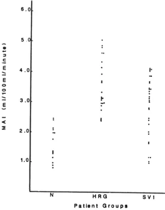
Measurements oí maximum arterial inflow. The measurements of arterial inflow obtained in the three groups of limbs investigated are shown in Fig. 6, and the mean and range are given in Table l. These measure-ments are not normally distributed, so the Mann-Whitney U test was used for statistical comparison be-tween the three groups. This showed that the maximum arterial inflow is significantly higher in the high-risk limbs (p < 0.01) and in the limbs with primary varicose veins (p < 0.005) than in the normallimbs, but no sig-nificant difference was found between the Corroer
(Ta-ble 11).
Etiologic findings. The resultsobtained are summa-rized in Table 1, giving the incidence of valvular incom-petence, the mean and range of K, and the median and range of the maximum arterial inflow in the three gróups of normal, high-risk, and primary varicose veins. The statistical significances of these findings are sum-marized in Table 11.
These results indicate that there is a significant dif-ference in the overall findings of all three investigations between the normallimbs and the limbs with primary varicose veins. The results fron} the high-risk limbs demonstrate that although K and the maximum arterial inflow measured in these limbs ~re comparable with
Volume 111 Number 4
Venous wal(function and varicose veins 407
6. 100.0 5 00 I
1-~.
I-.
~ c: E E o o ~ E o(~
::.4.
1" a..
i a a -N E z1,
)( ~.
~ " ~ o ~9-.
.! UJ 3..
-2i.
(: ,.l.
lo I..
l.
¡.
j 1. 0.1 HRG SVI Patient GroupsFig. 6. Maximum arterial inflow (MAl) for the three patient groups. N, Normal; HRG, high risk; SVI, superficial vein incompetence.
N HRG SVI
Pallenl Groupa
Fig. 5. Elastic modulus (K) for the three patient gl"oups. N, Normal; HRG, high risk; SVI, superficial vein incompetence.
The other aspect of this study that should be consid-ered is the sensitivity of the methods of investigation used in detecting early physiologic changes. The tech-niques used fall into two categories: duplex scanning provides an anatomically specific and localized assess-ment of the presence of reflux in individual veins within the limb, and the other two techniques provide an over-all and generalized assessment of the limbo
The localized nature of the duplex scanning tech-nique indicates the importance of an extensive and thorough examination to minimize the possíbility of missing any localized incompetence. The dupl~x exam-ination performed in this study was both careful and extensive, checking the deep and superficial veins at multilevels for sites of reflux. This examination cannot exclude histologic changes in the venous val ves, but in terms of the etiologic theories it is the function of the val ve that is important, beca use in the presence of val-vular competence there is no increased pressure distally to produce localized dilatation of the vein wall.
The generalized nature of the plethysmographic techniques indicates that the assessment of venous elas-ticity and arterial inftow mar be less sensitive to sma!! or localized changes, particularly if they are localized proximal to the calCo
The results obtained show increased arterial inftow and decreased elasticity in the high-risk limbs but no corresponding increase in the incidence of valvular in-competence. The thoroughness of the duplex examina-tion precludes the explanaexamina-tion of this finding in terms of undetected localized incompetence. These results do not differentiate between the roles of the vein wall and the arterial inftow. This mar indicate that the selected high-risk limbs have venous disease that has progressed beyond a stage in which only one physiologic pwame-ter has changed. Alpwame-ternatively this mar indicate that the changes in these two physiologic parameters are inter-active and effectively occur simultaneously.
The theory proposed to explain the development of varicose veins in terms of arteriovenous malformations4 is based on the premise that the anastomoses are mul-tiple and localized and produce localized changes in the
10.0