Review Article
Effects of Physical Exercise on Neuroplasticity and Brain Function:
A Systematic Review in Human and Animal Studies
Matheus Santos de Sousa Fernandes
,
1Tayrine Figueira Ordônio,
2Gabriela Carvalho Jurema Santos
,
3Lucas Eduardo R. Santos
,
1Camila Tenório Calazans,
4Dayane Aparecida Gomes
,
1,5and Tony Meireles Santos
1 1Neuropsquiatry and Behavior Science Postgraduate Program, Federal University of Pernambuco, Recife, PE, Brazil2Laboratório de Imunopatologia Keizo Asami-LIKA, University of Pernambuco, Recife, PE, Brazil 3Academic Center of Vitoria de Santo Antão, Federal University of Pernambuco, PE, Brazil 4School of Physical Education, University of Pernambuco, Recife, PE, Brazil
5Department of Physiology and Pharmacology, Center of Biosciences, Federal University of Pernambuco, Recife, PE, Brazil Correspondence should be addressed to Tony Meireles Santos; [email protected]
Received 29 August 2020; Revised 2 November 2020; Accepted 30 November 2020; Published 14 December 2020 Academic Editor: Grzegorz Hess
Copyright © 2020 Matheus Santos de Sousa Fernandes et al. This is an open access article distributed under the Creative Commons Attribution License, which permits unrestricted use, distribution, and reproduction in any medium, provided the original work is properly cited.
Background. Physical exercise (PE) has been associated with increase neuroplasticity, neurotrophic factors, and improvements in
brain function.Objective. To evaluate the effects of different PE protocols on neuroplasticity components and brain function in a
human and animal model.Methods. We conducted a systematic review process from November 2019 to January 2020 of the
following databases: PubMed, ScienceDirect, SciELO, LILACS, and Scopus. A keyword combination referring to PE and neuroplasticity was included as part of a more thorough search process. From an initial number of 20,782 original articles, after reading the titles and abstracts, twenty-one original articles were included. Two investigators evaluated the abstract, the data of
the study, the design, the sample size, the participant characteristics, and the PE protocol.Results. PE increases neuroplasticity
via neurotrophic factors (BDNF, GDNF, and NGF) and receptor (TrkB and P75NTR) production providing improvements in
neuroplasticity, and cognitive function (learning and memory) in human and animal models.Conclusion. PE was effective for
increasing the production of neurotrophic factors, cell growth, and proliferation, as well as for improving brain functionality.
1. Introduction
Environmental stimuli throughout life can result in
struc-tural and functional changes in organs and tissues (Westneat
et al. [1]). These changes are more propitious in structure
that has plastic characteristics, as for example the brain,
sus-ceptible to changes from development to aging
(Martínez-Morga et al. [2]). This process is called neuroplasticity and
is de
fi
ned as the capacity of the central nervous system to
promote the neurogenesis and connections due to
psycho-physiological and environmental factors (Gulyaeva [3]). In
this sense, neuroplasticity occurs with an increase in the
pro-duction of neurotrophins that generate changes in the
growth and di
ff
erentiation of cell signaling (Kempermann
et al. [4]).
Neurotrophins are a family of proteins closely related to
the survival, development, and functionality of the central
and peripheral nervous systems (Kozorovitskiy and Gould
[5]; Yamaguchi et al. [6]). The main neurotrophins involved
in the neuroplasticity process are neurotrophic factor derived
from the glial cell line (GDNF), nerve growth factor (NGF),
neurotrophin 3 (NT3), neurotrophin 4 (NT4), and
brain-derived neurotrophic factor (BDNF). Studies show that reduced
levels of these neurotrophins, especially BDNF, are responsible
for decreased brain functions, such as memory, concentration,
and learning (Bekinschtein et al. [7]; Parrini et al. [8]).
Volume 2020, Article ID 8856621, 21 pagesThe activation process of the BDNF signaling pathway is
mainly regulated by tropomyosin-related receptor kinase B
(TrkB). A previous study showed that increased expression
of TrkB was able to reduce the appearance of brain changes,
such as depression (Zborowski et al. [9]). It has also been
observed that the activation of TrkB a
ff
ects neuronal dendritic
a
ff
orestation, spinogenesis, dendritic growth, and spinal
mor-phogenesis (Guo et al. [10]). Faced with such mechanisms
responsible for neuroplasticity, we seek to understand which
environmental factors can act to modulate this process.
In this context, PE has been described as an e
ffi
cient
modulator of the health status through increased
mitochon-drial bioenergetics, adenosine triphosphate (ATP) synthesis,
and reduced lipogenesis, reactive oxygen species (ROS)
pro-duction, endoplasmatic reticulum stress, and proin
fl
amma-tory cytokine production such as tumor necrosis factor
alpha (TNF-
α
) (Broxterman et al. [11]; Nakandakari et al.
[12]; Presby et al. [13]; Daou [14]; Tofas et al. [15]). In
addi-tion, recent works have shown that PE is able to promote
neuroprotection (Byun and Kang [16]; Martland et al.
[17]). A study conducted with elderly women found that 12
weeks of aerobic and resistance exercise improved cognitive
function and BNDF expression (Byun and Kang [16]). This
is due to the unique capability of the skeletal muscle to
increase activation to the cellular signaling pathways
con-nected to crosstalk between muscle and brain (Muchlinski
et al. [18]; Kato et al. [19]).
The brain is characterized by having a high plastic
capac-ity; it is necessary to elucidate how environmental factors,
such as PE, can in
fl
uence the production of neurotrophic
fac-tors providing improvements in brain functionality, through
signaling, growth, and cell di
ff
erentiation. The main aim of
this systematic review is to evaluate the e
ff
ects of di
ff
erent
PE protocols on neuroplasticity components in a human
and animal study. The second aim is to evaluate the e
ff
ects
of PE on the production of neurotrophic factors, signaling,
cell growth and di
ff
erentiation, and functional outcomes.
2. Material and Methods
The present study was performed following the guideline of
the PRISMA statement (Moher et al. [20]).
2.1. Strategy Search.
The researchers searched the scienti
fi
c
literature from November 2019 to January 2020, using the
following databases: PubMed (244), ScienceDirect (11,860),
SciELO (2), LILACS (96), and Scopus (104). The following
search terms were selected using Medical Subject Headings
(MESH): (
“
Physical Exercise
”
OR
“
Exercise, Physical
”
OR
“
Exercises, Physical
”
OR
“
Physical Exercises
”
AND
“
Plastic-ity, Neuronal
”
OR
“
Neuronal Plasticities
”
OR
“
Plasticities,
Neuronal
”
OR
“
Neuroplasticity
”
OR
“
Neuroplasticities
”
OR
“
Neural Plasticity
”
OR
“
Neural Plasticities
”
OR
“
Plasticities,
Neural
”
OR
“
Plasticity, Neural
”
). Reference lists of all
included studies were also reviewed for potentially eligible
articles.
2.2. Study Selection.
Two independent reviewers (MSSF) and
(GCJS) selected the articles according to the following
inclu-sion criteria: (1) written in English, (2) between the years
2010 and 2019, involving studies, (3) utilizing
“
physical
exer-cise
”
or its variations as intervention, (4) di
ff
erent brain
tis-sues, and (5) neuroplasticity, performed in animal and
human studies. Articles were included if they ful
fi
lled the
fol-lowing PICOS criteria (
P
articipants: animals and humans,
I
nterventions: physical exercise,
C
omparisons:
PE vs. No PE
groups
,
O
utcomes: neuroplasticity components,
S
tudy:
ani-mal models and human studies) (Yensen [21]). In the next
stage, a comparison was made between searches and
evalua-tion of titles and abstracts according to the eligibility criteria.
The selected abstracts were submitted to the second stage of
analysis, in which two other independent researchers
reviewed the articles completely and, by consensus, excluded
articles that did not meet the criteria. The data regarding the
characteristics of the samples, methodology, and main
out-comes found were extracted from the selected articles.
2.3. Data Extract.
The reviewers (MSSF) and (GCJS)
extracted the data studies on a preestablished database. The
data extracted from each study included: (1) study design,
(2) sample characteristics, (3) physical exercise intervention,
and (4) e
ff
ects on the neuroplasticity of di
ff
erent brain
tis-sues. All extracted data was entered into a spreadsheet in
Excel by the primary researcher and veri
fi
ed by another
researcher. Discrepancies were resolved by consensus.
2.4. Risk of Bias Assessment.
Two independent authors
per-formed an analysis of the risk of bias in the selected studies.
The methodological judgment provided by the Revman
5.3.0 program of the Cochrane Handbook program was used
(Higgins et al. [22]). The following are among the criteria of
the structure of the bias assessment: (1) generation of
ran-dom sequence, (2) concealment of allocation, (3) masking
of participants and personnel, (4) masking of the result
eval-uation, (5) result data incomplete, (6) selective reporting, and
(7) other bias. The studies were classi
fi
ed as low, medium, or
high risk of bias.
3. Results
3.1. Study Selection.
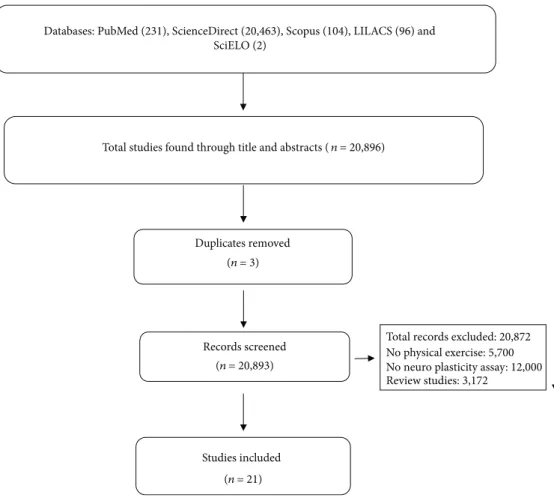
The
fl
owchart in (Figure 1) shows the
successive steps taken to select studies in this systematic
review. A total of 12,306 titles and abstracts were selected
ini-tially; 11,414 were excluded because they did not comply with
the eligibility criteria or were duplicated. Tables 1 and 2
pro-vide the information of the included articles.
3.2. Risk of Bias.
After this critical evaluation, the twenty-one
included studies were classi
fi
ed with low risk of bias
(Figures 2 and 3).
3.3. Description of Included Studies.
We identi
fi
ed 12,306
studies in the databases. Next, 11,414 were removed because
they had no data on PE (5201), neuroplasticity analysis
(4025), and because they were revision studies (2167). In
the end, 21 studies were included (Figure 1). Among the
selected, 15 articles were conducted on animals and six in
humans.
In animal studies, only one used female exclusively; 13
studies used male animals and one study used both female
and male animals. Among eleven studies used, three strains
of
Rattus norvegicus
including Wistar (six studies),
Sprague-Dawley (four studies), and spontaneous
hyperten-sive rats (SHR) (one study) were used. Four studies were
per-formed with C57BL6 (three studies) and BALB/CJ (one
study) mice. The animals were also exposed to models of
high-fat diet, diabetes, chemo brain, corticosterone
adminis-tration, transient middle cerebral artery occlusion,
posttrau-matic stress disorder, and transgenerational e
ff
ects of PE.
Among the brain area evaluated, 12 studies evaluated the
hippocampus, and the others evaluated the prefrontal,
orbi-tofrontal, and entorhinal cortex. Fourteen studies used
aero-bic PE protocol, of which 11 were on a treadmill and three
were voluntary exercise on a running wheel. Only one study
used the nonaerobic and strength exercise protocol.
In human studies, only one study used a sample
com-posed exclusively of women. The others used both sexes.
The participants
’
ages ranged from 18 to 80 years old. Among
the brain areas evaluated, three studies used magnetic
reso-nance imaging (MRI) to analyze the hippocampus; one
eval-uated the dorsolateral prefrontal cortex, posterior cingulate,
precuneus cortex, hand motor area, occipital lobe, and
cere-bellum. One of the studies did not report the assessed brain
area (Eftekhari and Etemadifar [39]). Of the six selected
stud-ies, two studies used strength training, two studies performed
dance activities, one study utilizes cycling, one used
com-bined exercise (aerobic, balance, weightlifting, and yoga),
and one study used balance and relaxation exercises.
3.4. E
ff
ects of Physical Exercise on Neurotrophic Factors.
In
some animal studies (Table 3) using aerobic exercise
(tread-mill and running wheel training, respectively), increased
expression of BDNF protein, receptor levels, and mRNA in
the hippocampus was observed (Aguiar et al. [24]; Gomes
da Silva et al. [23], Aguiar et al. [25]; Kim et al. [29]; Park
and Kim [32]; Vilela et al. [30]; Park et al. [34]). Likewise, a
study using resistance training showed an increase in BNDF
levels after training sessions (Vilela et al. [30]). In studies
with humans, the subjects were submitted to di
ff
erent PE
protocols, including pilates, dance, and sports that
demon-strated elevated serum and plasma BDNF levels that were
evaluated by the ELISA method (Müller et al. [40]; Eftekhari
and Etemadifar [39]; Rehfeld et al. [42]) (Table 4). However,
when di
ff
erent cycling intensity patterns were used, no di
ff
er-ences were observed on BNDF serum levels (Woost et al.
[43]).
Six works evaluated TrkB receptor expression in animal
studies (Table 3). Five studies observed increased TrkB
expression in the hippocampus after aerobic training (Gomes
da Silva [23]; Kim et al. [29]; Park and Kim [32]; Park et al.
[33]; Park et al. [34]). Only one study identi
fi
ed a reduction
in TrkB mRNA level when compared to control after exercise
Records screened Duplicates removed Studies included (n= 21) (n= 20,893) (n= 3)Total studies found through title and abstracts (n= 20,896)
Databases: PubMed (231), ScienceDirect (20,463), Scopus (104), LILACS (96) and SciELO (2)
Total records excluded: 20,872 No physical exercise: 5,700 No neuro plasticity assay: 12,000 Review studies: 3,172
Table 1: Description of included animal studies. Auth or (year) Aim Animal species/experimental groups Brain tissues Analysis Protocol of physical exercise Gomes da Silva et al. [23] To investigate the morphological and functional hippocampal changes in adult rats submitted to daily treadmill exercise during the adolescent period. Male Wistar rats at 21 days of age were divided into two groups: exercise ( n =2 7 ) and control ( n =2 7 ). Hippocampus Immuno fl uorescence; ELISA; immunoblotting An aerobic physical exercise protocol (running on a treadmill) was performed. Only animals that were classi fi ed as medium, above average, and good runner were included in the procedure. The physical exercise protocol was performed betwe en days 21 and 60 of life. Each session started with a 5-minu te warm-up at 8-10 m/minute. The running time and speed were gradually increased, reaching a maximum of 18 m/min for 60 min. Animals in the control group were treated in the same way as animals in the exercise group. Aguiar et al. [24] Investigate whether long-term light physical exercise on a running wheel or treadmill improves spatial learning, memory, and plasticity of the hippocampus in elderly rats. Female Wistar rats aged 24 months were divided into two groups: exercised ( n =1 7 animals) and sedentary controls ( n =1 8 animals). Hippocampus Western blotting; RT-PCR Animals underwent an adaptation period for 1 week on a treadmill, with a daily session of 3 minutes at a speed of 2 m/min. Then, the aerobic exercise protocol was started, which was performed for 4 consecutive weeks, with 4 sessions per week. Each exercise session consisted of a warm-up session (3 min, 2 m/min), followed by two sessions of running sessions (4-6 min) at a constant belt speed 10 m/min. Each session was spaced by a 1-minute rest interval. Aguiar et al. [25] To analyze the e ff ects of ph ysical exercise on mitochondrial physiology, anxiodepressive-like behaviors and neuroplasticity in mice. Male C57BL/6 mice aged 8-10 weeks old were divided into two groups: voluntary exercise ( n =1 6 ) and control gro up ( n =1 6 ). Hippocampus RT-PCR; HP LC; Western blotting; spectrophotometrical assays for assessing A voluntary physical exercise protocol was carried out in individual cages equi pped with a steering wheel to stimulate voluntary exercise for 6 weeks (Aguiar et al. [26]). Bhattach arya et al. [27] To determine the e ff ects of EGCG ( ∼ 250 mg/kg/day), B-ALA ( ∼ 550 mg/kg/day), and their combination with voluntary wheel running exercise. 91 mal e BALB/cJ mice at 10 weeks of age were di vided into eight groups: control ( n =1 1 sedentary and 11 runners), B-ALA ( n =1 1 sedentary and 12 runners), EGCG ( n =1 2 sedentary and 11 runners), or EGCG and B-ALA combined ( n =1 2 sedentary and n =1 1 runners). Hippocampus Immunohistochemistry A voluntary physical exercise protocol was carried out for 39 days. Wheel rotations were monitored continuously in one-hour increments throughout the experiment via magn etic switches interfaced to the computer.
Table 1: Continued. Auth or (year) Aim Animal species/experimental groups Brain tissues Analysis Protocol of physical exercise Brockett et al. [28] To investigate whether running alters performance on cognitive tasks that require the prefrontal cortex and whether any such changes are associated with astr ocytic, as well as neuronal plasticity. Adult male Sprague-Dawley male rats are divided into two groups: sedentary controls ( n =1 8 ) and runners ( n =1 8 ) groups. Hippocampus; medial
prefrontal cortex; orbitofrontal cortex
Immunolabeling for astrocyte and synaptic markers; DiI impregnation A protocol of voluntary ph ysical exercise in a running wheel was performed for 12 days. The running distance was recorded daily digital counters mou nted on the racing wheels. Kim et al. [29] To investigate the e ff ect of treadmill exercise on impairment of cognitive function in relation with hippocampal neuroplasticity using high-f at diet-induced obese mice. C57BL /6 mal e mice at four weeks of age were divided into four groups: control group ( n =1 0 ), control and exercise group ( n =1 0 ), high-fat diet group ( n =1 0 ), and high-fat group and exercise ( n =1 0 ). Hippocampus Western blotting; immunohistochemistry An aerobic exercise protocol was performed in treadmill for 20 weeks. Vilela et al. [30] To inv estigate the e ff ect of aerobic and strength training on spatial memory and hippocampal plasticity in aging rats. Male Wistar rats of 24 months old were divided into three groups: ( n =6 /group) untrained, aerobic training, and strength training gro ups. Hippocampus Western blotting Two physical exercise proto cols were performed. The aerobic exercise protocol was performed on a running treadmill. Each session las ted 50 minutes and there was an interval of 48 hours betwe en sessions. The anaerobic exercise protocol was performed through strength training. Climbing with 1 m inclined at 85 ° was carried ou t with weight attached to the tail. The weight attached to the tail was gradually increased from 50 to 100% during the 8 weeks of training. Three to fi ve sets of 8 to 12 repetitions, with a 1-minute rest betwe en repetitions and a 2-minute rest between sets, were performed for 3 or 4 days/week. Each session lasted 40 to 50 minutes, with an interval of 48 hours betwe en sessions. de Senna et al. [31] To investigate the e ff ects of physical exercise to prevent or reverse spatial memory de fi cits produced by diabetes and some biochemical and immunohistochemical changes in hippocampal astrocytes of type 1 diabetes mellitus mod el. Three-month-old male Wistar rats were divided into four groups: nontrained control ( n =1 5 ), trained control ( n =1 5 ), nontrained diabetic ( n =1 3 ), and trained diabetic ( n =1 3 ). Hippocampus Immunohistochemistry; morphological analysis of astrocytes. The aerobic physical exercise protocol was performed on a running treadmill at moderate intensity. The protocol took place once a day for 5x a week for 5 weeks. Park and Kim [32] To assess the e ff ects of paternal physical exercise on spatial learning ability in relation with hippocampal Male and female Sprague-Dawley rats of 4 weeks old were divided into four gro ups: nonexercising mal e and normal female group ( n =5 ), exercising male and Hippocampus Immunohistochemistry; Western blotting An aerobic exercise protocol was performed on a running treadmill. The exercise was performed once a day and 6 days a week for 12 consecutive weeks
Table 1: Continued. Auth or (year) Aim Animal species/experimental groups Brain tissues Analysis Protocol of physical exercise neuroplasticity in the rat pups born from the obese maternal rats. normal female gro up ( n =5 ), nonexercise male and obese female group ( n =5 ), and exercise male and obese female group ( n =5 ). [ fi rst 3 weeks (30 minutes — speed: 10 min/min); 4-6 weeks (40 minutes — 10 m/min); 7-9 weeks (30 minutes — 15 m/min); 10 to 12 weeks (40 min — 15 m/min)]. Park et al. [33] To determine whether symptoms of chemo brain and disruptions in the neuroplasticity and functioning of hippocampal mitochondria can be prevented or relieved by exercise. Male Wistar rats of 6 weeks olds were divided into four groups: control ( n =1 5 ), control and exercise ( n =1 5 ), DOX-induced chemo brain ( n =1 5 ), and DOX-induced chemo brain and exercise ( n =1 5 ). Hippocampus Immuno fl uorescence; immunohistochemistry; Western blotting An aerobic exercise protocol was performed on a running treadmill. The exercise was performed once a day (30 min at 10 m/min) and six days per week for 4 consecutive weeks. Park et al. [34] To investigate whether the decline in cognitive function caused by a high-fat diet could be improved through exercise by examining insulin signaling pathways and neuroplasticity in the hippocampus. Male C57BL/6 mice of 4 weeks old were divided into fi ve groups: control ( n =2 0 ), control and exercise ( n =2 0 ), exercise ( n =2 0 ), high-fat diet ( n =2 0 ), and high-fat diet and exercise ( n =2 0 ). Hippocampus Immuno fl uorescence; immunohistochemistry; Western blotting An aerobic exercise protocol was performed on a running treadmill. The treadmill exercise started 20 weeks after taking the HFD. The exercise was performed once a day and six days per week for 12 consecutive weeks [1-2 weeks (30 min — 10 m/min); 3-4 weeks (40 min — 10 m/min); 5-6 weeks (30 min — 13 m/min); 7-8 weeks (40 min — 16 m/min); 9-10 weeks (40 min — 16 m/min); 10-12 weeks (50 min — 16 m/min)]. Yau et al. [35] Examine the e ff ects of training on the administration of corticosterone on the hippocampus neurogenesis, cell proliferation and di ff erentiation, synaptic protein expression, expression of neurotrophic factors, and behavioral analys is. Adult male Sprague-Dawley rats were divided into four groups ( n =4 ‐ 6 ): (1) control rats (CON); (2) CORT-treated rats that could run only during the 2-week CORT administration period (CR); (3) CORT-treated rats with the 2 weeks prior running only (PR); and (4) CORT-treated rats that could run both prior and concurrently with the COR T adm inistration period ( PR + CR ). Hippocampus Immunohistochemistry; Western blotting A protocol of voluntary ph ysical exercise in a running wheel was performed for 34 days. The wheels were then unlocked for the rats with running for 14 days prior to the 50 mg/kg corticosterone treatment. Pan et al. [36] To explore the speci fi c role of physical exercise in novel object recognition memory after stroke and the exact cortical regions in which memory is restored by physical exercise. Spontaneously hypertensive rats of 10-12 weeks old were divided into four groups ( n =2 0 rats/group): control groups; tMCAO (2 d) group, in which rats underwent tMCAO surgery and NOR tests were performed 2 days later; the tMCAO (28 d) group, in which NOR tests were performed at 28 days post-Entorhinal cortex Histochemistry; Western blotting The physical exercise protocol was performed on a motorized racing wheel for 26 days from day 3 after treatment with tMCAO. During the fi rst 12 days, the speed of 3 m/min was maintained for 20 minutes, twice a day. In the foll owing 14 days, the speed was maintained at 6 m/min.
Table 1: Continued. Auth or (year) Aim Animal species/experimental groups Brain tissues Analysis Protocol of physical exercise tMCAO; and a tMCAO (28 d)+PE group, in which tMCAO was established and rats exercised in a running wheel for 26 consecutive days starting at the third day post-tMCAO. Ra belo et al. [37] To evaluate whether the intrinsic capacity for physical exercise in fl uences dopamine neuroplasticity induced by physical training. Male Wistar rats of 2 months old were divided into three groups: low performance LP ( n =8 ), standard performance (SP) ( n =8 ), and high performance (HP) rats ( n =8 ) which were randomized into the SED and TR groups. Not informed PCR The aerobic exercise protocol was performed on a treadmill for 6 weeks, 5 times a week. The training time was obtained through an exercise test (40% of the maxi mum time obtained during the test of 60% Vmax ) and the speed was kept constant. The duration of the training sessions was increased by 10% per week (except for the transition from the 3rd to the 4th week). Seo et al. [38] To evaluate whether exercise can improve psychiatric status and cognitive functioning, increasing the mitochondrial function of the hippocampus and neuroplasticity in a mod el of rats with posttraumatic stress disorder. Ma le Sprague-Dawley of 4 weeks old were divided into four groups: control (CON) group, a control and exercise (CON+EX) group, a PTSD group, and a PTSD and exercise (PTSD+EX) group ( n =1 5 in each group). Hippocampus Immuno fl uorescence; Western blotting; mitochondrial analys is The aerobic exercise protocol was performed on a treadmill once a day, 6 days a week, for 4 consecutive weeks. In the fi rst two weeks, the exercise lasted 30 min with a speed of 10 m/min. In the third week, the rats performed 40 minutes of exercise at a rate of 12 m/min. In the fourt h week, the exercise lasted 50 minutes at 13 m/min. B-ALA : β -alanin e; COR T: corticoste rone; Dox: do xorub icin; ELISA : enz yme-li nked imm unosorb ent assay; ECGC : epigalloca techin-3-gallate; EX: exer cise; GFA P: glial fi br illary acidic protein; HPLC : high-perform ance liquid chroma togra phy; NOR : novel object rec ognitio n; PE: phy sical exer cise; PTSD : posttr aumatic stress disord er; RT-PC R: real -tim e polymera se cha in reaction; SED: sede ntary; tMCA O: transie nt middle cerebral artery occ lusion; TR: trained .
Table 2: Description of the included studies with human. Auth or (year) Aim Sample description Brain tissue Other analysis Study design/protocol of physical exercise
Eftekhari and Etemadifar [39]
To determine the chronic e ff ect of Mat Pilates on serum levels of interleukin-10 and brain-derived neurotrophic factor in women with multiple sclerosis. 25 women are su ff ering from relapsing-remitting multiple sclerosis with expanded disability status scale 2-6, based on McDonalds criteria. They were divided into two groups: control group ( n =1 2 ; age yearsðÞ =3 4 : 46 ± 7 : 29 ) and Pilates training group (PT) ( n =1 3 ; age yearsðÞ =3 1 : 41 ± 8 : 89 ). — ELISA This is a randomized controlled study, whi ch was carried out from April 2015 to June 2015. The anaerobic exercise protocol was performed through Ma t Pilates training and lasted eight weeks, three times a week, lasting 30-40 min/day. The exercises were performed at low to moderate intensity according to the patient ’ s performance. The intensity of the exercise gra dually increased with the inclusion of more repetitions (3-10), decreasing the re st time and increasing the number of sets (1-2). Blood colle ction was carried out between 8 and 9 am to determine the serum levels of pre-and po sttest. The posttest blood sample was collected 48 hours after the last Pilates se ssion. Müller et al. [40] To assess whether a dance training program that emphasizes constant learning of new movement patt erns is superior in terms of neuroplasticity to conventional cond itioning with repetitive exercise and wheth er the extension of the training duration has additional bene fi ts. 62 healthy (63 to 80 years old) were recruited through advertisements in local newspapers. The following exclusion criteria were adopted: claustrophobia, tinnitus, me tal implants, tattoos, diabetes mellitus, depression, cognitive de fi cits, neurological diagnosis, and regular physical exercise (1 h/week). 10 participants were excluded from the criteria. Only 22 participants completed the entire intervention. The se were divided into two groups: dancing gro up ( n =1 2 ) and sport group ( n =1 0 ). Gyrus and right parahippocampal ELISA; MRI This is a controlled intervention study with a total duration of 18 months. The intervention was carried out in two periods: fi rst period (twice a week in sessions of 90 minutes for 6 months) and second period (training once a week in se ssions of 90 minutes for 12 months). Two protocols were used: dan ce gro up (participants were asked to lear n new sequences of mov ement, which required coordination; each rhythm was changed after the fourth se ssion). Sport group (participants performed strength-resistance training, with low demand for coordination; each se ssion lasted 20 minutes). The two training programs were comparable in terms of intensity, duration, and frequency. Baseline assessments were made after 6 and 18 months of training. Ji et al. [41] To assess whether motor skill causes changes in plasticity after exercise in di ff erent modalities. 24 healthy individuals ( 70 ± 7 : 78 years old; 12 women) were recruited through advertisements. These were divided into two groups: control ( n =1 2 ; 7 women; 73 ±8 years old) and trained gro up ( n =1 2 ;5 women; 67 ± 6 : 4 years old). Dorsolateral prefrontal cortex, posterior cingulate, precuneus cortex, hand motor area, occipital lobe, and cerebellum MRI The study was a q uasiexperiment. A physical exercise protocol was carried out that had four domains (aerobic, balance, weight lifting, and yoga). Participants were instructed to practice at home for 30 min every day for 6 weeks.
Table 2: Continued. Auth or (year) Aim Sample description Brain tissue Other analysis Study design/protocol of physical exercise Reh feld et al. [42] To evaluate the e ff ects of a dance program in the elderly on brai n plasticity. 62 volunteers were selected through a local advertisement. After the exclusion cr iteria, 52 elderly people (25 men; 27 women) aged 63 ± 80 years were randomly assigned to the experimental dance group (DG) and the control group to the sports group (SG). In the end, 38 participants comp leted the intervention. Dance group ( n =2 0 ) and sport group ( n =1 8 ). Frontal, temporal, cortical, and cerebellar regions ELISA; MRI This is an intervention study that used two protocols: dance intervention (subjects were trained to accurately memorize and access di ff erent rhythms and step se quences in space, all under accuracy and time pressure) and sport intervention (each session included three di ff erent units: endurance training, strength-endurance training, and fl exibility training). The protocols happened twice a week for 90 minutes and six months lon g. Wo ost et al. [43] To investigate whether a se quential combination of physical and spatial training in young, healthy adults elicits an additive e ff ect on training and transfer gains. 99 volunteers aged between 18 and 35 participated in the stud y. Hippocampus ELISA; MRI The stud y presents an experimental design. Participants performed eight sessions: (1) three weeks of 20 minutes of cycling per day classi fi ed based on high-int ensity training between T0 and T1 (ERGO); (2) fi ve weeks with 16 sessions of 30 minutes of space training between T1 and T2 (MAZE); and (3) a sequential combination of both (COMBO) or re sted as passive controls. Rogge et al. [44] To test if balance training, challenging the sensory-motor system and vestibular self-motion perception, induces structural plasticity. Participants were recruited through public announcements. Healthy adults between 19 and 65 years of age who did not report regular exercise were eligible for the study. 59 participants were randomized between gro ups; however, only 38 completed the study (24 women/14 men). Participants were divided into two gro ups: balance group ( n =1 9 ) and relaxation group ( n =1 8 ). Hippocampus; basal ganglia MRI It is an intervention study, where the participants carry out 12 training sessions, with two sessions per week, each lasting 50 min. Two protocols were performed: balance training (eight di ff erent stations per session) and relaxation training (progressive muscle relaxation and autogenic training). ELISA: enzyme-linked imm unosorb ent as say; MRI : magne tic resona nce.
protocol intervention (Vilela et al. [30]). Regarding GDNF
level, two studies showed that aerobic exercise was able to
increase protein and mRNA levels (Rabelo et al. [37]; Vilela
et al. [30]). The p75 neurotrophin receptor (p75NTR) was
evaluated by Vilela et al. (Vilela et al. [30]) which showed
an increase in P75NTR expression in both aerobic and
resistance training protocols compared to untrained animals.
Aerobic exercise was also able to increase the levels of
postsynaptic density protein 95 (PSD-95), phosphorylated
N-methyl-D-aspartate receptor (pNMDA), and synapsin
(SYN) when compared to the control (Yau et al. [35];
Brock-ett et al. [28]; Vilela et al. [30]) and hypertensive groups (Pan
et al. [36]).
3.5. E
ff
ects of Physical Exercise on Cellular Signaling Factors
That Stimulate Neuroplasticity.
The expression of cellular
signaling factors was investigated only in animal studies
(Table 3). The di
ff
erent aerobic protocols (treadmill and
running wheel) increased expression and phosphorylation
of alpha serine/threonine kinase (AKT), cyclic adenosine
monophosphate-responsive element-binding protein (CREB),
and cyclic adenosine monophosphate (cAMP)
phosphoryla-tion (Aguiar et al. [24]; Aguiar et al. [25]; Vilela et al. [30])
in the hippocampus. CREB levels were also increased after
strength training (Vilela et al. [30]). Sprague-Dawley rats that
were treated with corticosterone, a glucocorticoid released by
the adrenal cortex, obtained increased levels of insulin-like
growth factor-1 (IGF-1) in the hippocampus after performing
aerobic and strength training (Yau et al. [35]).
3.6. E
ff
ects of Physical Exercise in Neuronal Growth and
Di
ff
erentiation.
Only protocols with aerobic exercises
evalu-ated cell growth and di
ff
erentiation of brain areas.
Neurogen-esis was evaluated in animal studies. In this sense, PE
promoted an increase in the number of cells in the dental
gyrus, hippocampus, prefrontal cortex, and orbital cortex
(Bhattacharya et al. [27]; Brockett et al. [28]; Kim et al.
[29]; de Senna et al. [31]; Pan et al. [36]; Park et al. [34]).
However, in the perirenal cortex, these responses after PE
were not observed (Brockett et al. [28]). In addition, PE
increased cell di
ff
erentiation, proliferation, and survival in
the hippocampus of Sprague-Dawley rats (Yau et al. [35];
Park and Kim [32]). PE also increases the density of neuronal
fi
bers and dendritic column in animal studies (Gomes da
Silva et al. [23]; Brockett et al. [28]). In humans, MRI was
used to assess neural structures (Table 4). An increase in
the mass of white and gray matter was observed in di
ff
erent
regions (hippocampus, cortex, cerebellum, and basal
gan-glia) (Ji et al. [41]; Müller et al. [40]; Rehfeld et al. [42];
Rogge et al. [44]).
3.7. E
ff
ects of Physical Exercise on Cognitive Abilities.
In
Wis-tar rats, a reduction in latency (Aguiar et al. [24]; Gomes da
Silva et al. [23]; Vilela et al. [30]) and length of the swimming
track (Aguiar et al. [25]) were observed through the water
maze test and Barnes
’
maze, in trained Wistar rats with
aer-obic exercise and of strength compared to control (Table 3).
Better spatial memory capacity (Kim et al. [29]) and both
short-term (Park et al. [33]) and long-term (Park et al.
[34]) were also observed in the Y-maze and water maze tests
after the aerobic exercise protocol in Wistar rats and
C57BL/6 mice. Wistar rats and C57BL/6 mice trained in
aer-obic exercise showed faster learning (Aguiar et al. [25]) and
Aguiar (2011) R ando m s eq uence g enera tio n (s elec tio n b ias) B lindin g o f pa rt ici pa n ts a n d p er so nnel (p er fo rm an ce b ias) B lindin g o f o u tc o m e ass essmen t (det ec tio n b ias) In co m p let e o u tco m e da ta (a tt ri tio n b ias) S elec ti ve r ep o rt in g (r ep o rtin g b ias) Ot her b ias Allo ca tio n co nce almen t (s elec tio n b ias) Aguiar (2014) Bhattacharya (2015) Brockett (2015) De Senna (2017) Eftekhari, Etemadifar (2018) Gomes da Silva (2010) Ji (2017) Kim, Choi Chung (2016) Muller (2017) Pan (2017) Park (2019) Park, Kim (2017) Park, Kim (2018) Rabelo (2017) Rehfeld (2018) Rooge (2018) Seo (2019) Woost (2018) Vilela (2017) Yau (2014)Figure2: Risk of bias graph: review authors’judgments about each
risk of bias item presented as qualitative analysis across all included studies.
increased spatial learning (Aguiar et al. [25]) after the water
maze and Morris water tests. When aerobic PE was
per-formed by the parents, an increase in the puppies
’
spatial
learning capacity (Sprague-Dawley rats) was also
demon-strated (Park and Kim [32]).
In humans, the tests to assess memory were verbal
learn-ing test, attentional performance test, immediate, delayed,
and recognition recall from Hopkins Verbal Learning
Test-Revised, immediate and delayed story recall from the
river-mead behavioral memory test, WAIS-III digit symbol
substi-tution modality test, WAIS-III digit span, trail making test,
Stroop Color and Word Test, digit span forward and
back-ward of the Wechsler Memory Scale, Rey-Osterrieth complex
fi
gure test, VVM, subtests
“fi
gures,
” “
dices,
”
and
“
matrices
”
from the Intelligence Structure Test 2000R, and
“
California
Verbal Learning Test
”
(Table 4). In humans, memory
improvements have been demonstrated in combined exercise
(Ji et al. [41]) and dance and strength training protocols
(Rehfeld et al. [42]). After strength training, dancing and
cycling improvements in cognition were also observed
(Müller et al. [40]; Woost et al. [43]). Both strength training
and dance cause better responses about a short-term recall,
long-term recall, verbal long-term recognition, and attention
reaction (Müller et al. [40]). Woost et al. did not observe
dif-ferences on cognition and memory after cycling training.
Two studies evaluated the e
ff
ect of aerobic exercise on
anxiety through the elevated plus maze test and open
fi
eld
test in the animal (Table 3). It was observed that the trained
animals had increased time of exploration with open arms,
locomotion, and time of exploration (Aguiar et al. [25]; Seo
et al. [38]). In the open
fi
eld test, there was an increase in
exploration in the central regions, in addition to an increase
in locomotion (Aguiar et al. [25]). Depression was evaluated
in three studies using the tall suspension test (Seo et al. [38])
and forced swim test (Rogge et al. [44]; Seo et al. [38])
proto-cols. In all studies, it was observed that trained animals had
reduced immobility after exercise compared to the control
groups (Yau et al. [35]; Rogge et al. [44]; Seo et al. [38]). In
models with corticosterone administration, aerobic exercise
was also able to reduce immobility time (Yau et al. [35]). In
humans, no study has evaluated anxiety and depression
parameters.
4. Discussion
The present review con
fi
rmed the hypothesis that di
ff
erent
modalities of PE (aerobic and resistance training) are capable
of potentiating neuroplasticity in di
ff
erent species (animals
and humans), through the high production of neurotrophic
factors, cell signaling, growth, and development, resulting
in improved cognition. In addition, it was also found that
PE improved functional abilities such as state of anxiety
and depression in animals. PE, especially aerobic exercise,
was responsible for the high expression of brain trophic
fac-tors. Sleiman et al. [45] observed the increase in BDNF levels
in the hippocampus after thirty days of free access to the
run-ning wheel in mice. The increase in this neurotrophic factor,
according to the authors, may occur due to endogenous
pro-duction and a
ffi
nity of
β
-hydroxybutyrate, by BDNF
pro-moter regions (Sleiman et al. [45]).
Park et al. [33] observed that paternal exercise was able to
increase BDNF levels in the hippocampus, demonstrating a
transgenerational e
ff
ect like that observed in the study by
Yin et al. [46]. In this study, it was demonstrated that
pater-nal exercise provided increased levels of BDNF in the o
ff
-spring (Yin et al. [46]). The changes promoted by exercise
can be transmitted by epigenetic mechanisms, as well as the
decrease in DNA methylation in the o
ff
spring
’
s hippocampus
(Dyer et al. [47]).
In humans, results like those in animal studies have been
observed. An increase in serum/plasma BDNF was observed
after aerobic (dance and sports) and anaerobic (Pilates)
exer-cise. Hakansson et al. [48] realized a study with healthy
vol-untaries
who
performed
moderate-intensity
aerobic
exercise. They observed an increase in BDNF levels in the
elderly
’
s bloodstream after thirty-
fi
ve sessions of aerobic
exercise analyzed by ELISA (Hakansson et al. [48]). BDNF
produced peripherally can act as a metabolite in response
to the contraction of skeletal muscle (Jiménez-Maldonado
et al. [49]). This compound is carried by the blood stream
crossing the blood-brain barrier, promoting its activation in
the central nervous system (Jiménez-Maldonado et al. [49]).
When performing a cycling protocol in high-intensity
train-ing (HIT), Woost et al. (2018) showed no increase in BDNF
levels.
Random sequence generation (selection bias)
Blinding of participants and personnel (performance bias) Blinding of outcome assessment (detection bias) Incomplete outcome data (attrition bias) Selective reporting (reporting bias) Other bias
0%
Low risk of bias Unclear risk of bias High risk of bias
25% 50% 75% 100%
Allocation concealment (selection bias)
Table 3: Description of the main re sults of anima l studies. Auth or (year) Neuroplasticity outcomes Functional outcomes Neurotrophins and receptors Cell signaling Cel l growth and di ff erentiation Gomes da Silva et al. [23] An increase in BDNF expression was observed in the hippocampus ( p =0 : 00 ) and TrkB ( p =0 : 03 ) in the exercise gro up when compared to the control group. There were no di ff erences for P75NRT receptor ( p =0 : 69 ). — No signi fi cant di ff erences in neuronal density were found between the exercise group and control groups [CA1 ( p =0 : 49 ); CA3 ( p =0 : 66 ); DG ( p =0 : 69 )]. However, the density of mossy fi bers was higher in the hippocampal formation of the exercise group ( p =0 : 01 ) when compared to the control group. Learning and memory of animals from exercise and control gro ups were analyzed in the water maze for fi ve days. Latency ( p =0 : 00 ) and length of the swimming path ( p =0 : 00 ) in the exercise group were signi fi cantly shorter than in the control group. Ho wever, there were no di ff erences in speed ( p =0 : 65 ), showing that the results were not in fl uenced by physical conditioning. On day 66, the platform was removed from the water maze to assess the time spent in each of the four imaginary quadrants. The results showed that both groups show ed preferences for the previous quadrant ( p <0 : 05 ). On day 96, the platform was replaced in the water maze to analyze long-term memory. It was observed that the latency in fi nding the platform was lower in the exercise group ( p =0 : 00 ) compared to the control group. Aguiar et al. [24] BDNF mRNA expression ( p =0 : 05 ) and BDNF protein levels ( p =0 : 05 ) were higher in the hippocampus of elderly rats after short li ght intensity exercises. Phosphorylation of Akt ( p =0 : 05 ) and CREB ( p =0 : 05 ) was greater in the hippocampus of trained elderly rats when compared to the control. — Learning and memory were analyzed in the water maze. A reduction in the escape latency of the exercised gro up was observed when compared to the control group ( p =0 : 05 ). In addition, more time was spent in the correct quadrant ( p =0 : 05 ) in the exercise vs. control group. There were no di ff erences in swimming speed ( p =0 : 48 ) and distance covered ( p =0 : 64 ). Aguiar et al. [25] Physical exercise increased the expression of BDNF ( p <0 : 05 ) and GDNF ( p <0 : 05 )i n the hippocampus. Physical exercise increased the phosphorylation of the element re sponsive to cAMP and CREB protein in the hippocampus ( p <0 : 05 ). — The e ff ects of physical exercise on depression-like and anxiety-like behaviors were evaluated. In the elevated plus maze (EPM) test, it was observed that PE promoted an anxiolytic e ff ect indicated by increased open arms by exercised rats comp ared to the control group [EPM open arms: % total arms entries ( p <0 : 05 ); time of investigation ( p <0 : 05 )]. In the open fi eld, there was an increase in the exploration of the central regions of the open fi eld in exercised animals. The increase in
Table 3: Continued. Auth or (year) Neuroplasticity outcomes Functional outcomes Neurotrophins and receptors Cell signaling Cel l growth and di ff erentiation locomotion ( p <0 : 05 ) and exploration time in the center of the open fi eld ( p <0 : 05 ) was greater in the trained vs. sedentary group. In the tail suspension test, the exercised group showed reduced immobility ( p <0 : 05 ). However, no di ff erences were found between groups regarding latency time ( p =0 : 53 ). Bhattach arya et al. [27] —— Exercise increased the total number of BrdU + cells in the granular layer of the dentate gyrus compared to the sedentary group ( p <0 : 0001 ). To assess the conditioning of contextual fear, physical exercise increased the duration of freezing in the original context ( p <0 : 00 ). In the assessment of fear conditioning, physical exercise promoted a lo nger duration of freezing behavior ( p =0 : 00 ). Brockett et al. [28] Runner animals showed an increase in PSD-95 in the hippocampus ( p =0 : 01 ), medial prefrontal cortex ( p =0 : 00 ), orbitofrontal cortex ( p =0 : 00 ), and perirenal cortex ( p =0 : 02 ). — An increase in body area of astrocyte cells was observed in the hippocampus ( p =0 : 02 ),medial prefrontal cortex ( p =0 : 03 ), and orbitofrontal cortex ( p =0 : 00 ) in the runners gro up when compared to the sedentary group. No di ff erences were observed in the perirenal cortex. Concerning the density of the dendritic column, runner animals had an increase in the apical ( p =0 : 0001 ) and basal dendrites of the 2/3-layer pyramidal neurons ( p =0 : 0001 ) in the medial prefrontal cortex. There was also a signi fi cant increase in apical ( p =0 : 001 ) and basal ( p =0 : 00004 ) trees in runner animals. Object memory testing was used to evaluate mem ory. Runners showed the highest discrimination ratio on the object in place task ( p =0 : 00 ). On the other hand, there was no di ff erence in discrimination rates betwe en groups on the novel object preference after 12 days of running ( p =0 : 4 ). To assess cognitio n, an attentional set-shifting task was performed. Runners showed an improvement in simple ( p =0 : 01 ), reversal ( p =0 : 00 ), and extradimensional ( p =0 : 01 ) discrimination, changing the attention-changing task in terms of the number of attempts to reach the criterion. However, sedentary mice also performed the task as expected. Kim et al. [29] Physical exercise increased the expression of BDNF and TrkB compared to the control and high-fat group ( p <0 : 05 ). — Treadmill exercise increased the number of DCX-p ositive and BRDU-positive cells in control and high-fat diet-induced obese mice ( p <0 : 05 ). Memory was assessed by the Y-maze test. Treadmill exercise alleviated deterioration of spatial and short-term memory in control mice [spontaneous alternation ( p <0 : 05 ), correct number ( p <0 : 05 ), and error number ( p <0 : 05 )] and obese pat ients induced by a high-fat diet.
Table 3: Continued. Auth or (year) Neuroplasticity outcomes Functional outcomes Neurotrophins and receptors Cell signaling Cel l growth and di ff erentiation Vilela et al. [30] Aerobic exercise showed an increase in PSD-95 levels when comp ared to the other gro ups ( p <0 : 05 ). However, the two physical exercise protocols showed improvements over the levels of pNMDA ( p <0 : 05 ), BN DF ( p <0 : 05 ), and p75NTR ( p <0 : 05 ) when compared to the control. TrkB levels were decreased after aerobic exercise ( p <0 : 05 ). CREB increased after training for both exercise protocols ( p <0 : 05 ). The Barnes maze test was used to assess memory and cognition. The latency to fi nd the escape ori fi ce was reduced and spatial memory (increase in the time spent in the destination quadrant) was improved after the strength and aerobic exercise ( p <0 : 05 ) comp ared to the sedentary gro up. de Senna et al. [31] —— A signi fi cant increase in the number of astrocytic rami fi cation in all directions was observed in trained di abetic rats when compared to the diabetic group ( p <0 : 05 ). Memory was assessed using the place recognition test. After physical training, the analysis of the exploration time of trained diabetic rats was greater than in the diabetic group ( p <0 : 05 ). Park and Kim [32] Paternal exercise enhanced BDNF ( p <0 : 05 ) and TrkB ( p <0 : 05 ) expressions in the male rat pups from the obese maternal rats. — Paternal exercise increased cell di ff erentiation (DCX-positive cells; p <0 : 05 ) and cell proliferation (BrdU-positive cells; p <0 : 05 ) in the hippocampus of male rat pups from the obese maternal rats. Spatial learning ability was evaluated using the Morris water maze task. Paternal exercise reduced the escape latency ( p <0 : 05 ) and time in probe quadrant ( p <0 : 05 ), improving the spatial learning capacity in male o ff spring of obese rats. Park et al. [33] Physical exercise reduced the e ff ects of CHEMO, increasing the levels of BDNF ( p <0 : 05 ) and TrkB ( p <0 : 05 ). — Exercise increased levels of cell proliferation in animals that received CHEMO [BRDU/Neun-positive cells in dentate gyrus ( p <0 : 05 )]. Short-term memory was assessed using the step-down avoidance task, and Morris water maze task was performed to assess spatial learning and working memory. A reduction in the step-dow n latency time ( p <0 : 05 ), escape latency ( p <0 : 05 ), and longer time in the quadrant probe ( p <0 : 05 ) was observed in CHEMO +EX compared to the CHEMO gro up. Park et al. [34] Exercise reduced the e ff ects of the high-fat diet, increasing the hippocampal levels of BDNF ( p <0 : 05 ) and TrkB ( p <0 : 05 ). Exercise reduced the e ff ects of the high-fat diet, increasing cell proliferation [number of Brdu/NeuN-positive cells in the dentate gyrus ( p <0 : 05 )] and cell di ff erentiation [number of DCX-positive cells in the dentate gyrus ( p <0 : 05 )]. The Morris water maze test was used to assess spatial memory. Exercise reduced the e ff ects of the high-fat diet, reduced escape latency ( p <0 : 05 ), stepped down latency time ( p <0 : 05 ), and increased time in probe quadrant ( p <0 : 05 ). Yau et al. [35] Treadmill exercise improved levels of PSD-95 ( p <0 : 05 ) and SYN ( p <0 : 05 ) reduced by treatment with costicosterone. Treadmill exercise improved IGF-1 levels ( p <0 : 05 ) reduced by treatment with costicosterone. Treadmill exercise improved levels of CIdU-positive cells ( p <0 : 05 ), number of proliferating cells in dentate gyrus ( p <0 : 05 ), CIdU-positive cells in the dentate gyrus ( p <0 : 05 ), IdU-labeled cells in dentate gyrys Depression-like behavior was measured according to the method of the forced swim test. Continuous running decreased immobility time ( p <0 : 05 ) and decreased
Table 3: Continued. Auth or (year) Neuroplasticity outcomes Functional outcomes Neurotrophins and receptors Cell signaling Cel l growth and di ff erentiation ( p <0 : 05 ), BrdU-positive cells ( p <0 : 05 ) and increased neuronal di ff erentiation (DCX cells, p <0 : 05 ) reduced by treatment with costico sterone. depression-like phenotypes compared to the CORT group. Pan et al. [36] Physical exercise increased the reduced levels of SYN ( p <0 : 001 ) and PSD-95 ( p <0 : 001 ) in the entorhinal cortex in tMCAO rats. — Physical exercise improved cell proliferation [nestin-positive cells ( p <0 : 01 ), Ki-67-positive cells ( p <0 : 001 ), and TUNEL-positive cells ( p <0 : 001 )] in transient rats with middle cerebral artery occlusion (tMCAO). Memory was assessed by the novel object recognition test. Physical exercise improved me mory (discrimination ratios, p <0 : 01 )o f the recognition of new objects in transient mice with occlusion of the middle cerebral artery (tMCAO). Ra belo et al. [37] Physical training elicited an increase in Gdnf mRNA levels in the CPu of low performance rats ( p <0 : 01 ), whereas it decreased Bdnf expression only in high-performance rats ( p <0 : 05 ). —— — Seo et al. [38] There was an increase in hippocampal expression of BDNF ( p <0 : 05 ) in animals trained with posttraumatic di sorder. However, there was no di ff erence in TrkB levels. — There was no di ff erence on positive cells for BrDU/NeuN and DCX in the dentate gyrus between the groups, with no di ff erences on neurogenesis and cell di ff erentiation. The open fi eld, elevated plus maze, forced swimming test, and Morris water test were performed to assess depressive-like behavior, anxiety, depressive state, and spatial learning and working memory, respectively. The exercise group showed an increase in open arms ( p <0 : 05 ) and time in open arms ( p <0 : 05 ) when compared to the PTSD group. There was a reduction in immob ility time ( p <0 ) and average distance in the centers ( p <0 ) in the PSTS +EX group. Thus, a reduction in anxiety, depression-like behavior, and a depressed state was observed in the trained anima ls. There was no e ff ect of exercise on escape latency and time in the probe quadrant, and average distance from the center demonstrating no improvement in memory and learning. AKT: pro tein kinase B; APE1: apur inic/apyrimidinic endo nuclease 1; BNDF : br ain-de rived neurot rophic factor ; CAM P: cyc lic adenosine 3 ′,5 ′-mo nopho sphat e; CREB : cyc lic amp-response elem ent binding protein; CR: CORT-treated rats that were allowe d to run only duri ng 2 weeks; CORT: cort icosteron e; CPu: cau date-putamen; DAT: dopa mine tr ansporter ; DO PAC: 3,4 -dihydro xyphenylacet ic acid; DA: dopamine; DCX: doub lecorti n; GDN F: glial cell-derived ne urotro phic factor ; HP: high pe rforman ce; LP: low perfo rmance; mRNA: messenger RNA; PDS-95: po stsyna ptic dens ity prot ein 95; p75N TR: p7 5 neurot rophin recepto r; PR : COR T-treated rats of 2 wee ks prio r to running only; PE: phy sical exer cise; Tr kB: tropom yosin rec eptor kinase B; TR: train ed ; tMC AO: tran sient midd le cerebral artery occlusion; 5-HT: se rotonin.
Table 4: Description of the main results of human studies. Auth or (year) Neuroplasticity outcomes Cognitive outcomes Neurotrophic factors Structural changes
Eftekhari and Etemadifar [39]
An increase in BDNF levels was observed after an 8-week Pilates training ( p =0 : 03 ). —— Müller et al. [40] Dancing group presented an increase in BDNF level ( p <0 : 05 ); after 6 months, there were no di ff erences in the sport groups. Through MRI, an increase in gray matter was observed on the left side of the precentral gyrus in the dance group after 6 months ( p <0 : 05 ). At 18 months, there was an increase in gray matter in the right parahippoc ampal gyrus in the dance group ( p <0 : 05 ). Neuropsychological tests were performed to assess short-and long-term memory (verbal learning test and attentional performance test). Both gro ups demonstrated improvements in short-term memory (early VLMT recovery, p =0 : 00 ) and long-term (long-term free verbal recall, p =0 : 04 ; long-term verb al re cognition, p =0 : 00 ) after 6 and 18 months of training in both tests. However, there was no di ff erence between groups. Ji et al. [41] — The trained group showed an increase in gray matter in the do rsolateral prefrontal cortex, posterior cingulate/precuneus cortex, hand motor area, occipital lobe, and cerebellum. There was no di ff erence in the striatum. The following tests were used to assess neuropsychological function: immediate, delayed, and recognition recall from Hopkins Verbal Learning Test-Revised (HVLT_Imm, HVLT_ Delay, and HVLT_Recog); immediate and delayed story recall from the rivermead behavioral mem ory test (RM_Imm and RM _Delay), WAIS-III digit symbol substitution mod ality test (DSST), WAIS -III digit span, trail making test (trails A and trails B), and Stroop color and word test. The ph ysical training gro up performed the cognitive tests before and after the 6-week training. All neuropsychological test scores increased after exercise, except HVLT_Recognition. An improvement was observed on memory ( p =0 : 01 ) and executive function ( p =0 : 01 ). The physical training group showed signi fi cantly great er improvement in the discrimination rate than the control group ( p =0 : 03 ) after 6 weeks. Reh feld et al. [42] The dance group showed an increase in plasma BDNF levels ( p <0 : 018 ) when compared to the sport group. After the intervention, the dance group show ed greater volume in gray matter in frontal and tem poral cortical areas, including the anterior cingulate and medial cortex, the left supplementary motor area, the left precentral gyrus, the left medial frontal gyrus, the left insula, the upper left temporal gyrus, and the left post-entral gyrus compared to the sport gro up. However, there was greater volume in the occipital and cerebellar regions in the sport group. In white mass, there was greater volume in the dance group in the truncus and splenium of the corpus callosum, in the right and left frontal and right parietal. In the sport group, there was a greater volume of white matter in the right tem poral and right occipital. Tests were used to assess cognition (alertness, go/no go, divided attention, and fl exibility ), processing speed (trail test), verbal fl uency of words, short-term memory and working memory (digit span forward and backward of the Wechsler Memory Scale), verbal episodic memory (verbal learning and memory task), and visuospatial memory (Rey-Osterrieth complex fi gure test). The visuospatial me mory improved after the two training se ssions ( p <0 : 00 ) as well as for late recovery ( p <0 : 001 ). The other analyses did not obtain di ff erences between the beginning and the end of the protocol. There were no di ff erences between groups.
Table 4: Continued. Auth or (year) Neuroplasticity outcomes Cognitive outcomes Neurotrophic factors Structural changes Wo ost et al. [43] BDNF levels did not di ff er over time ( p =0 : 09 ). — Cognitive performance was assessed using the following tests: spatial cognition assessment (FRS), “ Dresden Spatial Navigation Task, ” human analogue of the “ Morris wate r maze ” (huWMZ), “ lo cation memory ” subtest of the “ Berlin Intelligence Structure Test ” (BIS) 51 and memory evaluation: VVM, subtests through “ fi gures, ”“ data, ” and “ matrices ” of “ Intelligence Structure Test 2000R ” (IST), and “ Califor nia Verb al Learning Test ” (CVLT). The groups did not di ff er on the lo ngitudinal change in cognitive performance ( p >0 : 05 ). Rogge et al. [44] — There were no di ff erences between subcortical gray matter volume in the hippocampus of either group, in the right hemisphere ( p =0 : 226 ) or in the left ( p =0 : 743 ). The balance group showed a reduction in putamen gray matter volume bilatera lly compared to the relaxation group (left hemisphere, p =0 : 019 ; right, p =0 : 025 ). In addition, better balance performance caused greater precentral cortical thickness (left hemisphere, p =0 : 002 ; right, p =0 : 426 ). Participants who showed the greatest increase in balance performance showed a greater decrease in gray matter volume in the left putamen ( p =0 : 002 ). — BDNF : brain-d erived neur otrophi c factor ; HTLV: re cognitio n recall from Hop kins Ve rbal Learn ing Test -Revised; IGF-1 : insuli n-like grow th factor -1; VEGF : vascu lar endothe lial grow th fact or.