Facet Joint Pain in Chronic Spinal Pain: An Evaluation of
Prevalence and False-positive Rate of Diagnostic Blocks
Rajeev Manchukonda, BDS, Kavita N. Manchikanti, BA, Kimberly A. Cash, RT,
Vidyasagar Pampati, MSc, and Laxmaiah Manchikanti, MD
Study Design:A retrospective review.
Objectives:Evaluation of the prevalence of facet or zygapophy-sial joint pain in chronic spinal pain of cervical, thoracic, and lumbar origin by using controlled, comparative local anesthetic blocks and evaluation of false-positive rates of single blocks in the diagnosis of chronic spinal pain of facet joint origin.
Summary of Background Data:Facet or zygapophysial joints are clinically important sources of chronic cervical, thoracic, and lumbar spine pain. The previous studies have demonstrated the value and validity of controlled, comparative local anesthetic blocks in the diagnosis of facet joint pain, with a prevalence of 15% to 67% variable in lumbar, thoracic, and cervical regions. False-positive rates of single diagnostic blocks also varied from 17% to 63%.
Methods: Five hundred consecutive patients receiving con-trolled, comparative local anesthetic blocks of medial branches for the diagnosis of facet or zygapophysial joint pain were included. Patients were investigated with diagnostic blocks using 0.5 mL of 1% lidocaine per nerve. Patients with lidocaine-positive results were further studied using 0.5 mL of 0.25% bupivacaine per nerve on a separate occasion. Medial branch blocks were performed with intermittent fluoroscopic visualiza-tion, at 2 levels to block a single joint. A positive response was considered as one with at least 80% pain relief from a block of at least 2 hours duration when lidocaine was used, and at least 3 hours or longer than the duration of relief with lidocaine when bupivacaine was used, and also the ability to perform prior painful movements.
Results: A total of 438 patients met inclusion criteria. The prevalence of facet joint pain was 39% in the cervical spine [95% confidence interval (CI), 32%-45%]; 34% (95% CI, 22%-47%) in the thoracic pain; and 27% (95% CI, 22%-33%) in the lumbar spine. The false-positive rate with a single block in the cervical region was 45%, in the thoracic region was 42%, and in the lumbar region 45%.
Conclusions:This retrospective review once again confirmed the significant prevalence of facet joint pain in chronic spinal pain.
Key Words: facet joints, zygapophysial joints, medial branch blocks, controlled comparative local anesthetic blocks, cervical pain, thoracic pain, lumbar pain
(J Spinal Disord Tech2007;20:539–545)
T
he lifetime prevalence of spinal pain has been reported as 54% to 80%.1–9It is conventionally believed thatmost episodes of low back pain will be short-lived, with 80% to 90% of attacks resolving in about 6 weeks, irrespective of the administration or type of treatment, and with only 5% to 10% of patients developing persistent back pain. However, this belief is flawed as the condition tends to relapse and most patients will experience recurrent episodes. Modern evidence has shown that chronic persistent low back pain and neck pain are seen in up to 25% to 75% of patients, 1 year or longer after the initial episode.1,10–18In addition, chronic
pain with the involvement of multiple regions is a common occurrence in more than 60% of patients.19–22
Yet, even with its high prevalence and scientific advances, it has been suggested that a specific etiology of back pain can be diagnosed with certainty in only about 15% of patients based on clinical examination, radiologic evalua-tion, and nerve conduction studies in the absence of disc
herniation and radicular symptomatology.1,23,24
However, on the basis of precision diagnostic blocks, the diagnosis of the cause of spinal pain may be made in approximately 85% of the patients.1,24,25
Intervertebral discs, facet joints, ligaments, fascia, muscles, and nerve root dura have been identified as tissues capable of transmitting pain in the low back.26
Facet joint pain, discogenic pain, and sacroiliac joint pain also have been proven to be common causes of pain with proven diagnostic techniques.1,24,25 The facet or
zygapo-physial joints are paired diarthrodial articulations be-tween posterior elements of adjacent vertebrae. Spinal facet joints have been shown in normal volunteers to be a source of pain in the neck and referred pain in the head and upper extremities,27–30 upper back, mid back,
and referred pain in the chest wall,31,32and the low back
and referred pain in the lower extremity.33–37Facet joints
are well innervated by the medial branches of the dorsal rami.38–40
Copyrightr2007 by Lippincott Williams & Wilkins
Received for publication December 14, 2006; accepted March 1, 2007. From the Pain Management Center of Paducah, Paducah, KY. Conflict of Interest: None.
Disclaimer: There was no external funding in preparation of this manuscript.
Reprints: Laxmaiah Manchikanti, MD, Pain Management Center of Paducah, 2831 Lone Oak Road, Paducah, KY 42003 (e-mail: [email protected]).
Biomechanical studies have shown that lumbar and cervical facet joint capsules can undergo high strains
during spine-loading.41 Neuroanatomic studies have
demonstrated free and encapsulated nerve endings in facet joints, and also nerves containing substance P and calcitonin gene-related peptide.41 Neurophysiologic
studies have shown that facet joint capsules contain low-threshold mechanoreceptors, mechanically sensitive nociceptors, and silent nociceptors.41
On the basis of controlled diagnostic blocks of facet joints, in accordance with the criteria established by the International Association for the Study of Pain,42 facet
joints have been implicated as responsible for spinal pain in 15% to 45% of the patients with chronic low back pain,21,25,43–46 42% to 48% of patients with chronic
thoracic pain,21,47 and 54% to 67% of patients with
chronic neck pain.21,48–50
Multiple authors25,43–50 in the past have studied
individual regions and presented the results in various types of practices. Manchikanti et al21 evaluated 500
patients in an interventional pain management practice setting with comparative local anesthetic blocks in a prospective study and reported the prevalence of facet joint pain in patients with chronic cervical spine pain as 55%, chronic thoracic spine pain as 42%, and lumbar spine as 31%. They also reported false-positive rates with single blocks with lidocaine of 63% in the cervical spine, 55% in the thoracic spine, and 27% in the lumbar spine. Systematic reviews51,52 also have established the
preva-lence, false-positive rates, and the value and validity of diagnostic facet or zygapophysial joint blocks. Even then, all these studies were performed by 2 investigators of heterogenous settings and populations.
Generalizability is a measure of the extent to which others can achieve the same outcomes as those of original authors. Consequently, it is an important feature of any intervention, either therapeutic or diagnostic. In practical terms, generalizability may be reflected by the extent to which practitioners in a conventional practice can reproduce results obtained in academic research studies, or reproduce the results in a large population base.53
Generalizability is also achieved by multiple prospective or randomized trials. Consequently, in the case of prevalence of spinal facet joint pain, all the prospective studies have been produced by only the 2 groups. Thus, further trials by different groups are required. However, another approach is to conduct a retrospective review in a substantial series of patients. Such an evaluation would demonstrate whether the same or similar outcomes can be achieved in practical settings as have been reported in research studies.
Yet another method of confirming the results of a previous study and solving the problem of reproducibility is to conduct a clinical audit on the basis of available data to ensure this knowledge is being used to the best effect.
Outcomes for a large series of patients, formerly initiated in 1993 at the United Kingdom’s National Health Service (NHS), clinical audit was defined as, ‘‘a quality improvement process that seeks to improve
patient care and outcomes through systematic review of care against explicit criteria and the implementation of the change.’’54 A recently published clinical audit of
results of radiofrequency neurotomy for chronic neck pain provided evidence of efficacy and generalizability of the procedure in routine clinical practice.53
In this study, we sought to evaluate the prevalence of facet joint pain by spinal region in patients with chronic spinal pain presenting to an interventional pain management practice for diagnosis and treatment. We used the same protocol21 as was previously published to
determine the presence of facet joint pain using responses to controlled comparative local anesthetic facet joint nerve blocks, performed in accordance with International Association for the Study of Pain criteria.42
MATERIALS AND METHODS
In this retrospective evaluation, 500 consecutive patients receiving controlled comparative local anesthetic blocks were included. The patients presented with chronic neck, thoracic, or low back pain or a combination thereof. Patients were managed by one physician in a nonuniversity, private practice setting in the United States. The procedures were performed in an interven-tional pain management ambulatory surgery center. The practice provides comprehensive, interventional pain management services.
The chart review was performed by 3 investigators who were not involved in performing the procedures.
Inclusion criteria at the institution included: con-secutive patients undergoing controlled comparative local anesthetic blocks, of ages 18 to 90 years, who had pain for at least 6 months, which was nonspecific rather than radicular in nature. The treating physician excluded disc-related pain with radicular symptoms in all patients based on radiologic or neurologic testing, lack of a neurologic deficit, and radicular symptoms or pain that involved predominantly the upper or lower extremity or chest wall. All patients selected had failed conservative management including physical therapy, chiropractic manipulation, exercises, drug therapy, and bedrest.
Each of the patients had a work-up, which included comprehensive history, physical examination, and evalua-tion of the results of prior procedures and investigaevalua-tions. Of the 500 patients who had undergone controlled comparative local anesthetic blocks, 438 patients were eligible to be included in the retrospective review. The study period lasted from January 2004 to March 2006.
Facet joint pain was investigated in all patients starting with diagnostic blocks using 1% lidocaine. Patients with lidocaine-positive results were further studied using 0.25% bupivacaine on a separate occasion, usually 3 to 4 weeks after the first injection. The blocks were performed on the ipsilateral side in patients with unilateral pain or bilaterally in patients with bilateral or axial pain. Blocks were performed at a minimum of 2 levels to block a single joint. Target joints were identified by the pain pattern, local or paramedian tenderness over
the area of the facet joints, and reproduction of pain with deep pressure. Blocks were performed with intermittent fluoroscopic visualization using a 22-gauge, 2-inch spinal needle at each of the indicated medial branches in the cervical and thoracic spine, and with a 22-gauge, 3.5-inch spinal needle at each of the indicated medial branches at the L1-L4 levels and the L5 dorsal ramus at the L5 level of the lumbar spine.
Intravenous access was established and light seda-tion with midazolam was offered to all patients. Each facet nerve was infiltrated with 0.5 mL of 1% lidocaine or 0.25% bupivacaine. Following each block, the patient was examined and asked to perform previously painful movements. A positive response was defined as at least an 80% reduction of pain with ability to perform previously painful movements, as assessed using a verbal numeric pain rating scale. To be considered positive, pain relief from a block had to last at least 2 hours when lidocaine was used, and at least 3 hours, or longer than the duration of relief with lidocaine, when bupivacaine was used. Any other response was considered as a negative outcome.
All patients judged to have a positive response with lidocaine blocks underwent subsequent bupivacaine blocks. Patients who were determined not to have facet joint pain were offered other diagnostic or therapeutic interventions, including discography, epidural injections, or sacroiliac joint injections.
Data were recorded on a Microsoft Access 97 database. The SPSS version 9.0 Statistical Package was used to generate frequency tables. The prevalence and 95% confidence intervals (CI) were calculated. Differ-ences in proportions were tested using thew2test. Fischer
exact test was used whenever the expected value was less than 5. Results were considered statistically significant if thePvalue was <0.05.
RESULTS
Five hundred consecutive patients undergoing con-trolled comparative local anesthetic blocks for the evaluation of facet joint pain were reviewed. Of these, 438 patients met the inclusion criteria. Sixty-two patients were excluded from the review. Patient flow is illustrated in Figure 1.
Patient Evaluation Flow Pattern
All the patients were included in the analysis. As shown in Figure 1, follow-up was not available after the first block in 4 patients in lumbar, 8 patients in cervical, and 3 patients in thoracic regions, whereas follow-ups were not available after the second block in 10 patients in lumbar, 12 patients in cervical, and 2 patients in thoracic groups. The results were considered negative or false-positive for all the patients with lack of follow-up.
Demographic Characteristics
Demographic characteristics are illustrated in Table 1. Significant differences were identified in patients with involvement of 1 region and 2 regions with a greater proportion of female patients compared with male. An
additional variable in the demographic characteristics was height, which was greater in patients with involvement of 1 region compared with 2 regions or 2 to 3 regions.
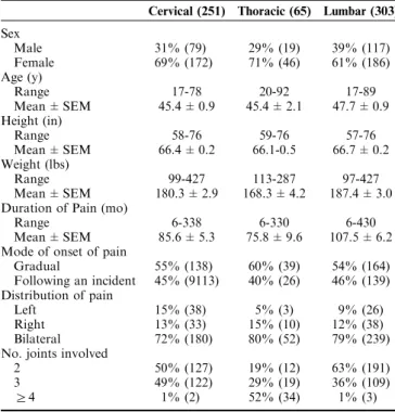
Salient features based on regional involvement of the spine are illustrated in Table 2. There were no significant differences noted in any of the variables.
Results of Comparative Local Anesthetic Blocks
Table 3 illustrates the results of diagnostic blocks evaluating facet joint pain in all 3 regions. In the cervical spine, lidocaine blocks were performed in 251 patients, in 65 patients in the thoracic spine, and 303 patients in the lumbar spine. Of these, 175 of 251 patients in the cervical spine reported a definite response with a positive result to initial lidocaine blocks, whereas 38 of the 65 patients in the thoracic spine reported a positive result, and 150 of 303 patients with lumbar spinal pain reported positive results with a single lidocaine block.
Of the 175 patients positive for single blocks with lidocaine, 97 patients reported positive response to bupivacaine blocks in the cervical spine, whereas 22 of 38 patients in the thoracic spine positive for single lidocaine blocks were also positive with bupivacaine blocks. Eighty-three patients in the lumbar spine showed positive results with bupivacaine from 150 of the lidocaine positive patients.
The controlled comparative local anesthetic blocks with positive results using double local anesthetic blocks provided a prevalence rate of facet joint pain in patients with chronic neck pain of 39% (95% CI, 32%-45%); 34% (95% CI, 22%-47%) in patients with chronic thoracic pain; and 27% (95% CI, 22%-33%) in patients with chronic low back pain.
Table 3 also illustrates false-positive rates. False-positive rates were calculated by assuming all patients with no response to lidocaine to be true negative, and all patients with a positive response to lidocaine and a negative response to bupivacaine or positive response to both lidocaine and bupivacaine as false-positives. The false-positive rates for cervical facet joints with a single block were 45% (95% CI, 37%-52%), for thoracic facet joints with a single lidocaine block were 42% (95% CI, 26%-59%), and for lumbar spine 45% (95% CI, 36%-53%).
Characteristics of Regional Involvement
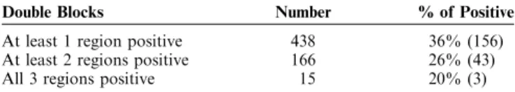
Table 4 illustrates facet joint involvement by region on the basis of controlled comparative local anesthetic blocks. Overall, at least one region was judged to be positive in 156 patients or 36% of the 438 patients. However, in 25% of the 181 patients were positive among patients with involvement of 2 and 3 regions.
Adverse Effects
No major adverse effects were noted in this study.
DISCUSSION
This retrospective evaluation of patients with chronic nonspecific spinal pain involving the cervical,
thoracic, and lumbar regions, alone or in combination, demonstrated by spinal region that the prevalence of cervical facet or zygapophysial joint pain in patients with neck pain is 39%, thoracic facet joint pain in patients with mid back or upper back pain is 34%, and involvement of lumbar facet joints in patients with chronic low back pain is 27%. The prevalence will be reduced if it is calculated on the basis of the total number of patients evaluated for the entire spine rather than regional. Thus, these numbers show regional involvement and evaluation of the patients for that particular region. These results are in overall agreement with the previous studies, although somewhat less than the ones published by the same group.21
The false-positive rates were quite high with single blocks: 45% for the cervical spine, 42% for the thoracic spine, and 45% for the lumbar spine. Once again, the results are similar to previous findings, even though there are some differences with the same group of authors with the results published in a prospective evaluation.21,25,43–52
Eligible PatientsAssessed 500 Patients included for evaluation= 438 Involvement of one region = 272 Involvement of two regions = 151 Involvement of three regions = 15 Lumbar pain = 303 Cervical pain = 251 Thoracic pain = 65 Patients included in
analysis = 303 Patients included in analysis = 251
Patients included in analysis = 65
Follow-up not available after 1 block = 4 Follow-up not available after 2 block = 10 Total = 14
Follow-up not available after 1 block = 8 Follow-up not available after 2 block = 12 Total = 20
Follow-up not available after 1 block = 3 Follow-up not available after 2 block = 2 Total = 5
Patients not meeting inclusion criteria = 62 36 patients did not meet at least 6 months duration of pain criteria
4 patients were less than 18 years of age 12 patients have undergone surgical intervention within the past 6 months in cervical, thoracic, or lumbar spine with disc procedures or vertebroplasty
10 patients had no information with evaluation for disc-related problems or had no information on conservative treatment prior to facet joint blocks
Results were considered negative = 4 Results were considered false-positive = 14
Results were considered negative = 8 Results were considered false-positive = 20
Results wereconsidered negative = 3 Results were considered false-positive = 5
FIGURE 1. Schematic representation of the review and patient flow.
TABLE 1. Demographic Characteristics
One Region (272) Two Regions (151) Three Regions (15) Total (438) Sex Male 45% (122) 26% (39) 33% (5) 38% (166) Female 55%*(150) 74%*(112) 67% (10) 62% (272) Age (y) Range 18-92 17-84 25-57 17-92 Mean ± SEM 48 ± 1.0 46 ± 1.2 42 ± 2.4 47 ± 0.7 Weight (lbs) Range 97-390 99-427 123-220 97-427 Mean ± SEM 187 ± 2.9 180 ± 4.1 173 ± 7.2 184 ± 2.3 Height (in) Range 57-76 58-76 60-72 57-76 Mean ± SEM 67.3w± 0.2 65.9 ± 0.3 65.1 ± 1.0 66.8 ± 0.2 *Significant difference compared to the male with involvement of 1 or 2 regions (P<0.01).
wSignificant difference compared to the group with involvement of 2 or 3 regions (P<0.05).
The study also demonstrated bilateral involvement in 72% of patients in the cervical spine, 80% in the thoracic spine, and 79% in the lumbar spine. Overall 77% of the patients presented with bilateral involvement. In addition, this study also showed involvement of multiple regions with 38% of the patients with involvement of more than one region.
This study once again reaffirms the evidence that involvement of facet joints, as a cause of chronic spinal pain is real as proven overwhelmingly by numerous studies. Facet joints have been shown to be a source of chronic spinal pain by means of diagnostic techniques of known reliability and validity.21,25,43–52,55–59Blocks of
facet joints are performed to test the hypothesis that the target joint is a source of the patient’s pain.55Once a facet
joint is anesthetized by the medial branches of the dorsal rami that innervate the target joint, if pain is relieved, the joint is considered to be the source of pain.55
However, true-positive responses are determined by performing controlled blocks, either in the form of placebo injection of normal saline or more commonly in the form of comparative local anesthetic blocks on 2 separate occasions, when the same joint is anesthetized using local anesthetics with different durations of action. The value and validity of medial branch blocks and comparative local anesthetic blocks in the diagnosis of facet joint pain has been demonstrated.21,25,43–52,55–67
In addition, because there are no clinical features or diagnostic imaging studies that can determine whether a facet joint is painful or not, controlled blocks seem to be a reliable tool in the diagnosis of chronic spinal pain.
The results noted in this study confirm the previous results, lending external validity to the primary findings. The study was performed in the setting of an interven-tional specialty practice in a private practice setting in the United States. Yet, it may be criticized that the authors performing the procedures in the retrospective review also was involved in the primary study. On the one hand, this may provide uniformity. On the other hand, it provides basis for the criticism that the results are the same. This criticism may be invalidated by the fact that the present retrospective study shows lesser prevalence of facet joint pain. Prevalence of facet joint pain was 55% in the cervical spine, 42% in the thoracic spine, and 31% in the lumbar spine compared with 39% in the present study in the cervical region, 34% in the thoracic region, and 27% in the lumbar region. This study also reinforces that the results of a retrospective review can provide valuable information similar to a prospective evaluation.
Even though the existence of facet joint pain is not universally accepted and the diagnostic methodology is not endorsed, it is the best available method to diagnose facet joint pain in chronic spinal pain. By confirming the results of the original research studies of diagnostic facet joint blocks, the results of the present study might encourage others to use appropriate diagnostic techniques in managing chronic spinal pain.
TABLE 2. Baseline Salient Features Based on Regional Involvement of the Spine
Cervical (251) Thoracic (65) Lumbar (303)
Sex Male 31% (79) 29% (19) 39% (117) Female 69% (172) 71% (46) 61% (186) Age (y) Range 17-78 20-92 17-89 Mean ± SEM 45.4 ± 0.9 45.4 ± 2.1 47.7 ± 0.9 Height (in) Range 58-76 59-76 57-76 Mean ± SEM 66.4 ± 0.2 66.1-0.5 66.7 ± 0.2 Weight (lbs) Range 99-427 113-287 97-427 Mean ± SEM 180.3 ± 2.9 168.3 ± 4.2 187.4 ± 3.0 Duration of Pain (mo)
Range 6-338 6-330 6-430
Mean ± SEM 85.6 ± 5.3 75.8 ± 9.6 107.5 ± 6.2 Mode of onset of pain
Gradual 55% (138) 60% (39) 54% (164) Following an incident 45% (9113) 40% (26) 46% (139) Distribution of pain Left 15% (38) 5% (3) 9% (26) Right 13% (33) 15% (10) 12% (38) Bilateral 72% (180) 80% (52) 79% (239)
No. joints involved
2 50% (127) 19% (12) 63% (191)
3 49% (122) 29% (19) 36% (109)
Z4 1% (2) 52% (34) 1% (3)
TABLE 3. Results of Single and Double Facet Joint Nerve Blocks (Single Blocks With Lidocaine and Double Blocks With Lidocaine and Bupivacaine)
Cervical (251) Thoracic (65) Lumbar (303) Double Blocks* Double Blocks* Double Blocks* Single Blocksw Positive Negative Positive Negative Positive Negative
Positive 97 78 22 16 83 67
Negative — 76 — 27 — 153
Prevalence 39% (95% CI, 32%-45%) 34% (95% CI, 22%-47%) 27% (95% CI, 22%-33%)
False-positive rate 45% (95% CI, 37%-52%) 42% (95% CI, 26%-59%) 45% (95% CI, 36%-53%)
*With double blocks, 97 patients with neck pain, 22 with thoracic pain, and 83 with lumbar pain had positive responses.
wWith single blocks in the cervical spine, 175 patients (ie, 97+78) had positive responses with lidocaine blocks, 38 (22+16) patients with thoracic pain had positive
CONCLUSIONS
This evaluation showed that patients with chronic, nonspecific spinal pain involving cervical, thoracic, and lumbar spine may be evaluated for the prevalence of facet joint pain. However, this should be performed with controlled, comparative local anesthetic blocks owing to the significant rate of false-positive response. Painful cervical facet joints were identified in 39% of the patients in the cervical spine, 34% of the patients in the thoracic spine, and 27% of the patients in the lumbar spine.
ACKNOWLEDGMENTS
The authors thank Tonie Hatton and Diane Neihoff, transcriptionists; Kim S. Damron, RN and Carla D. McManus, RN, BSN, clinical coordinators; and Doris E. Brandon, CST; for their assistance in preparation of this manuscript.
REFERENCES
1. Boswell MV, Shah RV, Everett CR, et al. Interventional techniques in the management of chronic spinal pain: evidence-based practice guidelines.Pain Physician.2005;8:1–47.
2. Elliott AM, Smith BH, Hannaford PC, et al. The course of chronic pain in the community: results of a 4-year follow-up study. Pain. 2002;99:299–307.
3. Bressler HB, Keyes WJ, Rochon PA, et al. The prevalence of low back pain in the elderly. A systemic review of the literature.Spine. 1999;24:1813–1819.
4. Lawrence RC, Helmick CG, Arnett FC. Estimates of the prevalence of arthritis and selected musculoskeletal disorders in the United States.Arthritis Rheum.1998;41:778–799.
5. Cassidy JD, Carroll LJ, Coˆte´ P. The Saskatchewan Health and Back Pain Survey. The prevalence of low back pain and related disability in Saskatchewan Adults. Spine. 1998;23: 1860–1867.
6. Guo HR, Tanaka S, Halperin WE, et al. Back pain prevalence in US industry and estimates of lost workdays.Am J Public Health.1999; 89:1029–1035.
7. Coˆte´ P, Cassidy JD, Carroll LJ. The Saskatchewan Health and Back Pain Survey. The prevalence of neck pain and related disability in Saskatchewan adults.Spine.1998;23:1689–1698.
8. Linton SJ, Hellsing AL, Hallden K. A population based study of spinal pain among 35–45-year old individuals. Spine. 1998;23: 1457–1463.
9. Miemelainen R, Videman T, Battie MC. Prevalence and character-istics of upper or mid-back pain in Finnish men. Spine. 2006;31:1846–1849.
10. Cassidy JD, Coˆte´ P, Carroll LJ, et al. Incidence and course of low back pain episodes in the general population. Spine. 2005;30: 2817–2823.
11. Hestbaek L, Leboeuf-Yde C, Manniche C. Low back pain: what is the long-term course? A review of studies of general patient populations.Eur Spine J.2003;12:149–165.
12. Croft PR, Lewis M, Papageorgiou AC, et al. Risk factors for neck pain: a longitudinal study in the general population.Pain.2001;93:317–325. 13. Coˆte´ P, Cassidy JD, Carroll LJ, et al. The annual incidence and course of neck pain in the general population: a population-based cohort study.Pain.2004;112:267–273.
14. Enthoven P, Skargren E, Oberg B. Clinical course in patients seeking primary care for back or neck pain: a prospective 5-year follow-up of outcome and health care consumption with subgroup analysis.Spine.2004;29:2458–2465.
15. Elders LA, Burdorf A. Prevalence, incidence, and recurrence of low back pain in scaffolders during a 3-year follow-up study. Spine. 2004;29:E101–E106.
16. Croft PR, Papageorgiou AC, Thomas E, et al. Short-term physical risk factors for new episodes of low back pain. Prospective evidence from the South Manchester Back Pain Study. Spine. 1999;24: 1556–1561.
17. Miedema HS, Chorus AMJ, Wevers CWJ, et al. Chronicity of back problems during working life.Spine.1998;23:2021–2028.
18. Thomas E, Silman AJ, Croft PR, et al. Predicting who develops chronic low back pain in primary care. A prospective study.Brit Med J.1999;318:1662–1667.
19. Yeung SS, Genaidy A, Deddens J, et al. Prevalence of musculoske-letal symptoms in single and multiple body regions and effects of perceived risk of injury among manual handling workers. Spine. 2002;27:2166–2172.
20. Manchikanti L, Pampati V. Research designs in interventional pain management: is randomization superior, desirable or essential?Pain Physician.2002;5:275–284.
21. Manchikanti L, Boswell MV, Singh V, et al. Prevalence of facet joint pain in chronic spinal pain of cervical, thoracic, and lumbar regions. BMC Musculoskelet Disord.2004;5:15.
22. Jacob T, Zeev A. Are localized low back pain and generalized back pain similar entities? Results of a longitudinal community based study.Disabil Rehabil.2006;28:369–377.
23. Bogduk N, McGuirk B. Causes and sources of chronic low back pain. In: Bogduk N, McGuirk B, eds.Medical Management of Acute and Chronic Low Back pain. An Evidence-Based Approach: Pain Research and Clinical Management. Amsterdam: Elsevier Science BV; 2002;13:115–126.
24. Bogduk N, McGuirk B. An algorithm for precision diagnosis. In: Bogduk N, McGuirk B, eds. Medical Management of Acute and Chronic Low Back pain. An Evidence-Based Approach: Pain Research and Clinical Management. Amsterdam: Elsevier Science BV; 2002; 13:177–186.
25. Manchikanti L, Singh V, Pampati V, et al. Evaluation of the relative contributions of various structures in chronic low back pain.Pain Physician.2001;4:308–316.
26. Kuslich SD, Ulstrom CL, Michael CJ. The tissue origin of low back pain and sciatica: a report of pain response to tissue stimulation during operation on the lumbar spine using local anesthesia.Orthop Clin North Am.1991;22:181–187.
27. Fukui S, Ohseto K, Shiotani M, et al. Referred pain distribution of the cervical zygapophyseal joints and cervical dorsal rami. Pain. 1996;68:79–83.
28. Dwyer A, Aprill C, Bogduk N. Cervical zygapophyseal joint pain patterns: a study in normal volunteers.Spine.1990;15:453–457. 29. Aprill C, Dwyer A, Bogduk N. The prevalence of cervical
zygapophyseal joint pain patterns II: a clinical evaluation. Spine. 1990;15:458–461.
30. Windsor RE, Nagula D, Storm S. Electrical stimulation induced cervical medial branch referral patterns. Pain Physician. 2003;6:411–418.
31. Dreyfuss P, Tibiletti C, Dreyer SJ. Thoracic zygapophyseal joint pain patterns: a study in normal volunteers.Spine.1994;19:807–811. 32. Fukui S, Ohseto K, Shiotani M. Patterns of pain induced by distending the thoracic zygapophyseal joints. Reg Anesth. 1997; 22:332–336.
33. Mooney V, Robertson J. The facet syndrome.Clin Orthop.1976; 115:149–156.
34. McCall IW, Park WM, O’Brien JP. Induced pain referral from posterior elements in normal subjects.Spine.1979;4:441–446. TABLE 4. Facet Joint Involvement by Region on the Basis
of Double Facet Joint Blocks
Double Blocks Number % of Positive
At least 1 region positive 438 36% (156)
At least 2 regions positive 166 26% (43)
35. Marks R. Distribution of pain provoked from lumbar facet joints and related structures during diagnostic spinal infiltration. Pain. 1989;39:37–40.
36. Fukui S, Ohseto K, Shiotani M, et al. Distribution of referred pain from the lumbar zygapophyseal joints and dorsal rami.Clin J Pain. 1997;13:303–307.
37. Windsor RE, King FJ, Roman SJ, et al. Electrical stimulation induced lumbar medial branch referral patterns.Pain Physician. 2002;5:347–353.
38. Bogduk N. The clinical anatomy of the cervical dorsal rami.Spine. 1982;7:319–330.
39. Bogduk N, Wilson AS, Tynan W. The human lumbar dorsal rami. J Anat.1982;134:383–397.
40. Chua WH, Bogduk N. The surgical anatomy of thoracic facet denervation.Acta Neurochir.1995;136:140–144.
41. Cavanaugh JM, Lu Y, Chen C, Kallakuri S. Pain generation in lumbar and cervical facet joints. J Bone Joint Surg Am. 2006; 88:63–67.
42. Merskey H, Bogduk N.Classification of Chronic Pain: Descriptions of Chronic Pain Syndromes and Definitions of Pain Terms. 2nd ed. Seattle: IASP Press; 1994.
43. Schwarzer AC, Aprill CN, Derby R, et al. Clinical features of patients with pain stemming from the lumbar zygapophysial joints. Is the lumbar facet syndrome a clinical entity? Spine. 1994;19:1132–1137.
44. Manchikanti L, Pampati V, Fellows B, et al. Prevalence of lumbar facet joint pain in chronic low back pain.Pain Physician.1999;2: 59–64.
45. Manchikanti L, Pampati V, Fellows B, et al. The diagnostic validity and therapeutic value of medial branch blocks with or without adjuvants.Curr Rev Pain.2000;4:337–344.
46. Schwarzer AC, Wang SC, Bogduk N, et al. Prevalence and clinical features of lumbar zygapophysial joint pain: a study in an Australian population with chronic low back pain. Ann Rheum Dis. 1995;54:100–106.
47. Manchikanti L, Singh V, Pampati V, et al. Evaluation of the prevalence of facet joint pain in chronic thoracic pain. Pain Physician.2002;5:354–359.
48. Barnsley L, Lord SM, Wallis BJ, et al. The prevalence of chronic cervical zygapophyseal joint pain after whiplash. Spine. 1995; 20:20–26.
49. Lord SM, Barnsley L, Wallis BJ, et al. Chronic cervical zygapo-physial joint pain with whiplash: a placebo-controlled prevalence study.Spine.1996;21:1737–1745.
50. Manchikanti L, Singh V, Rivera J, et al. Prevalence of cervical facet joint pain in chronic neck pain.Pain Physician.2002;5:243–249. 51. Sehgal N, Shah RV, McKenzie-Brown A, et al. Diagnostic utility
of facet (zygapophysial) joint injections in chronic spinal pain: a systematic review of evidence.Pain Physician.2005;8:211–224.
52. Boswell MV, Singh V, Staats PS, et al. Accuracy of precision diagnostic blocks in the diagnosis of chronic spinal pain of facet or zygapophysial joint origin: a systematic review. Pain Physician. 2003;6:449–456.
53. Barnsley L. Percutaneous radiofrequency neurotomy for chronic neck pain: outcomes in a series of consecutive patients.Pain Med. 2005;6:282–286.
54. National Institute for Clinical Excellence. Principles for Best Practice in Clinical Audit. Oxon, UK: Radcliffe Medical Press Ltd; 2002.
55. Bogduk N. International Spinal Injection Society guidelines for the performance of spinal injection procedures. Part 1: zygapophyseal joint blocks.Clin J Pain.1997;13:285–302.
56. Dreyfuss P, Schwarzer AC, Lau P, et al. Specificity of lumbar medial branch and L5 dorsal ramus blocks.Spine.1997;22:895–902. 57. Kaplan M, Dreyfuss P, Halbrook B, et al. The ability of lumbar
medial branch blocks to anesthetize the zygapophysial joint.Spine. 1998;23:1847–1852.
58. Lord SM, Barnsley L, Bogduk N. The utility of comparative local anesthetic blocks versus placebo-controlled blocks for the diagnosis of cervical zygapophysial joint pain.Clin J Pain.1995;11:208–213. 59. Lord S, Bogduk N. Comparative local anesthetic blocks in the
diagnosis of cervical zygapophysial joints pain. Pain. 1993;55: 99–106.
60. Schwarzer AC, Aprill CN, Derby R, et al. The false-positive rate of uncontrolled diagnostic blocks of the lumbar zygapophysial joints. Pain.1994;58:195–200.
61. Schwarzer AC, Derby R, Aprill CN, et al. The value of the provocation response in lumbar zygapophysial joint injections.Clin J Pain.1994;10:309–313.
62. Barnsley L, Lord S, Wallis B, et al. False-positive rates of cervical zygapophysial joint blocks.Clin J Pain.1993;9:124–130.
63. Barnsley L, Bogduk N. Medial branch blocks are specific for the diagnosis of cervical zygapophyseal joint pain. Reg Anesth.1993; 18:343–350.
64. Manchikanti L, Singh V, Pampati S. Are diagnostic lumbar medial branch blocks valid? Results of 2-year follow-up.Pain Physician. 2003;5:147–153.
65. Manchikanti L, Pampati V, Damron KS, et al. A randomized, prospective, double-blind, placebo-controlled evaluation of the effect of sedation on diagnostic validity of cervical facet joint pain. Pain Physician.2004;7:301–309.
66. Manchikanti L, Damron KS, Rivera J, et al. Evaluation of effect of sedation as a confounding factor in the diagnostic validity of lumbar facet joint pain: a prospective, randomized, double-blind, placebo-controlled evaluation.Pain Physician.2004;7:411–417.
67. Manchikanti L, Pampati V, Fellows B, et al. Influence of psychological factors on the ability to diagnose chronic low back pain of facet joint origin.Pain Physician.2001;4:349–357.